An MR conditional afterloader that can operate simultaneously with an MRI scanner could enable real-time high-dose-rate (HDR) brachytherapy source localization for treatment verification. Image guidance is critical for accurate and safe radiation dose delivery. Such an afterloader, which enables the patient to remain in the same position during dwell position reconstruction, treatment planning and irradiation, could increase the safety of HDR brachytherapy, as well as streamline clinical workflow.
Researchers at the University Medical Center Utrecht have developed a prototype MR conditional afterloader and a method for MR-based HDR brachytherapy source localization (Phys. Med. Biol. 63 085002). In their latest study, they perform a proof-of-concept demonstration showing that an MRI scanner and the prototype can function simultaneously without impacting each other’s performance (Int. J. Radiat. Oncol. Biol. Phys. 10.1016/j.ijrobp.2018.04.066).
This research, leading to eventual commercial availability of an MR conditional afterloader, could have a huge impact on HDR brachytherapy. A patient could remain in the MRI bore throughout the treatment process. MR-based localization could also be applied for a direct reconstruction of the source dwell positions after catheter insertion, using a dummy source. Upon completion of this treatment planning process, the dose could be delivered under MRI guidance.
A new design
Current commercial brachytherapy afterloaders interfere with and are affected by MR scanner operation. Radiofrequency (RF) signals generated by the scanner can impact the correct functioning and reliability of the afterloader. Its electronic system contains parts that are affected by the scanner’s strong magnetic field. In addition, RF signals generated by the afterloader can interfere with the RF signals emitted by nuclear spins of the object to be imaged. Finally, the steel source cable poses safety risks of RF-induced heating and torque, as well as acting as an RF antenna from inside the afterloader into the MR bore.
The prototype MR conditional afterloader, developed by Elekta in cooperation with researchers at the university’s department of radiotherapy and Image Sciences Institute, was RF-shielded with a copper/aluminium coating. The data cable connecting the afterloader with the treatment control station was shielded with a conductive hose and brought into contact with the Faraday cage of the MRI room.
A 2.65 m plastic test cable containing a piece of steel at its tip, which served as a dummy source, replaced the 1.4 m steel source cable. This enabled the source to be sent to the scanner’s isocentre while the afterloader remained at a sufficient distance outside the 20 mT line (which indicates the position where the magnet’s fringe field has a strength of 20 mT).
Proof-of-concept
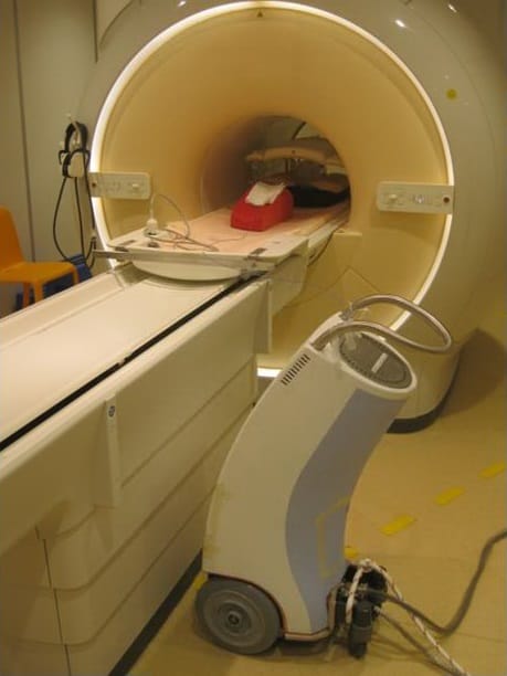
For the tests, the researchers placed a prototype afterloader next to a 1.5 T MRI scanner, 2.1 m from the isocentre. A plastic catheter placed inside a cylindrical Agar phantom via a prototype plastic transfer tube was connected to the afterloader. They conducted two tests with different programmed source positions and dwell times.
The researchers acquired MR images and performed localization of the dummy source for coronal and sagittal images, using a method based on MR artefact simulation and a phase correlation localization algorithm. They combined the two 2D positions into a 3D position of the dummy source and analysed the simultaneous functioning of the afterloader and the scanner. They also analysed RF interference, signal-to-noise ratio (SNR) and B0 homogeneity to evaluate the impact of the afterloader on the scanner’s performance.

Results of all tests showed that there was no deterioration of either system. The afterloader was able to send the dummy source to the predefined dwell positions through the catheter with fixed step size as the MR scanner was acquiring images. There was negligible RF interference, the SNR was not affected, and there were no significant distortions of the B0 magnetic field that would disturb the MRI performance.
A MR conditional source cable is required before the afterloader can be tested clinically. Lead author Ellis Beld tells Physics World that Elekta is developing such a cable. When this becomes available, they will conduct a patient study demonstrating the functioning of the MR conditional afterloader, to prove accurate treatment verification.
For now, the researchers are planning to test the source localization method as a first clinical test. They will insert a marker manually into a catheter in the prostate instead of using the afterloader and determine the position of this marker using the source localization algorithm.
“We also will need to perform safety tests, as well as all the tests required to obtain a CE Mark. However, we have now shown proof-of-concept of MR-based treatment verification of HDR brachytherapy with no degradation caused by either the MR scanner or the prototype MR conditional afterloader,” says Beld. “We believe that an MR conditional afterloader that enables source localization and treatment verification might have an enormous impact on the clinical workflow and practice of HDR brachytherapy.”


