
Proton therapy provides a means to target tumours with extreme precision. Steep dose gradients enable superior sparing of healthy tissue compared with photon-based treatments. But this steep dose fall-off also makes proton dose distributions highly sensitive to anatomical variations, patient set-up inaccuracies and tumour motion. As such, real-time image guidance during proton therapy delivery could prove invaluable.
One option could be to take the lead from photon radiotherapy and use MRI to visualize the tumour during treatment. At the recent ESTRO 38 conference in Milan, Aswin Hoffmann from OncoRay in Dresden took a look at the motivation for developing MR-integrated proton therapy and the current status of research efforts in this field.
“Image guidance in proton therapy is currently lagging behind image guidance in X-ray therapy,” Hoffmann explained, noting that proton therapy machines typically only have integrated 2D X-ray imaging. Some also have in-room CT or on-board cone-beam CT capability, but no in-room or on-board MRI is available. “Also, precise targeting is more important for proton therapy than X-ray therapy because of the steep dose gradient at the distal edge of the Bragg peak. This is why we still use relatively large margins and do not exploit the full dosimetric benefit of proton therapy.”
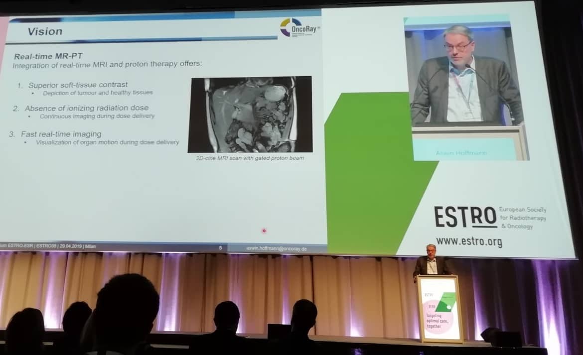
To address this, said Hoffmann, OncoRay has a vision to integrate real-time MRI with proton therapy. This would enable superior soft-tissue contrast for visualizing the tumour and surrounding healthy tissue. Meanwhile, the absence of ionizing radiation dose enables continuous real-time imaging during dose delivery. “Combining these two modalities could synchronize dose delivery with tumour position,” he pointed out.
Mutual impact
There are, however, many challenges when building such an integrated instrument, such as mutual electromagnetic interactions between the proton therapy and MRI systems, which may detrimentally impact the performance of each. Hoffmann first described the potential impact of the MR scanner’s magnetic field on the proton beam. Lorentz forces on the positively charged particles will deflect the beam from its straight trajectory, he explained.
Several research groups have used computer simulations to calculate the magnitude of this effect. And notably, recent measurements have validated these simulations. Hoffmann cited a study by OncoRay that used film dosimetry to determine beam deflection due to a magnetic field. Results showed that a 190 MeV beam deflects by about 1 cm when entering a 1T field, as predicted by the Monte Carlo simulations.
“We cannot ignore this effect, it has to be taken into account during treatment planning and dose delivery,” Hoffmann said. But because such dose effects can be accurately calculated using Monte Carlo simulations, he suggested that this represents a low risk.
And what about the effects of the magnets used for proton beam generation, transport and steering on MR image quality? Hoffmann shared the results of a magnetic survey performed in the experimental room at the Dresden proton therapy facility. This revealed that the cyclotron generates a magnetic gradient far smaller than the MR gradients, and can be compensated for by magnetic shimming of the MR scanner. Likewise, gantry rotation in the nearby therapy room causes small magnetic field effects that should not impact MR image quality.
The magnetic field of the beamline magnets, however, can be up to 100 µT, and could detrimentally affect the MR image quality. Thus, magnetic shielding may be necessary to enable simultaneous operation of the proton therapy and MRI systems.
Work in progress
To investigate these effects in more detail, OncoRay has built a prototype MR-integrated proton therapy system that integrates an open 0.22T MRI scanner with a horizontal fixed proton research beamline. Hoffmann and his team used the system to image the ACR small MRI knee phantom during proton beam irradiation.
When the beamline magnets to the gantry room were switched on during MR image acquisition, the image was distorted. But when the beamline magnets were switched on prior to MR image acquisition, image quality parameters did not change significantly due to irradiation. There was only a small uniform image shift in frequency encoding direction that can be corrected for via adequate pre-scan RF calibration. “We found that is important to synchronize MR image acquisition and operation of the proton therapy facility,” Hoffmann explained.
MRI guidance is feasible for proton therapy

The next step, he told the delegates, will be to combine MR imaging with a pencil-beam scanning proton system, to echo the clinical situation. Here, additional technical challenges include investigating the effect of the pencil-beam scanning magnets on MR image quality, and determining the impact of the MR fringe field on the beam steering system.
Hoffmann also examined the possibility of using MR imaging for online range detection. He described an experiment using a proton pencil beam to irradiate a water phantom, with beam energies of 190, 200, 210 and 225 MeV at very high dose rates. At each beam energy, MR images of the irradiated phantom contained either a hyperintense line artefact that exhibited a clear “wobble” at the expected residual range or showed an MR signature that mimics the full shape of the pencil-beam dose distribution, depending upon the MR pulse sequence chosen.
“For the first time, we have shown that you can use MRI to visualize the proton beam range in liquid water,” Hoffmann said. “This could be a valuable tool to perform quality assurance for MR-integrated proton therapy because you can see the beam in real time.” Hoffmann and his group are currently investigating whether these results can be translated into a clinical application.


