
Alzheimer’s disease is the most common form of dementia, but early diagnosis is challenging as many of the disease biomarkers also occur as part of normal aging. Once patients start to exhibit symptoms, such as cognitive decline, they are likely already in the mid to late stages of the disease.
A collaboration headed up at City University of Hong Kong (CityU) and Johns Hopkins University has now developed an MRI-based technique that dynamically monitors changes in cerebral glucose levels. Abnormal glucose uptake and clearance in the brain’s lymphatic system are a hallmark of early Alzheimer’s disease. As such, the team aims to apply this approach to diagnose Alzheimer’s at an early stage, enabling early intervention to halt or slow down disease progression.
Dynamic imaging of brain glucose levels, using a technique called dynamic glucose-enhanced (DGE) MRI, can provide information on glucose delivery, tissue transport and metabolism. To ease translation into clinical diagnosis, the method must be compatible with clinical MRI scanners with 3T field strength. To achieve this, the team designed an adjusted on-resonance variable delay multiple pulse (onVDMP) MRI approach, which can simultaneously measure dynamic glucose response curves for both brain parenchyma and cerebrospinal fluid (CSF) on a 3T MRI animal scanner.
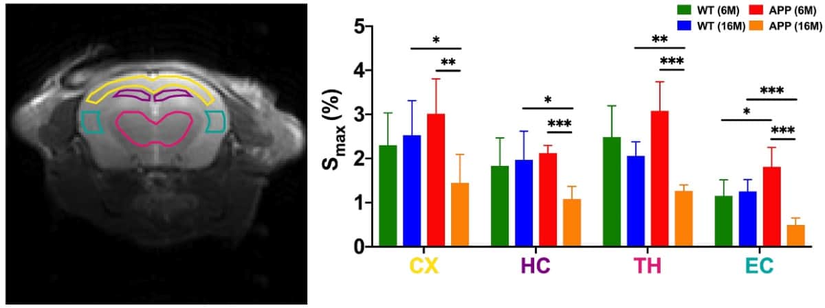
Using the 3T MRI animal scanner at CityU, the researchers tested their technique on six-month-old and 16-month-old transgenic mice with Alzheimer’s disease (AD), and healthy age-matched wild-type mice. Following a single injection of glucose, they used DGE MRI to monitor glucose uptake and clearance in the animals’ brain parenchyma (neurons and glial cells) and CSF.
In the younger mice, the team observed substantially higher glucose uptake in brain parenchyma in AD mice than in healthy mice. In the 16-month-old mice, the MRI results revealed notably lower glucose uptakes in both brain parenchyma and CSF of AD mice compared with age-matched healthy animals. In this older group, four typical brain regions studied (cerebral cortex, hippocampus, thalamus and entorhinal cortex) also had significantly lower maximum glucose uptake values after glucose injection.
Looking at glucose clearance, the team found that both young and old AD mice had significantly slower CSF clearance rates than age-matched healthy mice. This finding is consistent with recent reports of hampered CSF clearance, which leads to protein accumulation in the brain, and is attributed to abnormalities in the brain’s drainage system.
The team suggests that this reduced CSF clearance detected by non-invasive DGE could serve as an imaging biomarker to reveal the early pathology of Alzheimer’s disease, especially as its features are distinct from normal aging.
Eye scan can detect signs of Alzheimer’s disease
“By using glucose as a ‘tracer’, our imaging method can sensitively detect the distinctive changes of glymphatic system function at the molecular level at an early stage of the disease, helping us to differentiate [Alzheimer’s disease] from normal ageing,” says corresponding author Kannie Chan from CityU. “Besides, glucose is natural, biodegradable and commonly used in hospitals, such as the glucose tolerance test. Using it as a contrast agent for MRI is non-invasive and safe.”
The team published the results in Science Advances.


