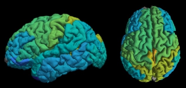
Aviv Mezer and colleagues at the Hebrew University of Jerusalem have developed a mathematical model that compares the water content of the brain with measures of physical properties obtained from MRI scans to calculate the molecular composition of lipids in the brain. They say that this technique could detect changes in the biological makeup of the brain over time and identify key changes associated with diseases like Alzheimer’s (Nature Communications 10.1038/s41467-019-11319-1).
MRI scans are a non-invasive way for doctors to view a patient’s brain and check for disease-related changes. The information provided is limited, however, as it only provides images of the brain tissues with little information on their molecular composition.
“When we take a blood test, it shows us the exact number of white blood cells in our body and whether that number is higher than normal due to illness. MRI scans provide images of the brain but don’t show changes in the composition of the human brain, changes that could potentially differentiate normal aging from the beginnings of Alzheimer’s or Parkinson’s,” explains Shir Filo, a co-author on the latest research.
To address this, the team turned to a technique known as quantitative MRI (qMRI).
MRI scanners use powerful magnets to align the protons (hydrogen atoms) in water molecules. Radiofrequency waves then excite the protons, which as they relax, produce weak radiofrequency signals that the scanner can detect. Images are created based on the varying relaxation times between different tissue types. qMRI also measures and records physical properties such as relaxation time, magnetization transfer rate and the water fraction. This information has been shown to be linked to biophysical properties of brain tissue.
To see whether the composition of lipids in brain tissue could affect these physical parameters, and therefore be measured by them, Mezer and colleagues conducted MRI scans on model lipid mixtures containing common brain lipids. This revealed unique relaxation signatures for different lipids. Next, they validated their results with MRI measurements and post-mortem data on the lipid composition of different brain tissues from a previous study.

“We found that many qMRI physical parameters are very sensitive to the water fraction and also affected by the environment,” Mezer tells Physics World. “In the brain, MRI is primarily a measurement of water hydrogen. We found that after accounting for this dependency – of the qMRI parameters on water fraction – we can characterize the interaction between water and the biological environment. In particular, we showed that changes in abundance of molecules such as membrane lipids can be monitored in this way using MRI.”
The researchers believe that their MRI technique could provide crucial insight into how our brains age. To test this, they conducted MRI scans on 23 adults in their late 20s and 18 adults in their late 60s and early 70s. In agreement with previous theories, they discovered distinct ageing patterns in different brain regions. They were able to identify region-specific patterns of molecular changes linked to brain ageing.
Mezer tells Physics World that the information they used can be obtained with current MRI machines and that calculating the molecular lipid signature from the qMRI measurements is relatively easy – the required code is available online. He adds that the main issue is the time that qMRI measurements take, compared with standard MRI scans. “Nowadays, there is a great effort in the MRI field to make it faster,” he says. “Nevertheless, time is a real issue for daily clinical use.”


