
Carbon-ion therapy is a promising radiotherapy technique that could enable dose escalation in tumours while reducing radiation dose to adjacent healthy tissue. Verification of the radiation dose after treatment delivery is key to optimizing treatment accuracy. To perform such dose verification, positron emission tomography (PET) of the positron emitters generated during irradiation is an increasingly popular approach, and currently the only method used clinically.
Researchers from Fudan University Shanghai Cancer Center and the Shanghai Proton and Heavy Ion Center have investigated the use of PET/CT imaging for in vivo 3D dose verification of carbon-ion irradiation. They determined that the tracer uptake had a roughly linear relationship with the effective dose, and suggest that this may prove a feasible clinical dose verification method, subject to additional research on the impact of blood circulation and tissue motion.
During irradiation, nuclear interactions between the carbon-ion beam and tissues in the patient generate positron emitters such as carbon-11 (11C), which has a half-life of approximately 20 minutes, mainly within the range of the Bragg peak. The distribution and radioactivity of these positron emitters can be detected by PET. The team hypothesized that this distribution should correspond to the prescription dose of the treatment plan.
For their study, reported in Frontiers in Oncology, first author Lining Sun and colleagues used a CT simulator to acquire a planning image of a Rando anthropomorphic head phantom. They then created three cubes in the treatment planning system, with volumes of 1, 4 and 10 ml, to represent different clinical target volumes for chordoma patients. Each target was prescribed six different doses, of between 2.5 and 10 Gy.
In total, the researchers created 18 virtual treatment plans for the various target volume and dose combinations. Dose–volume histograms created for each plan showed that a 100% prescription dose covered more than 95% of each virtual target volume.
The researchers delivered the plans to the phantom using raster scanned carbon-ion pencil beams with energies ranging from 174.5 to 248.6 MeV. After each of the 18 irradiations, they acquired PET/CT verification images of the head phantom within six minutes, to achieve a high signal-to-noise ratio. They then fused the treatment planning CT image with the PET/CT image.
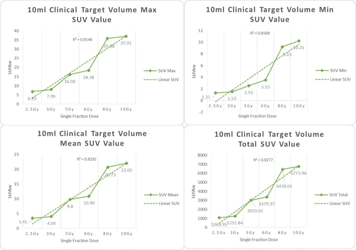
The team measured the maximum, minimum, average and total standardized uptake values (SUVs) for the three target sites after the various doses of carbon-ion irradiation. Histograms showing the SUVs for each of the six delivered doses revealed that the same effective dose generated statistically similar tracer uptake values in the different target volumes.
The findings indicate that the SUV in all targets had an approximately linear relationship with the effective dose. The team surmise that the SUV on a PET/CT image could be used for quantitative dose verification of carbon-ion therapy.
Simulated PET scans verify proton therapy delivery
“Accurate dose verification is an important prerequisite for the safe and effective implementation of radiation therapy,” the researchers conclude. “PET/CT after carbon-ion radiotherapy for clinical dose verification has been demonstrated to be a feasible method.”
The authors point out that their dose verification experiments were performed on a phantom, which did not replicate blood circulation or tissue motion. For use in patients, it will also be important to consider biological washout of the positron emitters, which can significantly affect the activity distribution.


