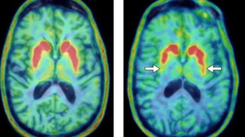
Researchers at the Wolfson Molecular imaging centre and the Sir Peter Mansfield Imaging Centre have reported on a new utility of the PET tracer florbetapir. They found that the tracer can distinguish frontotemporal dementia (FTD) from Alzheimer’s disease and the cognitively normal elderly. This ability removes the need for a separate scan to identify FTD, which can cause significant stress in such patients (Eur. J. Nucl. Med. Mol. Imaging 10.1007/s00259-018-4238-2).
FTD presents a unique diagnostic challenge. In particular, fibrous protein aggregates known as amyloid fibrils, which are closely associated with the onset and progression of Alzheimer’s and Parkinson’s disease, may or not be present in cases of FTD.
Current practice for distinguishing between different types of dementia usually requires two separate PET scans: a scan using a tracer such as florbetapir, which detects amyloid fibrils in Alzheimer’s disease, and a metabolism-sensitive scan using FDG. Patients with FTD often exhibit decreased cerebral blood flow, which can be detected using FDG.
Michael Asghar and colleagues tested whether the metabolic information could instead be obtained from a dual-phase florbetapir scan in patients with FTD, which would significantly reduce the patient burden. The researchers suspected that this metabolic information could be obtained from the activity distribution of florbetapir shortly after injection, and that the amyloid fibril information would become evident later.

The researchers re-examined data from a previous study (J. Nucl. Med. 10.2967/jnumed.114.147454), compiling PET scans from eight FTD patients, 10 cognitively normal controls and 10 patients with Alzheimer’s disease. Each subject had a florbetapir-PET scan and those with FTD received a second scan using FDG within 14 days.
Asghar and colleagues then compared the distribution of florbetapir during the first 2–5 minutes after injection with the FDG information and found significant correlation between the images. They also compared the early-stage florbetapir data from the FTD, cognitively normal and Alzheimer’s disease groups and found that analysis of the images by a clinical FDG expert matched the clinical diagnosis in 71% of cases.
“Early florbetapir frames provide important complementary diagnostic information to amyloid-beta deposition, shown by late florbetapir frames, which may be present not only in patients with Alzheimer’s disease, but also cognitively normal elderly and occasionally as secondary pathology in other neurodegenerative diseases, including FTD,” the researchers state.
This research places new value on the amyloid fibril biomarker, florbetapir, as a discriminative tool for neurodegenerative disorders.


