Mammography, the current gold standard for breast cancer screening, is a valuable but less than ideal imaging modality. The scans expose patients to X-ray radiation, are less sensitive in dense breast tissue and require breast compression – which can deter women from attending mammography appointments.
Now, researchers at Caltech Optical Imaging Laboratory have developed an alternative: a single-breath-hold photoacoustic computed tomography (PACT) system. The device, developed in the lab of Lihong Wang, can find tumours in as little as 15 s by shining pulses of near-infrared laser light into the breast (Nature Commun. 9 2352).
During a PACT scan, the incident light diffuses through the breast and is absorbed by haemoglobin in the patient’s red blood cells, causing the molecules to vibrate ultrasonically. These vibrations travel through the tissue and are detected by a 512-element ultrasonic transducer array. The recorded data are then used to construct an image of the breast’s internal structures.
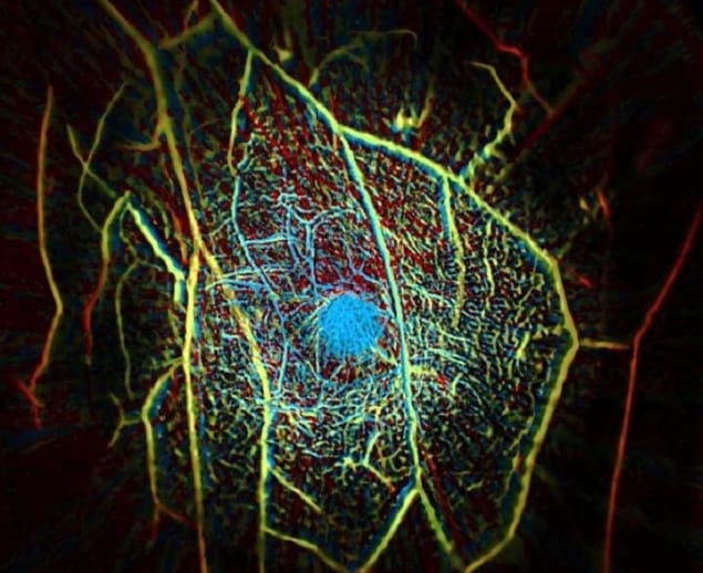
PACT creates images with a high in-plane spatial resolution of 255 µm, at a depth of up to 4 cm. Because the 1064 nm light is so strongly absorbed by haemoglobin, the images primarily show the blood vessels present in the tissue being scanned. This is useful for detecting cancer as many tumours induce the growth of new blood vessels, surrounding themselves with dense networks of vascular tissue.
A patient undergoing a PACT scan lies face down on a table with the breast to be imaged placed in a recess containing the ultrasonic sensors and laser. As the scan takes just 15 s, the patient can hold their breath while being scanned, resulting in a clearer image with negligible breathing-induced motion artefacts. “This is the only single-breath-hold technology that gives us high-contrast, high-resolution, 3D images of the entire breast,” says Wang.
In a pilot study, the team used PACT to image the breasts of one healthy volunteer and seven breast cancer patients. By assessing blood vessel density, PACT correctly identified eight of nine biopsy-verified breast tumours. Tumours were clearly revealed even in radiographically dense breasts, which could not be readily imaged by mammography.
Wang has founded a company to commercialize the PACT technology and conduct large-scale clinical studies. “Our goal is to build a dream machine for breast screening, diagnosis, monitoring and prognosis without any harm to the patient,” he says. “We want it to be fast, painless, safe and inexpensive.”


