Last week, the Institute of Physics in London hosted the meeting Physics-based Contributions to New Medical Techniques. A fascinating day of presentations saw 10 speakers describe physics technologies that enable a diverse range of medical applications, from biological and diagnostic imaging to radiation and proton therapies, as well as drug design and water purification.
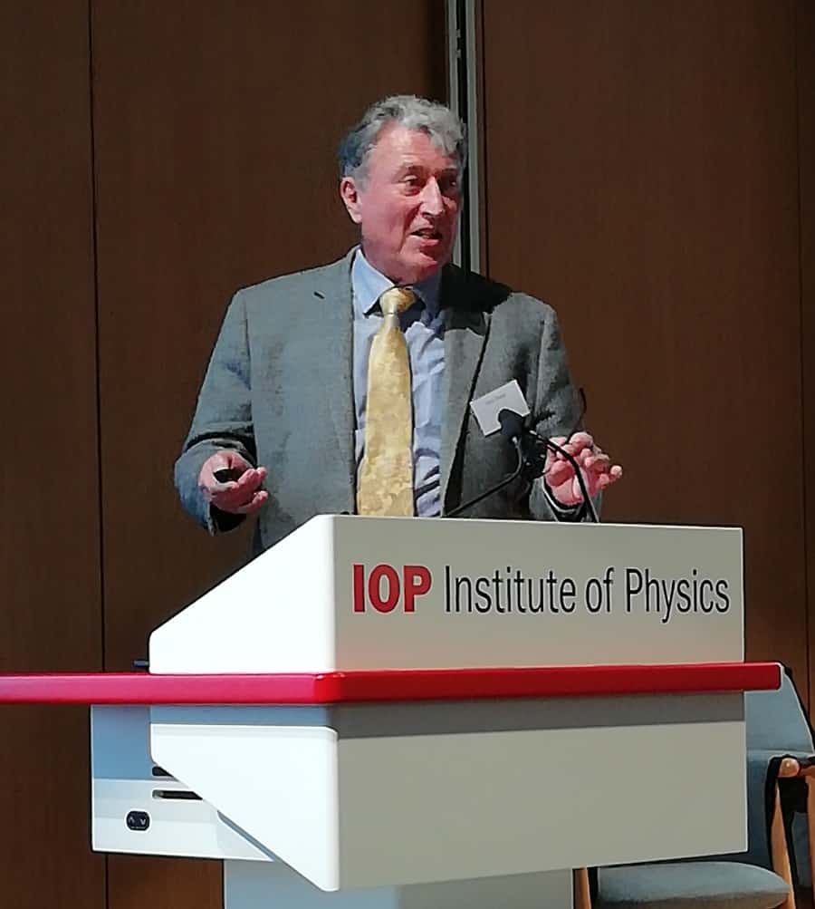
The first speaker, Xavier Golay from University College London, explained how MRI has evolved beyond simple anatomical imaging into a non-invasive method of choice for imaging physiology. Golay described how MR techniques can be used to measure brain metabolism. Perfusion imaging, for example, can assess cerebral blood flow, while blood oxygenation level dependent (BOLD) MRI can determine how much oxygen gets from the blood into the brain. Elsewhere, sodium imaging provides information on sodium levels in the brain, while in vivo MR spectroscopy can directly assess metabolites.
Golay described one project aiming to develop therapies for babies suffering from hypoxic ischemic encephalopathy, in which oxygen restriction damages the brain. The researchers found that the lactic acid signal in MR spectra provided the best predictive biomarker of outcome. And by performing lactate imaging on piglets, they found that melatonin could provide a potential neuroprotective agent.
The “holy grail of metabolic MR imaging”, says Golay, is direct assessment of glucose metabolism, as currently performed using FDG-PET. He explained that this could be achieved using a technique called chemical exchange saturation transfer (CEST) MRI of glucose, or glucoCEST, under development by the GLINT project, and presented some initial results using CEST to image gliomas.
Elekta’s Kevin Brown continued the theme of biomarkers, in this case, the potential for biomarker imaging using the Unity MR-linac. Brown began by describing the challenges of creating an integrated MRI-guided radiotherapy system and how its developers used physics to overcome problems that many thought insurmountable.
The Unity is now in clinical use at 14 sites worldwide, enabling users to adapt radiotherapy plans based on high-contrast soft-tissue imaging. In future, however, the system could also be used to image biomarkers that reveal the underlying biology of a patient’s tumour. Such information could be used, for example, to predict whether the patient will be a good or bad responder, or to tailor dose to their individual biology.
Brown explained that a radiotherapy session on the MR-linac, which currently involves scanning, adaptation and treatment, could also incorporate biomarker acquisition without adding to the treatment time or cost. Biomarker imaging could be performed during plan adaptation, for example, or even during radiation delivery. “I think imaging biomarkers have potential to make a massive change to the way we do radiotherapy,” he told the audience.
Radioimmunotherapy
Moving on to nuclear medicine, Steve Sugden from Harwell Consulting described a novel therapeutic radioisotope, along with a new way to create it. He noted that while 90% of nuclear medicine procedures are diagnostic, therapeutic applications – such as radioimmunotherapy (RIT), in which a radioactive payload is linked to a tumour-targeting biological agent – are on the increase.
Copper-67 is a beta emitter with ideal characteristics for therapy. It was used in some successful clinical trials in the late 1990s, but progress was stymied by the lack of availability. Copper-67 can be made on a research reactor or a very high-energy proton accelerator, but not with high purity, Sugden explained. As an alternative, his team is developing ways to create medical isotopes using a high-energy electron accelerator.
The technique involves bombarding an enriched zinc-68 target with high-energy photons to create copper-67. One big advantage of this approach, Sugden explained, is that the target and product are different elements and thus easy to separate. Using an enriched zinc target ups the yield five-fold (compared with a natural zinc target) and minimizes production of unwanted isotopes. He also noted that the extraction process enables target material recovery for reuse.
The isotope is now being produced by linacs at Idaho State University and Argonne National Laboratory, with production limited to about 2 Ci/day (enough to treat 10 patients). The team is also working with Clarity Pharmaceuticals to develop RIT for brain and prostate cancer, using copper-64 and copper-67 for diagnosis and treatment, respectively. This approach is currently in clinical trials in Australia.
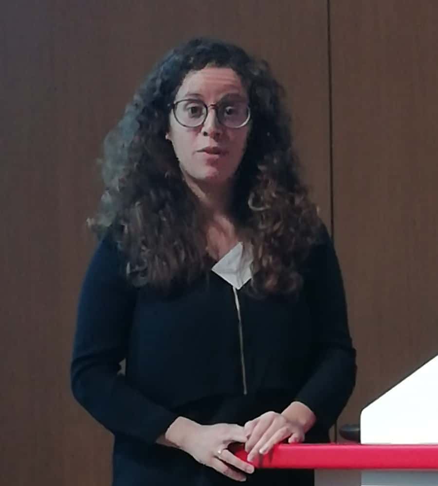
Next up, Catia Costa from the University of Surrey told delegates about the UK National Ion Beam Centre (UKNIBC), which provides ion beam technologies for use by researchers. She also introduced the RADIATE project that brings together several ion beam facilities around Europe. Costa described some of the biomedical projects being undertaken using ion beam analysis, including the use of PIXE (particle-induced X-ray emission) to identify unknown metal atoms within proteins and to determine the optimal approach for aerosol vaccine delivery.
Tackling bacteria
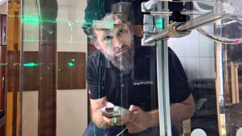
Kathryn Whitehead from Manchester Metropolitan University presented an entertaining, if slightly frightening, talk on the use of a microfluidic atmospheric-pressure plasma reactor to treat bacteria-contaminated water. “You think your water is clean? It’s not,” she declared, explaining that bacteria may lurk in pipes and taps, and emphasizing the particular danger if this occurs in a hospital setting.
Whitehead described tests demonstrating that the microfluidic plasma reactor could inactivate monocultures of E. Coli or Pseudomonas aeruginosa after a residence time of around 5 s. She noted, however, that it’s important to also study mixed bacterial samples, which may well be more resistant to plasma treatment. She also explained how bacteria remaining after such treatment may be in a small colony variant (SCV) or viable non-culturable (VBNC) state – persistent states that cause reinfections – and emphasized that this is a serious concern to consider in future studies.
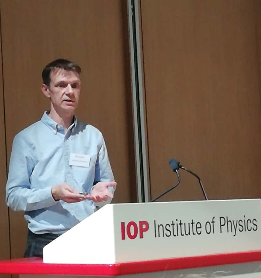
Rounding off the morning, Nick Lockyer from the University of Manchester described the use of nanoscale secondary ion mass spectrometry (nanoSIMS) to examine the intracellular distribution of the boron neutron capture therapy (BNCT) drug BPA. BNCT is a cancer treatment in which a drug containing boron-10 localizes within cancer cells, then the tumour is irradiated with low-energy neutrons that cause a fission reaction and create cell-killing alpha particles.
Lockyer and colleagues used nanoSIMS to examine boron-10 levels in biopsied human glioblastoma tumour cells cultured with BPA. They found that more BPA accumulated in cancer cells than in surrounding tissue, although both received a therapeutic BPA dose. They also saw that mean levels were slightly higher in cell nuclei than in cytoplasm, and that pre-treatment with tyrosine reduced BPA uptake in tumour, but not in surrounding cells. Lockyer also shared early results using SIMS to examine samples from two patients given BPA prior to tumour excision.
In the afternoon session, Ian Gilmore from the National Physical Laboratory explained how mass spectrometry can be used to help design better pharmaceuticals. In particular, he described how it can identify drug compounds that are unlikely to work by determining, for example, whether a drug reaches its target, binds to the target and, if so, whether it changes the behaviour of the disease. Gilmore presented the OrbiSIMS mass spectrometer, which enables 3D sub-cellular resolution imaging of metabolites and their response to potential drugs.
The remainder of the presentations focused on various aspects of particle therapy. Hywel Owen from the University of Manchester gave an introduction to UK proton therapy centres and described some novel projects aiming to improve the capabilities of such facilities. Simon Jolly from University College London described a new approach for proton therapy QA. And Dominik Perusko from Cosylab closed the day with a look at a next-generation dose delivery system for particle therapy. Keep an eye out for more detailed coverage of these talks coming up shortly on Physics World.