
Dehydration – defined as a total body water deficit – is associated with detrimental health outcomes across many populations. In patients admitted to hospital, dehydration can impair post-operative recovery and increases the risk of mortality by 80% on average. Elsewhere, soldiers are especially vulnerable to dehydration due to exposure to extreme environments, diminished thirst perception and limited availability of water.
But despite the serious consequences of unmanaged dehydration, there’s no quick and accurate diagnostic method available. Clinical symptoms are non-specific and easily confounded by other comorbidities, while existing diagnostic approaches require invasive bodily fluid sampling. To address this shortfall, researchers at Massachusetts Institute of Technology (MIT) are developing a portable MR sensor for non-invasive dehydration diagnosis (Magn. Reson. Med. 10.1002/mrm.28004).
“MRI provides an unparalleled non-invasive window into the body to study disease, physiology and fluid distribution,” says first author Ashvin Bashyam. “We felt that there was massive untapped clinical potential by bringing these systems out of centralized imaging facilities and towards the patient bedside and beyond.”
Measuring fluid shifts
Dehydration leads to relatively large volume depletion in the intramuscular fluid compartments (extracellular and intracellular), suggesting that measuring these tissue-specific fluid volumes could inform diagnosis. In particular, the extracellular fluid (ECF) compartment is highly responsive to fluid loss. The researchers propose that a portable MR sensor that measures ECF shifts could reliably identify clinically meaningful volume depletion.
To test this idea, Bashyam and colleagues examined fluid shifts in the skeletal muscle of dehydrated mice (exposed to high temperatures under fluid restriction) and control animals. They first used a benchtop NMR system to perform whole-animal multicomponent T2 relaxometry before and after dehydration, and showed that this MR measurement could identify dehydration.
Next, the researchers used a standard MRI scanner to perform multicomponent T2 relaxometry of skeletal muscle in the mice. They analysed the change in muscle ECF signal from before to after dehydration (to compensate for initial hydration variability) and observed significantly greater signal loss in dehydrated mice than in control animals. These findings demonstrate that the MR signal originating from muscle tissue alone is sufficient to identify and estimate the degree of dehydration.
Portable scanner
Using traditional MRI to diagnose dehydration is limited, however, by complexity and long measurement time, as well as the high cost and lack of portability of MR scanners. Thus, the team developed a portable, single-sided MR sensor – roughly 1000 cm3 in size and weighing 4 kg – that could perform comparable measurements.
“Traditional MRI systems involve a closed bore, so the sample must be placed inside the device where the magnetic fields are the strongest and most uniform,” explains Bashyam. “Our device utilizes a single-sided design, which means that the magnetic field is located outside of the sensor. This allows the sensor to be placed against a large sample and still perform a measurement, so the sensor can be miniaturized and highly portable.”
The sensor is based around a permanent magnet array that generates a static 0.28 T magnetic field. Permanent magnets benefit from low power consumption and far lower costs than superconducting coils used in conventional scanners. The resulting portable MR sensor can be used in environments including hospitals, outpatient facilities, sporting events and military operations.
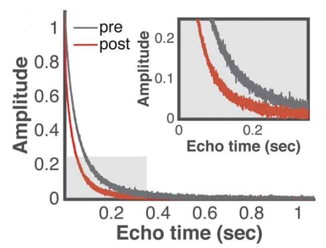
The team used the portable sensor to perform T2 relaxometry on skeletal muscle in the mouse leg, to characterize its ability to identify systemic fluid depletion in vivo. The relaxometry signal showed a decrease in decay time (from 67.1 to 45.4 ms) after dehydration, with the ECF component most responsive to fluid depletion.
Analysing the change in intramuscular ECF signal from before to after dehydration revealed significantly greater change in this signal among dehydrated versus control animals. The estimated change in fluid volume attributed to the ECF strongly correlated with weight loss in dehydrated mice, but not in the controls.
Bashyam says that the portable MR sensor can be adapted for use on human muscle tissue by increasing its size or applying novel pulse sequences to increase measurement depth. He also believes that, in future, it will be possible to measure dehydration changes from a single measurement through improved pulse sequence designs and establishing a range of normal hydration values.
“This work will move forward in two main directions,” Bashyam tells Physics World. “The first will be to develop more advanced sensors that can perform measurements more rapidly and deeper within tissue. The second will more robustly demonstrate the clinical utility of these sensors by testing them on healthy subjects as well as patients at nearby hospitals and medical centres. We are actively engaging with industrial partners to accelerate this work.”


