
A Raman spectroscopy guidance system integrated within a brain biopsy needle can help neurosurgeons identify malignant tissue for stereotactic biopsy sampling in real time. The ability to interrogate brain tissue at the tip of a biopsy needle, with minimal disruption to the surgical workflow, can help ensure that biopsy samples are collected in locations where cancer cell densities are high enough for reliable diagnoses. Such a system could help improve the diagnostic yield of biopsies and optimize treatment planning.
The guidance system was developed and validated by researchers from the Montreal Neurological Institute and Hospital of McGill University and the Polytechnique Montreal. The team successfully tested a second-generation Raman probe in the surgical biopsy procedure of one patient with glioblastoma and two patients with lymphoma. The probe was able to collect high-quality spectral data efficiently in a clinically acceptable time. These spectra contained the expected tissue features and exhibited similar spectral characteristics to in vivo and ex vivo spectra acquired previously under different experimental conditions (J. Biophotonics 10.1002/jbio.201800396).
The Raman probe
Raman spectroscopy is a non-destructive analysis technique that uses inelastic scattering of laser light to provide detailed information about a sample’s chemical structure. A Raman spectrum provides a distinct chemical fingerprint for a particular molecule or material. Raman spectroscopy instruments are used for surgical guidance, exhibiting high accuracy in identifying gliomas or metastases associated with colon, lung and skin cancer.
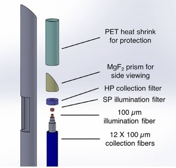
With an outer diameter of 900 μm, the Raman probe fits inside the internal cannula of a commercial biopsy needle. The probe consists of a central illumination fibre with a 100 μm core diameter surrounded by 12 collection fibres. The illumination fibre is coupled to a dual-wavelength spectrally stabilized laser that operates at 671 nm for high-wavenumber Raman spectroscopy (2000-4000 cm-1) and at 785 nm to probe the fingerprint region (500-2000 cm-1).
A navigation attachment positions the tip of the biopsy needle with reference to pre-operative MR images. A Y-shaped sealed plastic breakout enables the surgeon to create negative pressure inside the needle and obtain a tissue sample without removing the fibre-optic probe.
Clinical validation
For the clinical testing, co-principal investigator Frédéric Leblond and co-authors performed 13 Raman acquisitions in the three patients. They acquired 11 spectra in tumours and two spectra in normal brain tissue in the patients with lymphoma.
The researchers acquired spectral data from a single position on the target tissue, first recording a background spectrum with the laser off, followed by 10 measurements in both the fingerprint and high wavenumber regions with the laser turned on. The Raman system detects small wavelength shifts in the returning light, which create the spectral pattern. After processing the raw spectroscopic signal, the Raman spectra exhibited features associated with malignant and benign brain tissue.
The researchers compared the recorded spectra with 147 previously acquired in vivo spectra from 24 glioma patients, and with ex vivo acquisitions from a calf’s brain. They note that key Raman tissue bands were present for all measurements, suggesting that a classification model could be trained with one system and used for live classification with another system. They intend to develop an accurate statistical classification model to be used for real-time prediction of tissue type during a brain biopsy.
Leblond tells Physics World that the researchers have a new clinical study planned later in the spring, and that the probe will be commercialized by ODS Medical, headquartered in Montreal. He says the team is also testing the probe for use with lung and prostate cancers.


