
A new microscopy technique can image the mechanical properties of developing embryos at unprecedented speed while capturing spatial details on the scale of microns. The technique, which was developed by Robert Prevedel and colleagues at the European Molecular Biology Laboratory (EMBL) in Germany, does not damage delicate biological cells or tissues and could be used in several areas of research, including biomedicine, and cell and development biology.
The new technique relies on a phenomenon known as Brillouin scattering, which occurs when light interacts with the subtle sound vibrations (phonons, or collective vibrational modes) that are present in all matter. “As the scattered light interacts with the material, the light’s energy (that is, its colour/wavelength/frequency) and bandwidth are directly related to material properties such as elasticity and viscosity,” Prevedel explains. Monitoring the properties of the scattered light thus allows researchers to determine the properties of the material that scattered it.
Such information is important because the mechanical characteristics of biological cells and tissues are closely tied to their function. The physical forces that cells experience also play a critical role in processes such as embryo and tissue development and can even dictate how diseases like cancer evolve. Measuring these properties is no easy task, however, as most techniques for doing so are invasive and thus intrinsically disruptive.
This is where Brillouin scattering could come into its own. Indeed, researchers have already used it to observe and characterize tissue mechanics in a non-invasive way. The downside is that standard Brillouin microscopy relies on analysing one point in a sample at a time. This means that imaging speeds are slow, and the long light exposure times required can damage delicate biological cells.
An entire line of light at once
To overcome these problems, Prevedel and colleagues developed an improved method that increases the speed of imaging by a factor of more than 100. “We extended [the standard] approach from measuring from a single point at a time in a sample, as is currently the case, to measuring the same properties along an entire spatial line, that is, from more than 100 points in parallel,” he explains.
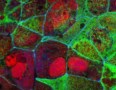
The researchers used this new approach to study mechanical changes in developing embryos in three animal species – fruit flies, mice and a marine organism with the scientific name Phallusia mammillata – over periods ranging from a few hours up to two days. Writing in Nature Methods, they describe how their technique, which they dub line-scanning Brillouin microscopy, can probe the sample to its entire depth in three dimensions with high resolution. This is in stark contrast to alternative techniques, which typically only probe the surface.
“Other techniques are also invasive as they need to exert forces on the biological samples being studied to measure their material properties,” Prevedel adds. “Brillouin microscopy overcomes these limits, and with our new approach, it has now become fast and gentle enough for longitudinal studies in biology and medicine,” he tells Physics World.
Brillouin scattering reveals tumour dynamics
The researchers suggest that line-scanning Brillouin spectroscopy could be employed in cell and development biology as well as biomedicine. Possible experiments might involve investigating the role of mechanics in morphogenesis during the development of embryos (as shown in this work) or studying the mechanics of cancer progression in artificially-grown organ-mimicking tissues known as organoids.
The EMBL team is now concentrating on improving the technique by, for example, increasing the weak interaction between the light and sound waves to generate more intense Brillouin scattering signals. “This will allow us to speed up imaging even further so that it is on a par with other (fluorescence) microscopy techniques,” Prevedel says. “We also need to measure other properties of our biological samples, such as their refractive index and material density, in three dimensions and at high resolution.”


