Microfluidic or “lab-on-a-chip” devices are commonly used to analyse blood and other fluid samples, which are pumped through narrow channels in a transparent chip the size of a postage stamp. These chips can be used to culture cells that are grown to mimic specific organ function and investigate the basic mechanisms of physiology and disease. For example, current efforts focus on studying how organs react to drugs and developing point-of-care diagnostic tools for various diseases. However, across these different applications, the design of the chip channels remains roughly the same, with innovation mainly coming from the composition of the surrounding cell layers or the addition of sensors.
Enter Khashayar Khoshmanesh and the interdisciplinary Mechanobiology & Microfluidics research group from the Royal Melbourne Institute of Technology (RMIT), who engineered a cavity the size of a grain of sand along the channel to enable new ways of analysing cells and particles (Adv. Funct. Mater. 10.1002/adfm.201901998).
Moulding a cavity
The team took advantage of gallium-based liquid metal alloys that can display properties of both metals and fluids. The researchers added droplets of the alloy Galinstan onto a chip that was then covered with the traditional polydimethylsiloxane (PDMS) layer and left to cure for 48 hours. Galinstan’s high surface tension means that it holds its form during the moulding process.

At the end of this process, they injected sodium hydroxide solution into the PDMS to release the Galinstan droplets and used a pipette to remove both the released droplets and the solution, producing an empty microfluidic structure with this novel cavity.
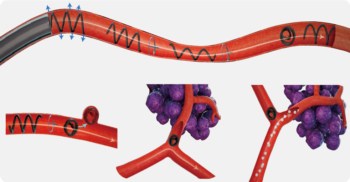
The spherical cavity left by the droplet acts as a mini-centrifuge — when a liquid is injected into the channel, it enters the cavity and spins around within. “This spinning creates a natural vortex, which just like a centrifuge machine in an analytics lab, spins the cells or other biological samples, allowing them to be studied without the need for capturing or labelling them,” reports Sara Baratchi, co-lead on the study.
The device only requires tiny samples, as little as 1 ml of water or blood, and can be used to study tiny bacterial cells measuring just 1 µm up to human cells as large as 15 µm.
Potential applications beyond blood sample analysis
The team performed numerical simulations to study the effects of various parameters — such as the size of cavity or flow rates — on the vortex magnitude and shear stress, as well on particles leaving the cavity to re-enter the channel. In this way, the researchers mimicked the response of blood cells under disturbed flow situations, similar to that found at junctions and curvatures of coronary and carotid arteries.
But mimicking human physiology on a chip is not the only potential application for this technology. It could also be used to identify parasites and other infections in waterways, especially in developing countries where options to detect and filter water impurities are scarce. “But with this device, the impurities will be captured and orbited by the vortex without any special sample preparation, saving time and money,” says Khoshmanesh.
Combined with a smartphone capable of capturing high-speed images, this new microfluidic device could prove to be a low-cost, self-sufficient and portable point-of-care diagnostic device.


