Nanoparticles deposited at tumour sites could enhance the effectiveness of radiation therapy by emitting UVC photons to augment cell killing by X-rays. The study, carried out by researchers in the US, reports that LuPO4:Pr3+-based radiosensitizers could reduce tumour recurrence and metastasis without increasing X-ray dose. Alternatively, such a system opens the door to similar tumour control with less radiation, to minimize damage to surrounding tissue (Radiother. Oncol. 10.1016/j.radonc.2018.06.016).
The scientists found that the local administered X-ray dose to human fibroblasts was effectively increased by a factor of at least two when cells had been incubated with LuPO4:Pr3+ nanoparticles. What’s more, the team point out that the resulting DNA damage in the irradiated cells is oxygen independent, which makes the approach particularly interesting for treating radio-resistant hypoxic tumours.
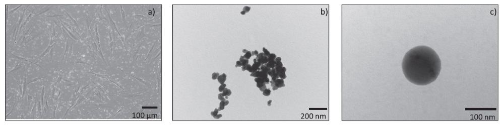
Although some agglomeration of the nanoparticles was detected, the majority of the material was sized in the range 50 – 200 nm and settled down in close proximity to the cells during the incubation period. The team adds that suspending the nanoparticles in complete cell culture medium with 10% serum was seen to reduce the agglomeration compared with suspending the material in water/phosphate-buffered saline.
Excitation of the nanomaterial by X-rays produced specific UV peaks between 220 and 285 nm to enhance cell lethality from X-ray irradiation. UV radiation produces two major types of DNA damage, cyclobutane dimers (CPDs) and 6–4 photoproducts, which can lead to permanent cell cycle arrest followed by cell inactivation.
In the study, the researchers confirmed the presence of such UV-induced DNA damage using a commercially available immunochemistry assay for CPDs.
Improved outcome
On its own, a 2 Gy dose of X-ray radiation decreased the surviving fraction of test cells to around 35%. However, when the same X-ray dose was applied to cells incubated with LuPO4:Pr3+ nanoparticles, the surviving fraction was reduced to as little as 2%.
As the researchers note, only cells in the immediate vicinity of the deposited nanomaterial are affected by the UVC-emission, as these photons are strongly absorbed within an extremely small distance of the LuPO4:Pr3+ particles, sparing normal tissue from exposure.
Pleased with the results of its bench testing, the group is keen to take its work further.
“We are currently making modifications to our nanoparticles to be able to use them in animals,” explains Martin Purschke, a researcher based at the Wellman Center for Photomedicine/Harvard Medical School. “This involves optimizing the size, shape and surface characteristics of the nanoparticles, and adding a coating and antibodies to make the particles more tumour-cell specific.”
The developments are designed to increase the efficacy of the formulation — enhancing its stability and longevity, for example — while lowering the risk of potential side effects.
Encouragingly, the team report no significant toxicity when low concentrations (less than 5 mg/ml) of LuPO4:Pr3+ nanoparticles were deposited in cell samples. In addition, no significant changes in toxicity were observed in fibroblasts for incubation periods up to 48 hours.
Once the scientists have succeeded in demonstrated the benefits in animal testing, they hope to start a clinical trial.