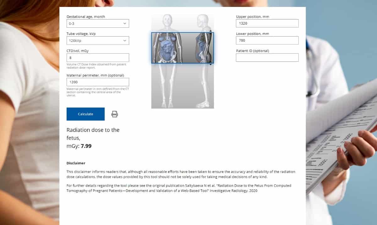
The number of CT examinations in pregnant patients has grown constantly during the past decades. But human embryos and foetuses are sensitive to ionizing radiation doses greater than 0.1 Gy. The potential health risks make it important to estimate radiation dose absorbed by the foetus when a pregnant woman has a CT scan. A free, web-based tool developed at the University Hospital Zurich’s Institute of Diagnostic and Interventional Radiology makes this calculation easier.
In addition to the type and technical parameters of the CT scan performed, the radiation dose absorbed by the foetus is also dependent upon the proximity of the uterus to the scanned body region, the anatomy of the patient and gestational age. Estimation tools exist, but because the developers consider these either expensive or difficult to use, they developed a user-friendly tool to make rapid foetal dose calculations, explaining their validation process in Investigative Radiology.

In this work, Hatem Alkadhi, together with medical physicist Natalia Saltybaeva and colleagues used hybrid computational phantoms to represent patients at the end of their third, sixth and ninth months of pregnancy. For each phantom, they simulated 47 axial CT scans with 15 mm width in the cranial direction to obtain radiation dose distributions from the upper chest to the lower pelvis.
To validate their findings, the researchers used patient-specific Monte Carlo (MC) dose simulations performed on 29 women who were imaged at two different hospitals using 64-slice GE Healthcare or 128-slice Siemens Healthineers CT scanners. The patients, aged between 20 and 42, and eight to 35 weeks pregnant, included 10 who underwent whole-body CT for polytrauma and 19 who had abdominal scans.
During the validation process, the researchers determined that the accuracy of their tool’s dose calculations could be improved by factoring in the maternal perimeter, defined from the CT section containing the central area of the uterus. With this addition, the average relative difference between foetal doses calculated by the computational algorithm and patient-specific MC simulations was just 11%.
The vendor-independent tool, accessible at www.fetaldose.org, can be used to calculate radiation dose exposure for any scanned body region, scan length and CT protocol. Its pull down-menu allows users to select gestational age and tube voltage. Additionally, users need to enter the volume CT dose index (CTDIvol), and select the upper and lower positions of the scan. The users can also add the maternal perimeter in millimetres to improve the accuracy of calculations, and the patient ID for their records. Calculations can be saved in PDF format to be added to the patient’s electronic health record.

Transabdominal oximetry offers non-invasive monitor of foetal health
“Comparing with previous literature and available tools in this field, our computational algorithm and web-based tools provide the advantages of realistic pregnant patient geometry, and the ability to take patient-specific size into account,” state the researchers. They say that high accuracy and the ability to make rapid calculations with only a few input parameters make the tool easy and appropriate for use in daily clinical routine.
“Our main goal was to make this tool as simple as possible, while guaranteeing a high accuracy of calculations,” Saltybaeva tells Physics World. “The current set of parameters requested by the tool for calculations can be easily provided by the referring physician. I think that this is our main advantage.”
She added that the tool potentially could be extended to other modalities, such as fluoroscopy, and that the research team may consider developing this in the future.


