When I was asked to review Unfinished Nature: Particle Physics at CERN, a new ethnography of CERN by Arpita Roy, an anthropologist at the University of California Berkeley, US, I was excited. Having recently completed a PhD in science communication where I studied the researchers at CERN – albeit from a very different, quantitative‐heavy, perspective – the subject is close to my heart.
Roy spent two and a half years doing fieldwork at CERN, around the time of the discovery of the Higgs boson in 2012. The book examines this event through an anthropological lens, asking questions such as how are scientific advances made and how do scientists understand their work? Unfortunately, although I read many books and papers of a similar nature for my doctoral studies, I struggled with Unfinished Nature.
A good book makes you pause to reflect. You may find yourself enlightened by the author’s perspectives or disagree with their arguments, but comprehension is key in either case. A book that has you stumbling through the pages without clarity, re-reading sentences over and over again in an effort to make sense of them, is frustrating. I may lack the expertise to appreciate the finer points of the subject, but I struggled despite repeated, earnest attempts to read the book with the care and attention the topic deserves.
Take the following snippet from the first page of the introduction, which sets the tone for what is to come: “But what has been lost to sight is the elucidation of how a science like particle physics may incorporate elements into its domain beyond what its epistemic assumption would lead us to expect, which deepens the mystery of what logic of classification it obeys. It is far from easy, however, to explicate the notion of classification, if only for the reason that it engenders notions of system, category, or context whose lucidity is hard to pinpoint in the scientific realm.” While I eventually understood (or at least think I did) what Roy is trying to say, the phrasing is unnecessarily convoluted.
None of this is criticism of Roy as a researcher but reflects the seemingly intentionally confusing language that academics – and my fellow social scientists in particular – are expected to use, despite increased calls to make research more accessible to those without specialist knowledge.
The ideas and stories Roy covers are no doubt interesting, even if the book itself isn’t an easy read. Unfinished Nature is more suited to the invested social scientist familiar with the particular flavour of academic prose adopted by anthropologists than physicists or physics enthusiasts indulging a more superficial interest in the lives of researchers at CERN.
Researchers at Google Quantum AI and collaborators have developed a quantum processor with error rates that get progressively smaller as the number of quantum bits (qubits) grows larger. This achievement is a milestone for quantum error correction, as it could, in principle, lead to an unlimited increase in qubit quality, and ultimately to an unlimited increase in the length and complexity of the algorithms that quantum computers can run.
Noise is an inherent feature of all physical systems, including computers. The bits in classical computers are protected from this noise by redundancy: some of the data is held in more than one place, so if an error occurs, it is easily identified and remedied. However, the no-cloning theorem of quantum mechanics dictates that once a quantum state is measured – a first step towards copying it – it is destroyed. “For a little bit, people were surprised that quantum error correction could exist at all,” observes Michael Newman, a staff research scientist at Google Quantum AI.
Beginning in the mid-1990s, however, information theorists showed that this barrier is not insurmountable, and several codes for correcting qubit errors were developed. The principle underlying all of them is that multiple physical qubits (such as individual atomic energy levels or states in superconducting circuits) can be networked to create a single logical qubit that collectively holds the quantum information. It is then possible to use “measure” qubits to determine whether an error occurred on one of the “data” qubits without affecting the state of the latter.
“In quantum error correction, we basically track the state,” Newman explains. “We say ‘Okay, what errors are happening?’ We figure that out on the fly, and then when we do a measurement of the logical information – which gives us our answer – we can reinterpret our measurement according to our understanding of what errors have happened.”
Keeping error rates low
In principle, this procedure makes it possible for infinitely stable qubits to perform indefinitely long calculations – but only if error rates remain low enough. The problem is that each additional physical qubit introduces a fresh source of error. Increasing the number of physical qubits in each logical qubit is therefore a double-edged sword, and the logical qubit’s continued stability depends on several factors. These include the ability of the quantum processor’s (classical) software to detect and interpret errors; the specific error-correction code used; and, importantly, the fidelity of the physical qubits themselves.
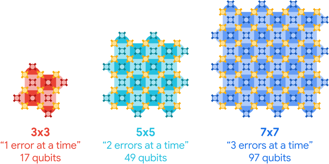
In 2023, Newman and colleagues at Google Quantum AI showed that an error-correction code called the surface code (which Newman describes as having “one of the highest error-suppression factors of any quantum code”) made it just about possible to “win” at error correction by adding more physical qubits to the system. Specifically, they showed that a distance-5 array logical qubit made from 49 superconducting transmon qubits had a slightly lower error rate than a distance-3 array qubit made from 17 such qubits. But the margin was slim. “We knew that…this wouldn’t persist,” Newman says.
“Convincing, exponential error suppression”
In the latest work, which is published in Nature, a Google Quantum AI team led by Hartmut Neven unveil a new superconducting processor called Willow with several improvements over the previous Sycamore chip. These include gates (the building blocks of logical operations) that retain their “quantumness” five times longer and a Google Deepmind-developed machine learning algorithm that interprets errors in real time. When the team used this new tech to create nine surface code distance-3 arrays, four distance-5 arrays and one 101-qubit distance-7 array on their 105-qubit processor, the error rate was suppressed by a factor of 2.4 as additional qubits were added.
How it works: Surface code logical qubits for processors of increasing size. Each larger processor can correct more errors than its predecessor. The encoded quantum state is stored on the array of data qubits (gold). Measure qubits (red, cyan, blue) check for errors on the neighbouring data qubits. (Courtesy: Google Quantum AI)
“This is the first time we have seen convincing, exponential error suppression in the logical qubits as we increase the number of physical qubits,” says Newman. “That’s something people have been trying to do for about 30 years.”
With gates that remain stable for hours on end, quantum computers should be able to run the large, complex algorithms people have always hoped for. “We still have a long way to go, we still need to do this at scale,” Newman acknowledges. “But the first time we pushed the button on this Willow chip and I saw the lattice getting larger and larger and the error rate going down and down, I thought ‘Wow! Quantum error correction is really going to work…Quantum computing is really going to work!’”
Mikhail Lukin, a physicist at Harvard University in the US who also works on quantum error correction, calls the Google Quantum AI result “a very important step forward in the field”. While Lukin’s own group previously demonstrated improved quantum logic operations between multiple error-corrected atomic qubits, he notes that the present work showed better logical qubit performance after multiple cycles of error correction. “In practice, you’d like to see both of these things come together to enable deep, complex quantum circuits,” he says. “It’s very early, there are a lot of challenges remaining, but it’s clear that – in different platforms and moving in different directions – the fundamental principles of error correction have now been demonstrated. It’s very exciting.”
People working in industry, biology and geology are all keen to understand when particles will switch from flowing like fluids to jamming like solids. With rigid particles, and even for foams and emulsions, scientists know what determines this crunch point: it’s related to the number of contact points between particles. But for squishy particles – those that deform by more than 10% of their size – that’s not necessarily the case.
“You can have a particle that’s completely trapped between only two particles,” explains Samuel Poincloux, who studies the statistical and mechanical response of soft assemblies at Aoyama Gakuin University, Japan.
Factoring that level of deformability into existing theories would be fiendishly difficult. But with real-world scenarios – particularly in mechanobiology – coming to light that hinge on the flow or jamming of highly deformable particles, the lack of explanation was beginning to hurt. Poincloux and his University of Tokyo colleague Kazumasa Takeuchi therefore tried a different approach. Their “easy-to-do experiment” sheds fresh light on how squishy particles respond to external forces, leading to a new model that explains how such particles flow – and at what point they don’t.
Pinning down the differences
To demonstrate how things can change when particles can deform a lot, Takeuchi holds up a case containing hundreds of rigid photoelastic rings. When these rings are under stress, the polarization of light passing through them changes. “This shows how the force is propagating,” he says.
As he presses on the rings with a flat-ended rod, a pattern of radial lines centred at the bottom of the rod lights up. With rigid particles, he explains, chains of forces transmitted by these contact points conspire to fix the particles in place. The fewer the contact points, the fewer the chains of forces keeping them from moving. However, when particles can deform a lot, the contact areas are no longer points. Instead, they extend over a larger region of the ring’s surface. “We can already expect that something will be very different then,” he says.
The main ingredient in Takeuchi and Poincloux’s experimental study of these differences was a layer of deformable silicone rings 10 mm high, 1.5 mm thick and with a radius of 3.3 mm, laid out between two parallel surfaces. The choice of ring material and dimensions was key to ensuring the model reproduced relevant aspects of behaviour while remaining easy to manipulate and observe. To that end, they added an acrylic plate on top to stop the rings popping out under compression. “There’s a lot of elastic energy inside them,” says Poincloux, nodding wryly. “They go everywhere.”
By pressing on one of the parallel surfaces, the researchers compressed the rings (thereby adjusting their density) and added an oscillating shear force. To monitor the rings’ response, they used image analysis to note the position, shape, neighbours and contact lengths for each ring. As they reduced the shear force amplitude or increased the density, they observed a transition to solid-like behaviour in which the rings’ displacement under the shear force became reversible. This transition was also reflected in collective properties such as calculated loss and storage moduli.
Unexpectedly simple
Perhaps counterintuitively, regular patterns – crystallinity – emerged in the arrangement of the rings while the system was in a fluid phase but not in the solid phase. This and other surprising behaviours make the system hard to model analytically. However, Takeuchi emphasises that the theoretical criterion for switching between solid-like and fluid-like behaviour turned out to be quite simple. “This is something we really didn’t expect,” he says.
The top row in the video depicts the fluid-like behaviour of the rings at low density. The bottom row depicts the solid-like behaviour of the rings at a higher density. (Courtesy: Poincloux and Takeuchi 2024)
The researchers’ experiments showed that for squishy particles, the number of contacts no longer matters much. Instead, it’s the size of the contact that’s important. “If you have very extended contact, then [squishy particles] can basically remain solid via the extension of contact, and that is possible only because of friction,” says Poincloux. “Without friction, they will almost always rearrange and lose their rigidity.”
Jonathan Bares, who studies granular matter at CNRS in the Université de Montpellier, France, but was not involved in this work, describes the model experiment as “remarkably elegant”. This kind of jamming state is, he says, “challenging to analyse both analytically and numerically, as it requires accounting for the intricate properties of the materials that make up the particles.” It is, he adds, “encouraging to see squishy grains gaining increasing attention in the study of granular materials”.
As for the likely impact of the result, biophysicist Christopher Chen, whose work at Boston University in the US focuses on adhesive, mechanical and biochemical contributions in tissue microfabrication, says the study “provides more evidence that the way in which soft particles interact may dominate how biological tissues control transitions in rigidity”. These transitions, he adds, “are important for many shape-changing processes during tissue assembly and formation”.
Full details of the experiment are reported in PNAS.
Medical physics techniques play a key role in all areas of cardiac medicine – from the use of advanced imaging methods and computational modelling to visualize and understand heart disease, to the development and introduction of novel pacing technologies. At a recent meeting organised by the Institute of Physics’ Medical Physics Group, experts in the field discussed some of the latest developments in cardiac imaging and therapeutics, with a focus on transitioning technologies from the benchtop to the clinic.
Monitoring metabolism
The first speaker, Damian Tyler from the University of Oxford described how hyperpolarized MRI can provide “a new window on the reactions of life”. He discussed how MRI – most commonly employed to look at the heart’s structure and function – can also be used to characterize cardiac metabolism, with metabolic MR studies helping us understand cardiovascular disease, assess drug mechanisms and guide therapeutic interventions.
In particular, Tyler is studying pyruvate, a compound that plays a central role in the body’s metabolism of glucose. He explained that 13C MR spectroscopy is ideal for studying pyruvate metabolism, but its inherent low signal-to-noise ratio makes it unsuitable for rapid in vivo imaging. To overcome this limitation, Tyler uses hyperpolarized MR, which increases the sensitivity to 13C-enriched tracers by more than 10,000 times and enables real-time visualization of normal and abnormal metabolism.
As an example, Tyler described a study using hyperpolarized 13C MR spectroscopy to examine cardiac metabolism in diabetes, which is associated with an increased risk of heart disease. Tyler and his team examined the downstream metabolites of 13C-pyruvate (such as 13C-bicarbonate and 13C-lactate) in subjects with and without type 2 diabetes. They found reduced bicarbonate levels in diabetes and increased lactate, noting that the bicarbonate to lactate ratio could provide a diagnostic marker.
Among other potential clinical applications, hyperpolarized MR could be used to detect inflammation following a heart attack, elucidate the mechanism of drugs and accelerate new drug discovery, and provide an indication of whether a patient is likely to develop cardiotoxicity from chemotherapy. It can also be employed to guide therapeutic interventions by imaging ischaemia in tissue and assess cardiac perfusion after heart attack.
“Hyperpolarized MRI offers a safe and non-invasive way to assess cardiac metabolism,” Tyler concluded. “There are a raft of potential clinical applications for this emerging technology.”
Changing the pace
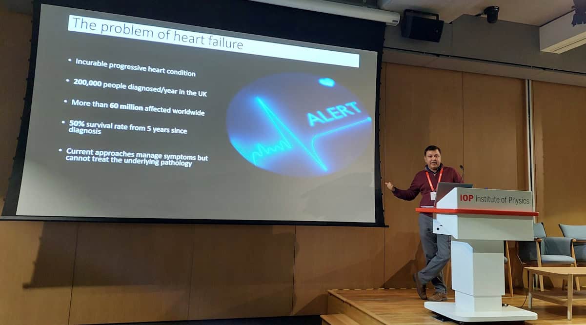
Alongside the introduction of new and improved diagnostic approaches, researchers are also developing and refining treatments for cardiac disorders. One goal is to create an effective treatment for heart failure, an incurable progressive condition in which the heart can’t pump enough blood to meet the body’s needs. Current therapies can manage symptoms, but cannot treat the underlying disease or prevent progression. Ashok Chauhan from Ceryx Medical told delegates how the company’s bio-inspired pacemaker aims to address this shortfall.
In healthy hearts, Chauhan explained, the heart rate changes in response to breathing, in a mechanism called respiratory sinus arrythmia (RSA). This natural synchronization is frequently lost in patients with heart failure. Ceryx has developed a pacing technology that aims to treat heart failure by resynchronizing the heart and lungs and restoring RSA.
Heart–lung synchronization Ashok Chauhan explained how Ceryx Medical’s bio-inspired pacemaker aims to improve cardiac function in patients with heart failure.
The device works by monitoring the cardiorespiratory system in real time and using RSA inputs to generate stimulation signals in real time. Early trials in large animals demonstrated that RSA pacing increased cardiac output and ejection fraction compared with monotonic (constant) pacing. Last month, Ceryx begun the first in-human trials of its pacing technology, using an external pacemaker to assess the safety of the device.
Eliminating sex bias
Later in the day, Hannah Smith from the University of Oxford presented a fascinating talk entitled “Women’s hearts are superior and it’s killing them”.
Smith told a disturbing tale of an elderly man with chest pain, who calls an ambulance and undergoes electrocardiography (ECG) that shows he is having a heart attack. He is rushed to hospital to unblock his artery and restore cardiac function. His elderly wife also feels unwell, but her ECG only shows slight abnormality. She is sent for blood tests that eventually reveal she was also having a severe heart attack – but the delay in diagnosis led to permanent cardiac damage.
The fact is that women having heart attacks are more likely to be misdiagnosed and receive less aggressive treatment than men, Smith explained. This is due to variations in the size of the heart and differences in the distances and angles between the heart and the torso surface, which affect the ECG readings used to diagnose heart attack.
To understand the problem in more depth, Smith developed a computational tool that automatically reconstructs torso ventricular anatomy from standard clinical MR images. Her goal was to identify anatomical differences between males and females, and examine their impact on ECG measurements.
Using clinical data from the UK Biobank (around 1000 healthy men and women, and 84 women and 341 men post-heart attack), Smith modelled anatomies and correlated these with the respective ECG data. She found that the QRS complex (the signal for the heart to start contracting) was about 6 ms longer in healthy males than healthy females, attributed to the smaller heart volume in females. This is significant as it implies that the mean QRS duration would have to increase by a larger percentage for women than men to be diagnosed as elevated.
She also studied the ST segment in the ECG trace, elevation of which is a key feature used to diagnose heart attack. The ST amplitude was lower in healthy females than healthy males, due to their smaller ventricles and more superior position of the heart. The calculations revealed that overweight women would need a 63% larger increase in ST amplitude to be classified as elevated than normal weight men.
Smith concluded that heart attacks are harder to see on a woman’s ECGs than on a man’s, with differences in ventricular size, position and orientation impacting the ECG before, during and after heart attacks. Importantly, if these relationships can be elucidated and corrected for in diagnostic tools, these sex biases can be reduced, paving the way towards personalised ECG interpretation.
Prize presentations
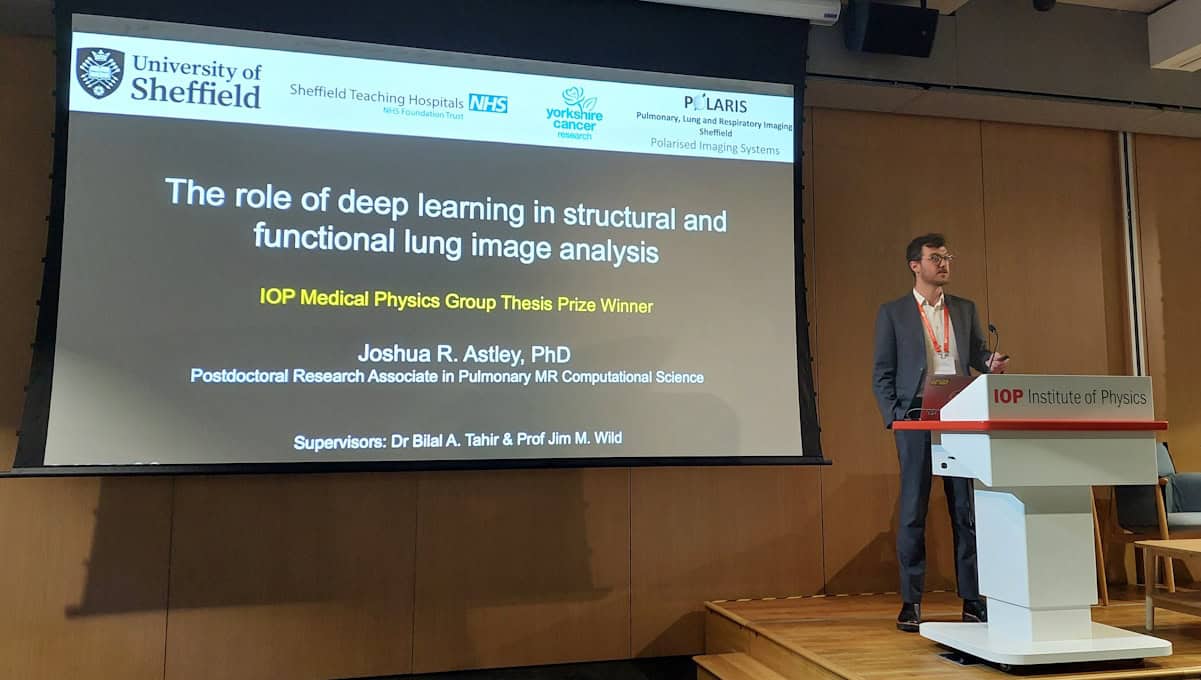
The meeting also included a presentation from the winner of the 2023 Medical Physics Group PhD prize: Joshua Astley from the University of Sheffield, for his thesis “The role of deep learning in structural and functional lung imaging”.
Prize presentation Joshua Astley from the University of Sheffield is the winner of the 2023 Medical Physics Group PhD prize.
Shifting the focus from the heart to the lungs, Astley discussed how hyperpolarized gas MRI, using inhaled contrast agents such as 1He and 129Xe, can visualize regional lung ventilation. To improve the accuracy and speed of such lung MRI studies, he designed a deep learning system that rapidly performs MRI segmentation and automates the calculation of ventilation defect percentage via lung cavity estimates. He noted that the tool is already being used to improve workflow in clinical hyperpolarized gas MRI scans.
Astley also described the use of CT ventilation imaging as a potentially lower-cost approach to visualize lung ventilation. Combining the benefits of computational modelling with deep learning, Astley and colleagues have developed a hybrid framework that generates synthetic ventilation scans from non-contrast CT images.
Quoting some “lessons learnt from my thesis”, Astley concluded that artificial intelligence (AI)-based workflows enable faster computation of clinical biomarkers and better integration of functional lung MRI, and that non-contrast functional lung surrogates can reduce the cost and expand use of functional lung imaging. He also emphasized that quantifying the uncertainty in AI approaches can improve clinician’s trust in using such algorithms, and that making code open and available is key to increasing its impact.
The day rounded off with awards for the meeting’s best talk in the submitted abstracts section and the best poster presentation. The former was won by Sam Barnes from Lancaster University for his presentation on the use of electroencephalography (EEG) for diagnosis of autism spectrum disorder. The poster prize was awarded to Suchit Kumar from University College London, for his work on a graphene-based electrophysiology probe for concurrent EEG and functional MRI.
Imagine a smartphone that charges faster, lasts longer and is more eco-friendly – all at a lower cost. Aluminium-ion batteries (AIBs) could make this dream a reality, and scientists are working to unlock their potential as a more abundant, affordable and sustainable alternative to the lithium-ion batteries currently used in mobile devices, electric cars and large-scale energy storage. As part of this effort, Dmitrii A Rakov and colleagues at the University of Queensland, Australia recently overcame a technical hurdle with an AIB component called the solid-electrolyte interphase. Their insights could help AIBs match, or even surpass, the performance of their lithium-ion counterparts.
Like lithium-ion batteries, AIBs contain an anode, a cathode and an electrolyte. This electrolyte carries aluminium ions, which flow between the positively-charged anode and the negatively-charged cathode. During discharge, these ions move from the anode to the cathode, generating energy. Charging the battery reverses the process, with ions returning to the anode to store energy.
The promise and the problem
Sounds simple, right? But when it comes to making AIBs work effectively, this process is far from straightforward.
Aluminium is a promising anode material – it is lightweight and stores a lot of energy for its size, giving it a high energy density. The problem is that AIBs are prone to instabilities as they cycle between charging and discharging. During this cycling, aluminium can deposit unevenly on the anode, forming tree-like structures called dendrites that cause short circuits, leading to battery failure or even safety risks.
Researchers have been tackling these issues for years, trying to figure out how to get aluminium to deposit more evenly and stop dendrites from forming. An emerging focus of this work is something called the solid-electrolyte interphase (SEI). This thin layer of organic and inorganic components forms on the anode as the battery charges, and like the protective seal on a jar of jam, it keeps everything inside fresh and functioning well.
In AIBs, though, the SEI sometimes forms unevenly or breaks, like a seal on a jar that doesn’t close properly. When that happens, the aluminium inside can misbehave, leading to performance issues. To complicate things further, the type of “jam” in the jar – different electrolytes, like chloroaluminate ionic liquids – affects how well this seal forms. Some electrolytes help create a better seal, while others make it harder to keep the aluminium deposits stable.
Cracking the code of aluminium deposition
In their study, which is published in ACS Nano, the Queensland scientists, together with colleagues at the University of Southern Queensland and Oak Ridge National Laboratory in the US, focused on how the aluminium anode interacts with the liquid electrolyte. They found that the formation of the SEI layer is highly dependent on the current running through the battery and the type of counter electrode (the “partner” to the aluminium anode). Some currents and conditions allow the battery to work well for more cycles. But under other conditions, aluminium can build up in uneven, dendritic structures that ultimately cause the battery to fail.
Work in progress: Assembling a cell for testing. (Courtesy: Dmitrii Rakov)
To understand how this happens, the researchers investigated how different electrolytes and cycling conditions affect the SEI layer. They discovered that in some cases, when the SEI isn’t forming evenly, aluminium oxide (Al2O3) – which is normally a protective layer – can actually aggravate the problem by causing the aluminium to deposit unevenly. They also found that low currents can deplete some materials in the electrolyte, leading to parasitic reactions that further reduce the battery’s efficiency.
To solve these issues, the scientists recommend exploring different aluminium-alloy chemistries. They also suggest that specific conditioning protocols could smooth out the SEI layer and improve the cycling performance. One example of such a conditioning protocol is pre-cycling, which is a process where the battery is charged and discharged in a controlled way before regular use to condition it for better long-term performance.
“Our research demonstrates that, like in lithium-ion batteries, aluminium-ion batteries also need pre-cycling to maximize their lifetime,” Rakov tells Physics World. “This is important knowledge for aluminium-ion battery developers, who are rapidly emerging as start-ups around the world.”
By understanding the unique pre-cycling needs of aluminium-ion batteries, developers can work to design batteries that last longer and perform more reliably, bringing them closer to real-world applications.
How far are we from having an aluminium-ion battery in our mobile phones?
As for when those applications might become a reality, Rakov highlights that AIBs are still in the early stages of development, and many studies test them under conditions that aren’t realistic for everyday use. Often, these tests use very small amounts of active materials and extra electrolyte, which can make the batteries seem more durable than they might be in real life.
In this study, Rakov and colleagues focused on understanding how aluminium-ion batteries might degrade when handling higher energy loads and stronger currents, similar to what they would face in practical use. “We found that different types of positive electrode materials lead to different types of battery failure, but by using special pre-cycling steps, we were able to reduce these issues,” Rakov says.
The 2024 Nobel prizes in both physics and chemistry were awarded, for the first time, to scientists who have worked extensively with artificial intelligence (AI). Computer scientist Geoffrey Hinton and physicist John Hopfield shared the 2024 Nobel Prize for Physics. Meanwhile, half of the chemistry prize went to computer scientists Demis Hassabis and John Jumper from Google DeepMind, with the other half going to the biochemist David Baker.
The chemistry prize highlights the transformation that AI has achieved for science. Hassabis and Jumper developed AlphaFold2 – a cutting-edge AI tool that can predict the structure of a protein based on its amino-acid sequence. It revolutionized this area of science and has since been used to predict the structure of almost all 200 million known proteins.
The physics prize was more controversial, given that AI is not traditionally seen as being physics. Hinton, with a background in psychology, works in AI and developed “backpropagation” – a key part of machine learning that enables neural networks to learn. For the work, he won the Turing award from the Association for Computing Machinery in 2018, which some consider the computing equivalent of a Nobel prize. The physics part mostly came from Hopfield who developed the Hopfield network and Boltzmann machines, which are based on ideas from statistical physics and are now fundamental to AI.
While the Nobels sparked debate in the community about whether AI should be considered physics or chemistry, I don’t see an issue with the domains and definitions for subjects having moved on. Indeed, it is clear that the science of AI has had a huge impact. Yet the Nobel Prize for Physiology or Medicine, which was awarded to Victor Ambros and Gary Ruvkun for their work in microRNA, sparked a different albeit well-worn controversy. This being that no more than three people can share each science Nobel prize in a world where scientific breakthroughs are increasing highly collaborative.
No-one would doubt that Ambros and Ruvkin deserve their honour, but many complained that Rosalind Lee, who is married to Ambros, was overlooked for the award. She was the first author of the 1993 paper (Cell75 843) that was cited for the prize. While I don’t see strong arguments for why Lee should have been included for being the first author or married to the last author (she herself also stated such), this case highlights the problem of how to credit teams and whether the lab lead should always be given the praise.
What sounded alarm bells for me was rather the demographics of this year’s science Nobel winners. It was not hard to notice that all seven were white men born or living in the UK, the US or Canada. To put this year’s Nobel winners in context, the number of white men in those three countries make up just 1.8% of the world’s population. A 2024 study by the economist Paul Novosad from Dartmouth College in the US and colleagues examined the income rank of the fathers of previous Nobel laureates. It found, instead of a uniform distribution, that over half come from the top 5% in terms of wealth.
This is concerning because, taken with other demographics, it tells us that less than 1% of people in the world can succeed in science. We should not accept that such a tiny demographic are born “better” at science than anyone else. The Nobel prizes highlight that we have a biased system in science and little is being done to even out the playing field.
Increasing the talent pool
Non-white people in western countries have historically been oppressed and excluded from or discouraged from science, a problem that continues to be unaddressed today. The Global North is home to a quarter of the world’s population but claims 80% of the world’s wealth and dominates the Global South both politically and economically. The Global North continues to acquire wealth from poorer countries through resource extraction, exploitation and the use of transnational co-operations. Many scientists in the Global South simply cannot fulfil their potential due to lack of resources for equipment; are unable to attend conferences; and cannot even subscribe to journals.
Moreover, women and Black scientists worldwide and even within the Global North are not proportionally represented by Nobel prizes. Data show that men are more likely to receive grants than women and are awarded almost double the funding amount on average. Institutions like to hire and promote men more than women. The fraction of women employed by CERN in science-related areas, for example, is 15%. That’s below the 20–25% of people in the field who are women (at CERN 22% of users are women), which is, of course, still half of the expected percentage of women given the global population.
AI will continue to play a stronger and more entangled role in the sciences, and it is promising that the Nobel prizes have evolved out of the traditional subject sphere in line with modern and interdisciplinary times. Yet the demographics of the winners highlight a discouraging picture of our political, educational and scientific system. Can we as a community help reshape a structure from the current version that favours those from affluent backgrounds, and work harder to reach out to young people – especially those from disadvantaged backgrounds?
Imagine the benefit not only to science – with a greater pool of talent – but also to society and our young students when they see that everyone can succeed in science, not just the privileged 1%.
Being sociable, switching topics in an instant and making judgements.
Being sociable may sound trivial, but collaboration has been vital in all the roles I have had, especially now that I work in such a large and complex organization. No single person has the answer to the challenges we face (although occasionally you meet people who think they do). By working together, humans accomplish amazing things.
A key feature of seniority – managerial seniority anyway – is juggling multiple topics each day; from the bogs and bike sheds; to finance; to investment decisions; to technical review; to people – it has few limits. With the fast pace of our work, especially with a new government coming in, we need to quickly adapt and reprioritize. I have several teams reporting to me at any one time, so it’s important to allocate time and focus effectively – this is a core skill I’m constantly working on.
What do you like best and least about your job?
Even though I am officially part of the Department for Science, Innovation and Technology (DSIT), as the national technology adviser, I love that my work spans all government departments. We have a fantastic network of departmental chief scientific advisers (CSAs), led by Dame Angela McLean, the government’s chief scientific adviser. This network lets me see the amazing work my colleagues are doing. Anyone who has worked in government knows how tricky it can sometimes be to work through the barriers between departments. But the CSA network is open, allowing us to have honest and productive conversations, which is crucial for effective collaboration.
I’m also incredibly lucky to have a wonderful, efficient and supportive private office. They help me connect with the right people across government to push our key projects forward.
What do you know today, that you wish you knew when you were starting out in your career?
I don’t spend time on regrets, but I do try to learn. Learning is part of the journey and the joy, so I am not sure that I would give my younger self any advice. There have been big highs and deep lows but it has turned out ok so far. I have had three career plans in my life; they made me feel secure, but I didn’t complete any of them because something more interesting cropped up. Since then, I have stopped having plans.
I would say two things to others, however. The first is advice that was given to me, which is to do the right things to make yourself useful in the first half of your career, then the second half will look after itself – don’t chase glory, just get good. The second is that whilst some might dismiss diversity as a buzzword, I see it as crucial to success, so value a wide range of views and skills when forming teams.
“We’ve been doing MR-guided radiotherapy at the University of Iowa for about five and a half years now,” says senior author Daniel Hyer, a medical physicist and professor of radiation oncology at the University of Iowa. “We want to treat as many patients as possible in MR-guided radiotherapy and improve the access to technology, but we also want to do it efficiently so that we don’t have intra-fraction motion.”
MR-guided radiotherapy combines magnetic resonance imaging (MRI) with radiation therapy to treat cancer. By providing real-time images of internal organs during treatment, clinicians can more accurately target radiation to a tumour and spare healthy tissues.
MR-linacs in clinics today support IMRT, which delivers radiation in a “step-and-shoot” manner. For many treatment sites, VMAT is often preferred over IMRT. VMAT delivers radiation continuously through one or more arcs around a patient and has advantages in target coverage, organs-at-risk sparing, and planning and delivery times. To date, however, VMAT isn’t available on commercial MR-linacs.
Incorporating VMAT delivery into the MR-linac could improve plan quality and efficiency and reduce patient discomfort. Current MR-guided radiotherapy workflows require the patient to lie on the scanner bore throughout MR imaging, contour registration, plan optimization, dose checks and a verification scan prior to plan delivery, which can take over 20 min.
“VMAT is one of the top asks from physicians when it comes to desired – but currently missing – MR-linac functionality. The future availability of VMAT on the MR-linac will allow access to highly precise MRI-guided treatments for more patients,” says co-author Martin Fast, an associate professor at University Medical Center Utrecht.
In 2022, Fast’s group had shown that VMAT deliveries were possible on the Elekta Unity, a 1.5 T MR-linac; however, they lacked clinical-quality VMAT plans with fluence modulation. A serendipitous meeting helped both groups overcome hurdles in their research.
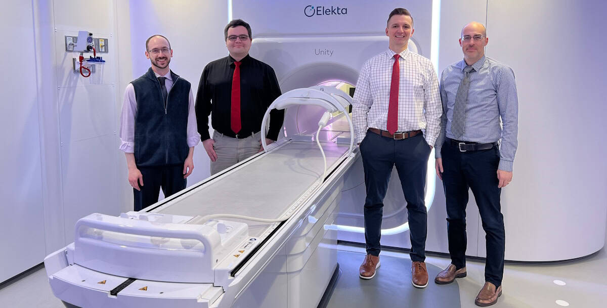
University of Iowa researchers The current MR-linac team at Iowa. From left to right: Blake Smith, Sam Rusu, Daniel Hyer and Joel St-Aubin. (Courtesy: Jillian York)
“Dan’s group and our group in Utrecht were independently working on MR-linac VMAT. Dan from the planning side, us from the delivery side,” Fast explains. “Dan’s limitation was that he couldn’t prove that his plans were deliverable, and we didn’t have the high-quality clinical-grade VMAT plans available for delivery. The collaboration started in July 2023 through a chance encounter in Houston, where Dan and I happened to present at the same session during an AAPM pre-meeting course.”
In their current study, the researchers demonstrated that VMAT deliveries are possible on Unity, without requiring changes to clinical hardware. The retrospective study showed that combined optimization and delivery time were shortened by up to 7.5 min compared with standard step-and-shoot IMRT.
“VMAT nearly doubles the delivery speed compared to conventional step-and-shoot IMRT, which means faster treatments (i.e., better patient comfort) and better accuracy (due to less chance for unexpected motion),” says Fast.
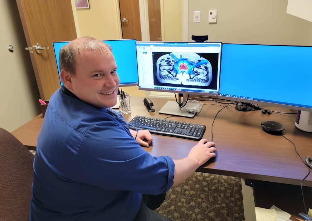
Plan analysis First author Jeffrey Snyder, now at Yale School of Medicine, working on a Monaco treatment plan. (Courtesy: James Laird)
In collaboration with Elekta, the researchers developed a modified version of software that allowed them to deliver a VMAT-like plan with Unity. For 10 prostate cancer patients previously treated on a 1.5 T MR-linac, they replanned treatments to deliver to 36.25 Gy in five fractions, using three techniques: step-and-shoot IMRT and a clinical optimizer; the same optimizer with a VMAT technique; and a research-based optimizer with VMAT.
The plans were adapted onto MRI datasets using two optimization strategies to assess adapt-to-position planning. The team assessed plan quality by evaluating organs-at-risk sparing and evaluated treatment efficiency by measuring the optimization time, delivery time and total (optimization plus delivery) time. Delivery accuracy was assessed via a gamma analysis (2%/2 mm).
Results showed that optimization time plus delivery time yielded savings of up to 7.5 min for the VMAT optimization and the research-based optimizer with VMAT, compared with the clinical optimizer with IMRT. Adapt-to-position planning showed a similar reduction in total time. All VMAT plans had gamma passing rates greater than 96%, and the delivery efficiency of VMAT plans was nearly 90%, compared with 50% for clinical IMRT.
“We’ve shown [that this technology] is feasible. We delivered it, we’ve done the quality assurance, we’ve made the plans. I think that is a huge milestone to pushing this towards clinical implementation. And there’s no major physics or technical hurdles that still need to be cleared – it’s mostly engineering…so that’s great news,” says Hyer.
Next steps for the researchers include providing physics guidance and quality assurance tests so that when VMAT becomes clinically available on MR-linacs, medical physicists have recommendations for implementation. They are also looking at VMAT for gated treatments, motion management strategies for VMAT deliveries, and other treatment sites.
This episode of the Physics World Weekly podcast explores the science and commercial applications of metamaterials with Claire Dancer of the University of Warwick and Alastair Hibbins of the University of Exeter.
They lead the UK Metamaterials Network, which brings together people in academia, industry and governmental agencies to support and expand metamaterial R&D; nurture talent and skills; promote the adoption of metamaterials in the wider economy; and much more.
According to the network, “A metamaterial is a 3D structure with a response or function due to the collective effect of meta-atom elements that is not possible to achieve conventionally with any individual constituent material”.
In a wide-ranging conversation with Physics World’s Matin Durrani, Hibbins and Dancer talk about exciting commercial applications of metamaterials including soundproof materials and lenses for mobile phones – and how they look forward to welcoming the thousandth member of the network sometime in 2025.
A team of scientists based in the US has developed a non-invasive headset device designed to track changes in blood flow and assess a patient’s stroke risk. The device could make it easier to detect early signs of stroke, offering patients and physicians a direct, cost-effective approach to stroke prevention.
The challenge of stroke risk assessment
Stroke remains the leading cause of death and long-term disability, affecting 15 million people worldwide every year. In the United States, someone dies from a stroke roughly every 3 min. Those who survive are often left physically and cognitively impaired.
About 80% of strokes occur when a blood clot blocks an artery that carries blood to the brain (ischaemic stroke). In other cases, a blood vessel can rupture and bleed into the brain (haemorrhagic stroke). In both types of stroke, deprived of oxygen from the loss of blood flow, millions of brain cells rapidly die every minute, causing devastating disability and even death.
As debilitating as stroke is, current methods for assessing stroke risk remain limited. Physicians typically use a questionnaire that assesses factors such as demographics, blood test results and pre-existing medical conditions to estimate a patient’s risk. While non-invasive techniques exist to detect changes after the onset of a stroke, by the time a stroke is suspected and patients are rushed to the emergency room, critical damage may have already been done.
Consequently, there remains an acute need for tools that can proactively monitor and quantify stroke risk before an event occurs.
Blood flow dynamics as proxies for stroke risk
Seeking to bridge this gap, in a study published in Biomedical Optics Express, a research team, led by Charles Liu of the Keck School of Medicine at the University of Southern California and Changhuei Yang of California Institute of Technology, developed a headset device to monitor changes in the brain’s blood flow and volume while a patient holds their breath.
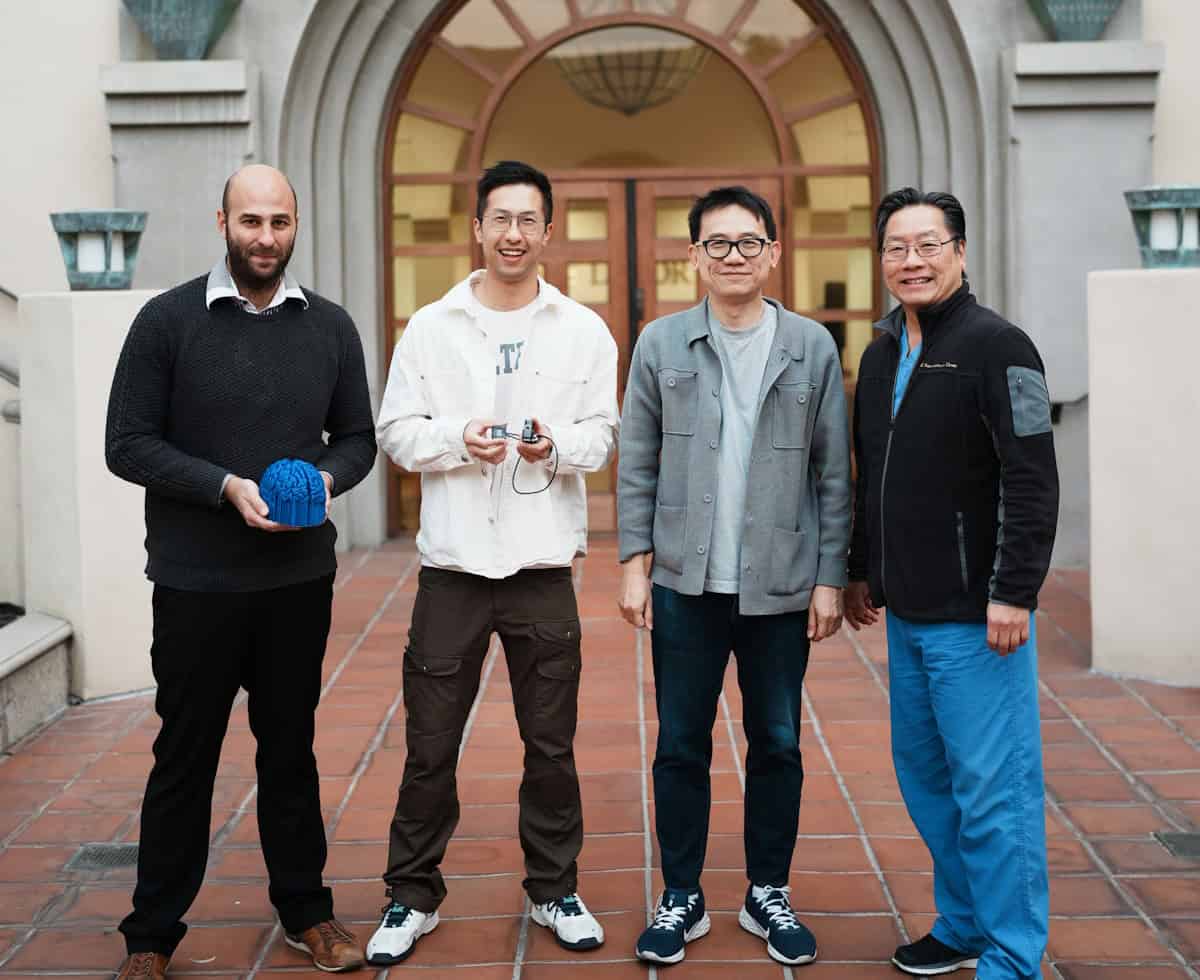
Team work From left to right: Simon Mahler, holding his own 3D printed brain from comparative MRI scans; graduate student Yu Xi (Max) Huang holding the SCOS device; Changhuei Yang; and Charles Liu. (Courtesy: Siyu (Steven) Lin)
“Stroke is essentially a brain attack. The stroke world has been trying to draw a parallel between a heart attack and a brain attack,” explains Liu. “When you have a heart disease, under normal circumstances – like sitting on the couch or walking to the kitchen – your heart may seem fine. But if you start walking uphill, you might experience chest pain. For heart diseases, we have the cardiac stress test. During this test, a doctor puts you on a treadmill and monitors your heart with EKG leads. For stroke, we do not have a scalable and practical equivalent to a cardiac stress test.”
Indeed, breath holding temporarily stresses the brain, similar to the way that walking uphill or running on a treadmill would stress the heart in a cardiac stress test. During breath holding, blood volume and blood flow increase in response to lower oxygen and higher carbon dioxide levels. In turn, blood vessels dilate to mitigate the pressure of this increase in blood flow. In patients with higher stroke risk, less flexible blood vessels would impede dilation, causing distinct changes in blood flow dynamics.
Researchers have long had access to various imaging techniques to measure blood dynamics in the brain. However, these methods are often expensive, invasive and impractical for routine screening. To circumvent these limitations, the team built a device comprising a laser diode and a camera that can be placed on the head with no external optical elements, making it lightweight, portable, and cost-effective.
The device transmits infrared light through the skull and brain. A camera positioned elsewhere on the head captures the transmitted light through the skull. By tracking how much the light intensity decreases as it travels through the skull and into the camera, the device can measure changes in blood volume.
When a coherent light source such as a laser scatters off a moving sample (i.e., flowing blood), it creates a type of granular interference pattern, known as a speckle pattern. These patterns fluctuate as blood moves through the brain – the faster the blood flow, the quicker the fluctuations. This technique, called speckle contrast optical spectroscopy (SCOS), enables the researchers to non-invasively measure the blood flow rate in the brain.
The researchers tested the device on 50 participants, divided into low- and high-risk groups based on a standard stroke-risk calculator. During a breath-holding exercise, they found significant differences in blood dynamic changes between people with high stroke risk and those at lower risk.
Specifically, the high-risk group exhibited a faster blood flow rate but a lower volume of blood in response to the brain’s oxygen demands, suggesting restricted blood flow through the stiff vessels. Overall, these findings establish physiological links between stroke risk and blood dynamics measurements, highlighting the technology’s potential for stroke diagnosis and prevention.
The future of stroke prevention
The team plans to expand these studies to a broader population to reinforce the validity of the results. “Our goal is to further develop this concept to ensure it remains portable, compact, and easy to operate without requiring specialized technicians. We believe the design is scalable, aligning well with our vision of accessibility, allowing diverse and underrepresented communities to benefit from this technology,” says co-lead author Simon Mahler, a postdoctoral scholar in the Yang lab at Caltech.
The researchers also aim to integrate machine learning into data analysis and conduct clinical trials in a hospital setting, testing their approach’s effectiveness in stroke prevention. They are also excited about the applications of their device in other neurological conditions, including brain injuries, seizures, and headaches.