

Fancy some more? Check out our puzzles page.
Pigeons’ immune system can sense Earth’s magnetic field, study suggests
Some animals could sense Earth’s magnetic field by using the superparamagnetic properties of white blood cells in their livers, a new interdisciplinary study has revealed. Led by Clivia Lisowski at the University of Bonn, the team’s insights into homing pigeon navigation could help scientists resolve the decades-old mystery of how animals can sense magnetic fields. The research also shines new light on the inner workings of the immune system.
Animals ranging from sharks to bats appear to use Earth’s magnetic field to navigate during long migratory voyages. However, the nature of this magnetoreception is poorly understood.
“How they sense Earth´s magnetic field has been a mystery,” Lisowski explains. “Several hypotheses were suggested, but these have often resulted from laboratory experiments. They were hardly ever done in the field, and so couldn’t explain how migration would work at night or in dark environments.”
In their study, Lisowski’s team investigated the origins of magnetoreception in homing pigeons. These birds are famous for finding their way back home after being released in unfamiliar locations. “We combined expertise from research fields that usually are not intersected, namely immunology, physics and animal behaviour,” Lisowski explains. “[This gave] us a truly interdisciplinary approach to solve the mystery of magnetoreception in homing pigeons.”
Searching for magnetism
As part of this approach, the researchers searched for magnetism in several different tissues of the bird – including the liver, spleen, muscle, and beak. They used vibrating-sample magnetometry (VSM), whereby samples are placed in a constant magnetic field and vibrated up and down. If the sample is magnetic, it will induce an electric field that is proportional to its magnetization.
The team also did field studies of homing pigeon flight patterns and studied the birds’ anatomy. The latter involved using dyes to visualize microscopic structures embedded in tissues along with genomic assays of the tissues.
This combined analysis revealed the presence of superparamagnetic macrophages in homing pigeon livers. Macrophages are specialized white blood cells whose primary function is to engulf and digest pathogens. They are a cornerstone of the immune system. Superparamagnetic means that the macrophages are very easily magnetized.
“The macrophages are superparamagnetic due to their physiological function of degrading old and damaged red blood cells,” Lisowski explains. “They sequester and store the iron from red blood cells as oxide nanoparticles in ferritin proteins.”
Connecting to the brain
Within homing pigeon livers, the researchers found that these macrophages were often in the vicinity of – and sometimes even in direct contact with – nerve fibres in liver tissue. This could allow magnetic information to be passed from macrophages to the brain.
At the atomic level, Lisowski’s team proposes that the effect emerges from unpaired electrons in ferritin proteins, which interact with each other through magnetic dipole–dipole coupling to enhance their magnetic susceptibility. This subatomic effect could in turn explain the distinctive circling behaviour observed in pigeons after they take off. This circling could allow these unpaired electrons to be imprinted with magnetic information, before the birds settle into a more uniform flight.
Honey bees navigate using magnetic abdomens
Read more

The research could also improve our understand of the immune system. The team suspects that the sensing abilities imparted by their superparamagnetic properties could enable macrophages to better gauge their surrounding environments: distinguishing between what to fight, and what to tolerate.
“Our finding that the immune system can also sense the Earth´s magnetic field is a completely new layer in this concept of ‘immuno-sensation’, and opens the door to new research,” Lisowski says.
The research is described in Science.
Physics-based models still beat AI for predicting extreme weather events
Artificial intelligence (AI)-based weather models are not as good as physics-based forecasting systems at predicating extreme weather events, say researchers at the University of Geneva, Switzerland and the Karlsruhe Institute of Technology, Germany. After comparing the outputs of different models on the same dataset of extreme events, the team found that AI models systematically erred on the side of normality, underestimating temperatures for extremely hot events while overestimating them for cold ones. This could be because the models learn from what has already happened and struggle to forecast events outside their training data.
With extreme weather events growing more common and intense due to our rapidly warming climate, being able to predict them is becoming ever more important. In recent years, meteorologists have sought to address this by developing a new generation of AI models that vie with physics-based numerical weather prediction (NWP) systems in the accuracy and extent of their forecasts – at least for everyday weather events.
Black or grey swans
In the new work, researchers led by Zhongwei Zhang and Sebastian Engelke sought to understand whether AI could also be competitive when forecasting extreme weather episodes. These episodes are usually defined by variables such as wind, atmospheric pressure or temperature falling well outside norms for location and season, and are sometimes termed “black swans” or “grey swans” depending on how extreme they are.
“In the past, only relatively moderate extremes were documented,” Zhang and Engelke observe. “But given the current rate of high rate of global warming, record-breaking events now sometimes exceed previous record levels by large margins.”
That’s potentially a problem for AI, they add, because several recent studies have shown that such models come up short when asked to extrapolate beyond their training data. For example, in 20205, researchers at the US National Oceanic and Atmospheric Administration (NOAA) and the Allen Institute for Artificial Intelligence in Washington, US, found that a seasonal AI forecasting model could not predict values for the North Atlantic Oscillation, which plays a crucial role in Europe’s weather and climate. Another study found that models tend to underpredict the intensity of the most extreme storms (as measured by mean sea-level pressure) or other high-impact events such as heat waves.
A large sample of record-breaking events
To test this hypothesis, Zhang and Engelke constructed a large dataset of record-breaking events for heat, cold, and wind extremes during 2018 and 2020. This dataset included several well-known events, such as the Siberian heatwave in early 2020 and the US heatwave of August 2020, but also tens of thousands of less-heralded ones. The 2020 dataset, for example, included 162,751 heat, 32,991 cold, and 53,345 wind records spread across different seasons and climatic zones.
The researchers assessed how well the three leading deterministic AI weather models –GraphCast, Pangu-Weather (and operational variants) and Fuxi – performed when extrapolating from this dataset of record-breaking events. They then compared these models’ performance to that of the physics-based High RESolution forecast (HRES) model developed by the European Centre for Medium-Range Weather Forecasts (ECMWF), which is widely acknowledged as today’s best physics-based NWP model.
In line with previous studies, the researchers found that both GraphCast and Fuxi were better than HRES at forecasting normal weather events. However, for record-breaking temperature and wind events in 2020, the situation was reversed, with the physics-based HRES model consistently outperforming all AI models for hot and cold temperature records as well as wind speed records.
A difficult study
The researchers report that the most difficult aspect of the study was the sheer length of computation time required to analyse huge AI and numerical forecast datasets. To complicate things further, Zhang notes that some of the most recent AI weather models are being developed by big tech companies and are not publicly available.
As well as showing that purely data-driven AI models struggle to forecast record-breaking extreme weather events, Engelke says the team’s work also provides a protocol for systematically evaluating forecasts of extreme events. “We hope this will motivate the research community to thoroughly evaluate the next generations of AI forecasts to advance our understanding of their advantages and their limitations compared to conventional physics-based models,” he says.
In this study, which appears in Science Advances, the Geneva/Karlsruhe researchers focused on evaluating the forecasts of deterministic AI weather models. They are now doing the same for forecasts made by recent probabilistic AI weather models, which they suspect will face similar extrapolation limitations.
“We are also working on building AI models ourselves that have improved forecast performance on extreme events,” Zhang and Engelke reveal. “This might be achieved this by creating hybrid models, which are a smart combination of physical and AI-based weather models.”
Word flower puzzle no. 5
How did you get on?
12 words Warming up nicely
17 words Getting hot, hot, hot
22 words Top dog!
Fancy some more? Check out our puzzles page.
Inside EPSRC: Charlotte Deane on funding the future of UK physics
This episode features Charlotte Deane, who is executive chair of the UK’s Engineering and Physical Sciences Research Council – or EPSRC.
In conversation with Physics World’s Matin Durrani, Deane talks about her career in both academia and industry – and her leadership of the University of Oxford’s Protein Informatics Group.
Deane explains EPSRC’s role in funding research in physics and other sciences in the UK. She talks about the long-term strategy behind changes in funding priorities that were announced earlier this year. She also explains how EPSRC is supporting students and highlights the importance of quantum technologies and artificial intelligence to the future of the UK.
Experiment that may or may not disprove Bohmian mechanics continues to spark debate
In the event of a nuclear holocaust, the only life remaining on Earth may be cockroaches, Keith Richards and physicists arguing about philosophical interpretations of quantum mechanics. One especially rich source of arguments is a deterministic alternative to the standard Copenhagen interpretation that dates back to the American (and later British, after Princeton University fired him for alleged communist sympathies) theoretical physicist David Bohm. According to Bohm, the position of a quantum particle is well defined everywhere and guided by a “pilot wave” which, alas, cannot be measured directly.
Despite these philosophical differences, Bohmian mechanics makes the same predictions as the Copenhagen interpretation. Except maybe it doesn’t. Occasionally, a bright theorist hypothesizes that, under very specific circumstances, one could distinguish them. Very occasionally, a bright experimentalist conducts an experiment that claims to actually do so.
This is what happened last year when Jan Klärs and colleagues at the University of Twente in the Netherlands sent photons from a laser down one of two coupled waveguides towards a potential step. When the photons reached the step, they could pass through it by quantum tunnelling. They could also pass into the other waveguide. The researchers interpreted the distance the photons travelled through the barrier before tunnelling into the other waveguide as a measurement of their speed.
The key result was that, when the wave functions on both sides of the barrier were the same, the photons still tunnelled at, ahem, light speed – matching the Copenhagen notion that tunnelling occurred equally in both directions. However, the Twente team calculated that Bohmian mechanics predicted that photons inside the step – where the guiding equation didn’t have a real-valued frequency – would be at rest and get stuck. Interferometric measurements showed that wasn’t happening.
Not so fast
Game, set and match to Copenhagen? Er, no. Proponents of Bohmian mechanics immediately disputed the team’s definition of velocity. “It’s just an operational definition,” says Aurélien Drezet of the CNRS University of Grenoble-Alps in France. “It has the units of velocity…but that doesn’t mean that Bohmian mechanics can interpret the result.”
In a “Matters Arising” article in Nature, Drezet and two colleagues at the Technion–Israel Institute of Technology in Haifa now add an experimental qualm. The fact that the Twente team was able to produce an image shows that radiation must be leaking out of the cavity, Drezet claims: “If you go to higher approximations and include cavity losses, you explain the experiment completely using Bohmian mechanics,” he says.
Klärs, who is preparing a formal response, is unconvinced. On the first point, he sees no reason “why the same speed measurement, which correctly captures the Bohmian velocity in the propagating regime, should cease to be a speed measurement in the evanescent regime”. On the second, he says his group did not ignore radiative leakage from the cavity: they measured it and found it “does not have a significant impact on the scattering physics we investigate or on the interpretation of our results”.
This could go on for some time – regardless of how many seconds it is to midnight.
X-ray velocimetry study earns Ronan Smith the PMB Early Career Researcher Award
Ronan Smith, a postdoctoral research fellow at Adelaide University, has been chosen as the winner of this year’s Physics in Medicine & Biology (PMB) Early Career Researcher Award. The award is presented to the author of the “best paper” in PMB’s 2025 Early Career Researcher Focus Collection, as selected by the journal’s editorial board.
Smith’s research involves implementing X-ray velocimetry (XV), a novel imaging method that uses X-rays to track lung motion during breathing and create 3D maps of local ventilation. In his award-winning paper, Visualising ventilation changes following endobronchial valve placement with x-ray velocimetry functional lung imaging, Smith investigates the potential of XV imaging to detect changes in lung function after insertion of an endobronchial valve (EBV).
EBVs are one-way valves that are placed into the lung to help treat emphysema – a condition that damages air sacs in the lungs, causing air to get trapped inside and making breathing difficult. The EBV, which in some cases can be used in place of surgery, prevents airflow into damaged lung areas so that the rest of the lung can function more effectively.
Successful valve placement causes the targeted area of lung to collapse, which can be imaged using CT. Smith proposed that use of XV functional lung imaging to non-invasively measure regional and local changes in airflow could more accurately assess the clinical impact of EBV placement.
“The lungs are a dynamic organ, their job is to be constantly moving,” he explains. “Because X-ray velocimetry looks at lung motion, it lets us see exactly where the air is or isn’t flowing, so you can instantly see that airflow has changed within the lungs. CT only measures structural changes, which may not necessarily be correlated to changes in lung function.”
In vivo demonstration
To investigate this potential advantage, Smith and colleagues carried out a pilot study on healthy sheep, which have a similar lung size to humans. They performed XV imaging on two anaesthetized and ventilated animals, before and after placing EBVs in their lungs.
The XV scanning process involves recording fluoroscopic videos of individual breaths at various angles around the lung, with anatomic positioning provided by an accompanying breath-hold CT scan. To analyse the data, the researchers used XV LVAS software from 4DMedical, the MedTech company that developed and commercialized the XV technology.
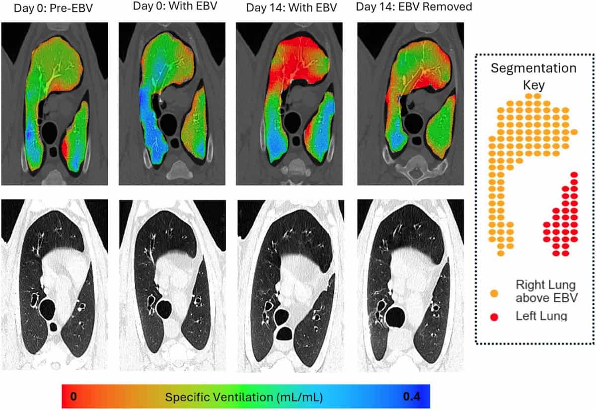
The software correlates motion in the XV videos with CT data to measure the lung’s expansion and contraction during a breath cycle. It creates a 3D map of specific ventilation (the change in voxel volume during a breath, divided by its starting volume) in small voxels throughout the lungs. This map can then be used to calculate mean specific ventilation and ventilation heterogeneity across a given lung region.
As soon as the EBVs were inserted into the animals’ lungs, XV imaging could visualize and quantify a reduction in airflow to areas downstream of the valves. This effect was seen both in regions where collapse was visible in CT scans, as well as those where collapse could not be detected by CT. Ventilation changes were also clearly observed in the remainder of the lungs.
“The main finding of our study was that X-ray velocimetry imaging can detect airflow changes from endobronchial valve placement in the lungs,” says Smith. “Our research could be really important for people [with emphysema], as tools to help with better placement and verification will lead to improved treatment options.”
Future prospects
Since the publication of this paper, Smith has focused on further applications of pre-clinical and clinical XV imaging. “I’ve been working as part of a great interdisciplinary team looking at how lung function changes in a range of diseases, to both understand the diseases, and as an outcome measure when we test treatments,” he says.
This work includes the world’s first paediatric clinical trial of XV imaging, which is examining the feasibility of using the technology in children with cystic fibrosis. The researchers have imaged around 30 children to date and aim to publish their findings later this year. They are currently planning future studies to see how XV imaging could enhance clinical decision making and improve outcomes for these children, as well as looking at other childhood diseases where it could be of relevance.
“As an early-career researcher, I’m also focussing on developing my own research, looking at another novel X-ray imaging method called dark-field X-ray imaging,” Smith adds.
The perfect award
Smith tells Physics World that he was excited to receive the PMB Early Career Researcher Award, acknowledging the efforts of everyone involved in this “hugely collaborative project”, including clinicians, scientists, 4DMedical and the staff in the preclinical imaging facility where the study was performed.
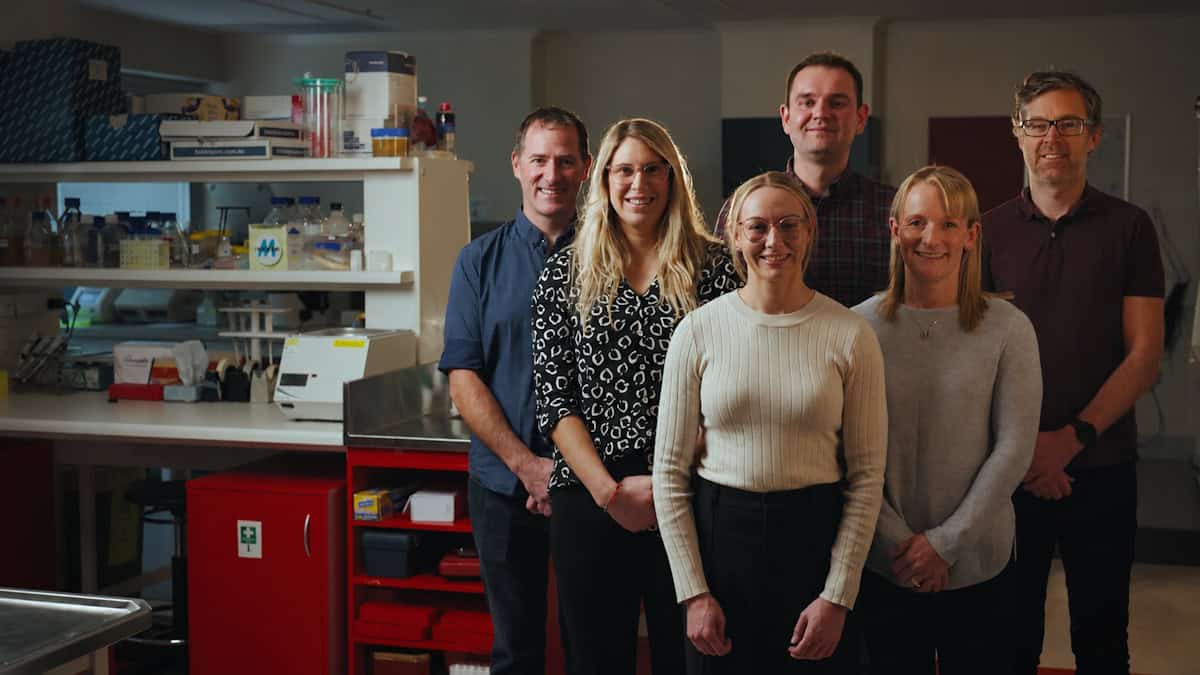
“As a physicist working in medicine/biology, it feels like the perfect award to get,” he says. “It’s great to see interest in the work we are doing, and fantastic evidence we can use to convince the funding bodies it’s worth continuing this work.”
- The PMB Early Career Researcher Focus Collection 2025 publishes papers from early-career researchers (defined for this collection as a postgraduate student or someone who completed their PhD in 2019 or later) in biomedical physics, with the aim of highlighting research excellence from emerging leaders. The Early Career Researcher Award was introduced to further recognise an outstanding contribution from one of the early-career authors, selected according to the quality of scientific content, number of citations and downloads, and peer review ratings.
Quantum influencers gather to celebrate London’s role in quantum tech
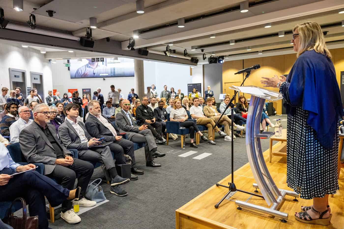
“There are two types of people when it comes to quantum,” joked Howard Dawber, deputy mayor of London for business and growth at a meeting at the Institute of Physics last night to celebrate the first anniversary of the London Quantum Cluster.
“There are those who understand quantum mechanics. There are those who don’t. And there are those who are in superposition of understanding and not understanding until they are observed.”
It was a light-hearted remark that matched the mood of what was essentially an evening of boosterism for quantum technology in London ahead of London Tech Week next week.
As chair of London and Partners – the growth agency for London – Dawber told the gathering of more than 100 “quantum influencers” that the organization was “100% behind the London Quantum Cluster”.
Founded in 2025 by University College London, King’s College London and Imperial College London, with support from the Mayor of London and the UK government, the cluster seeks to establish the capital as a powerhouse of quantum tech.
Georgia Siora from Warwick Economics and Development presented data to show that London is already doing well in the sector, being home to more than 160 quantum companies, with seven of the top 10 UK quantum firms based in the city. Small- and medium-sized quantum firms in the capital, she added, contribute an estimated £153m annually to the economy.
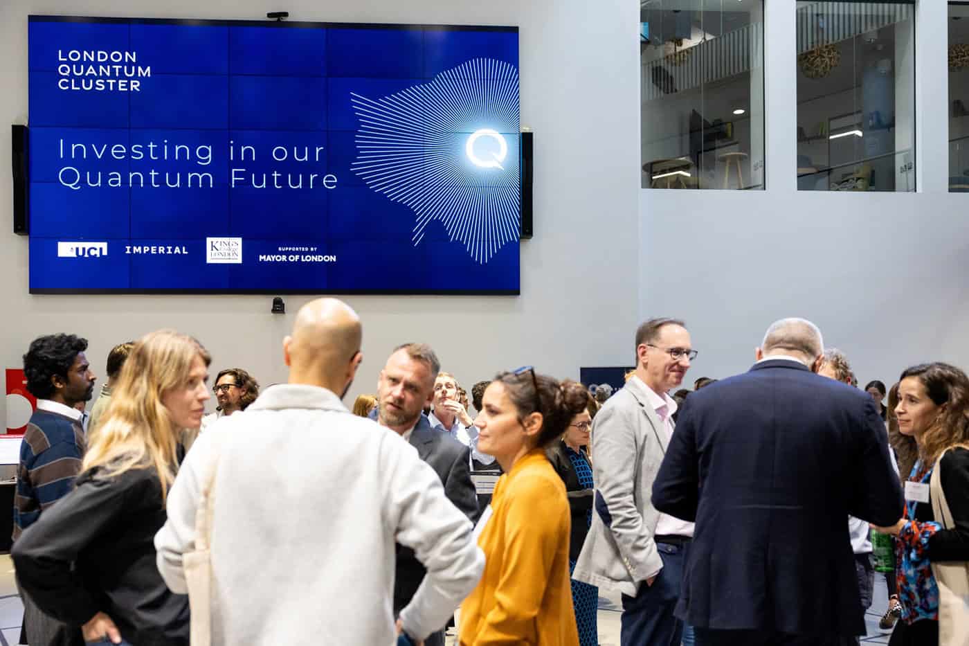
“Quantum and deep tech are at the heart of the capital’s 10-year growth plan,” added Janet Coyle, managing director of London and Partners.
Geraint Rees, vice-provost for research, innovation and global engagement at UCL, said his aim was “to make London the best place on the planet for serious quantum companies”. He pointed to UCL spin-out Quantum Motion, which has just won $160m of venture-capital funding, as an example of the kind of firm making a name in the city.
The evening ended with a panel debate chaired by Jess Wade from Imperial College London, featuring Maria Maragkou from Nu Quantum as well as Richard Murray, founder of ORCA Computing, who drew a distinction between universities being all about expanding the frontiers of knowledge, whereas for start-ups “the aim is to win”.
Also on the panel was physicist Alejandro Montblanch – head of quantum communication and networking at banking group HSBC – who made it clear that what he wanted to know was: “How can your quantum company help HSBC make more money?”
A welcome note of caution came from London-based venture-capital investor Eloisa Angeles, who pointed out that while the UK has a good track record of government-funded research, the UK is weak at “follow through”, with the government not focused on procurement and not having the end customer of the research in sight.
- For more about the commercial impact of quantum economy, check out this feature by Philip Ball.
The wonderful world of microfluidics
Confine a liquid to a region a millimetre or less in size, and you’re in the weird world of “microfluidics” – where surface tension and capillary action dominate in ways we easily overlook from our macro-scale vantage point. Despite being a term that was coined only in 1992, the underlying phenomena have been with us for millennia, as Albert Folch argues in How the World Flows: Microfluidics From Raindrops to COVID Tests.
Folch, who is a professor of bioengineering at the University of Washington in the US, has spent his career developing microfluidic devices that exploit the peculiar properties of flows at this scale. In this book, he now steps back to admire the full scope and impact of microfluidics, revelling in droplets that form rainbows, deliver inhalable asthma medications, make up salad dressings and cosmetics, and print scaffolds of living tissue to aid patients’ healing.
I particularly enjoyed Folch’s ode to the Olmec – Indigenous people who lived in Central America from about 1200 BCE to 400 BCE. They would harvest latex from Panama rubber trees by tapping the bark and collecting the liquid drop by drop before the day’s heat caused the latex to coagulate and seal the cut. The Olmec even had their own version of vulcanization – the process that makes rubber elastic – using the juice of morning glory vines to process the sap into bouncy elastic balls, stretchy bands, shoe soles and raingear.
The Olmec spread their knowledge to neighbours as well. In fact, the name “Olmec” comes from the Aztec language and literally means “the rubber people”. Like the maple sugar industry – which gets its own historical treatment from Folch – the Olmec latex harvest relied on a tree’s vascular system, made up of narrow 25 µm capillaries that carry liquids like water, sap, resin and latex to nourish and defend the tree. Although the Olmec civilization eventually declined, their technology and ingenuity lived on, impacting the entire world.
Folch walks readers not just through the physics and biology of these systems – capillary rise, photosynthesis, transpiration – but through their human impact, too. He includes wonders, such as natural rubber powering a Victorian-era craze that brought us tyres, inflatable boats, children’s dummies (pacifiers) and other objects. Folch also covers darker stuff, such as the British businessman Henry Wickham (1846–1928) who smuggled thousands of rubber tree seeds out of the Brazilian Amazon to establish plantations in Africa and Asia, where he could exploit cheaper labourers.
Another chapter begins in the mountains near Granada, Spain, where villagers are rebuilding acequias – open-air waterways originally built by the Moors in medieval times. For nearly 1000 years, acequias turned the arid slopes into terraced fruit gardens by diverting snowmelt toward agriculture and recharging the groundwater. Water seeps downward through the acequia’s dirt bottom, protecting it from evaporating in the hot Sun and feeding aquifers.
As Folch notes, aquifers contain nearly 30% of Earth’s freshwater – far more than is found in rivers and lakes, which make up less than 1% of the total. But in the US alone, industrial agriculture has been draining aquifers at rates as high as 60 cm per year, far exceeding the slow percolation of rainfall into these underground reservoirs.
In fact, aquifers take hundreds or thousands of years to recharge, which, Folch notes, means a depleted aquifer effectively ceases to exist for those of us living now. Without acequias and other dedicated efforts to replenish aquifer levels through microfluidic flow, today’s corn fields will soon turn to dust.
Candles to kidneys
How the World Flows charts the history of other unexpectedly microfluidic technologies too. Candles, for example, carry their fuel to the flame by capillary action along the wick. Then there’s paper, which soaks up ink through capillary action. Among more recent microfluidic inventions, Folch offers special laurels to the ballpoint pen, of which BIC alone has sold over 100 billion units since 1950.
Folch also takes care to introduce readers to many microfluidic medical devices, which might not be as widespread as ballpoint pens, but are perhaps more important. They range from dialysis machines that clean blood for failing kidneys, to COVID tests and continuous glucose monitors that let diabetics manage their blood glucose levels – a microfluidic technology that’s critical in my own home.
Folch describes the microfluidic devices that enable the Human Genome Project, as well as prototype instruments that could one day catch cancer cells through a simple blood screening. He also covers devices that could pick the perfect personalized drug cocktail for treating a patient’s tumour by studying their biopsied cells.
The most stunning microfluidic device Folch describes may be us
But the most stunning microfluidic device Folch describes may be us. As he argues, we ourselves are microfluidic. From our lungs to our cardiovascular system, from our lymphatic system to our sweat glands, our bodies rely on flow through tiny vessels.
Our bodies make up for diffusion’s slow pace by operating in parallel. Like the many co-operating central-processing units in a supercomputer, our bodies absorb oxygen through 500 million micro-sized alveoli in our lungs. We hear through ears equipped by microfluidics to act as both microphones and accelerometers. Our kidneys clean some 200 litres of blood a day – sending two litres of urine to our bladders as they do – through massively parallelized filtration.
Throughout How the World Flows, Folch’s enthusiasm for his subject shines. His approach is that of a storyteller, rather than a scientist, and the book is all the better for it. Whether readers are microfluidics experts or not, they will walk away with new stories to tell. (Don’t skip the footnotes…)
Although Folch’s stories are wide-ranging and entertaining, he stumbles at times with the overarching narrative. Some transitions are rough, and it’s not always clear why he’s chosen to order the stories in the way they’re presented. Nevertheless, his book is a fun and highly readable introduction to microfluidics that’s sure to entertain lay readers and excite a new generation of microfluidic engineers.
- 2025 Oxford University Press 306pp £22.31hb £19.16ebook
PET scan that detects dangerous blood clots is SNMMI ‘Image of the Year’
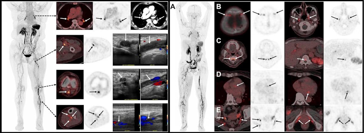
A PET radiotracer that can detect deep vein thrombosis (DVT) in the legs and clots that have travelled to the lungs has been chosen as the “Image of the Year” at the Society of Nuclear Medicine and Molecular Imaging (SNMMI) 2026 annual meeting. Developed by Sangwon Han and colleagues at the Asan Medical Center, University of Ulsan College of Medicine, in Korea, the novel tracer enables whole‑body imaging of blood clots (thrombi) in the legs and lungs in a single scan.
A DVT is a blood clot that forms in a deep vein, usually in the legs. It’s a common condition, with an incidence of roughly half that of all cancers, and it can lead to serious complications. Clots can break off and travel to the lungs, which could cause a potentially life‑threatening pulmonary embolism (a blockage in the artery supplying blood to the lungs). Early detection of DVT is therefore critical for determining the most appropriate treatment for each patient.
Currently, the standard imaging method for diagnosing DVT is venous ultrasonography (VUS). But while this works well for detecting clots in the thigh-to-knee region, whole-leg VUS requires skilled operators and advanced machines, takes longer, and has lower diagnostic sensitivity in the calf. In addition, conventional imaging techniques such as VUS and CT rely on indirect structural changes rather than directly visualizing the clot.
Aiming to enable faster and more efficient DVT diagnosis, Han and his research team are studying fluorinated GP1 (18F-GP1) a novel thrombus-targeted PET tracer. The tracer selectively binds to specific receptors on activated platelets (the cell fragments that cause blood to clots), allowing direct visualization of active thrombus formation.
“In our Phase 1 study, 18F-GP1 PET/CT showed 100% detection rate in 20 patients with confirmed DVT or pulmonary embolism,” Han told the SNMMI delegates. “But that study was limited by its small sample size and an absence of negative groups so specificity could not be assessed at that time.”
So in this latest work, Han and his team performed a phase 2, non-randomized study investigating the ability of 18F-GP1 PET/CT to identify acute lower-extremity DVT in 46 symptomatic patients. This included 22 patients with proximal DVT and 24 with none or distal DVT, as diagnosed using VUS.
The researchers acquired chest-to-feet PET/CT scans approximately 2 h after intravenous administration of 250 MBq of the radiotracer. The images were assessed by three blinded nuclear medicine physicians from different institutions, who assigned focal 18F-GP1 uptake higher than background activity as positive for thrombosis. They classified proximal DVT as clots involving the iliac (pelvic), femoral (thigh) and popliteal veins (behind the knee), and distal DVT as clots confined to the calf veins.
“Our primary objective was to assess the sensitivity and specificity of qualitive 18F-GP1 PET/CT interpretation for proximal DVT,” Han explained. “Secondary objectives included assessing the agreement between PET/CT and VUS for distal DVT, inter-reader reproducibility, exploring the detection of pulmonary embolism and assessing safety.”
When evaluated against VUS as a reference standard, 18F-GP1 PET/CT exhibited high diagnostic accuracy for detecting clots, demonstrating a sensitivity of 95% and a specificity of 92% for proximal DVT. “For distal DVT, both positive and negative agreement between PET/CT and VUS were strong,” added Han. “Inter-reader agreement was also excellent.”
The scans also identified concomitant pulmonary emboli in some patients, as confirmed by CT pulmonary angiography, illustrating the advantage of simultaneously assessing DVT and pulmonary embolism in a single scan. The researchers noted that the radiotracer was well tolerated, with no drug-related adverse events observed.
Speaking in the plenary session when his award was announced, Han shared a “striking image” recorded using 18F-GP1 PET/CT, which showed extensive blood clots, not only in the leg and lungs, but also in many unusual sites, including cranial and spinal vessels, cardiac valves, and vessels in the pelvic region. “This image clearly shows the remarkability ability of fluorinated GP1 to visualize thrombi throughout the body,” he explained.
“We believe this represents an important step towards thrombus-specific imaging,” Han concluded. “The potential of GP1 PET can expand beyond DVTs to many other thrombotic diseases such as embolic stroke or other cardiovascular diseases.”
The SNMMI Image of the Year is the society’s highest award, and the most anticipated, given out in recognition of an image that’s truly cutting-edge and representative of the future of nuclear medicine. This year’s winning image was chosen from nearly 1500 abstracts submitted for the meeting.
“It is truly a great honour to receive the Image of the Year award,” said Han.
Copyright © 2026 by IOP Publishing Ltd and individual contributors