Earlier this week, this year’s award was given to two independent teams for entangling two macroscopic vibrating drumheads, thereby advancing our understanding of the divide between quantum and classical systems.
Appearing in this podcast are Mika Sillanpää and Laure Mercier de Lepinay representing the team from Finland’s Aalto University and the University of New South Wales, Australia. Also on hand are John Teufel and Shlomi Kotler, who led the team at the US National Institute of Standards and Technology (NIST).
This quartet of applied physicists explain the motivation behind their experiments and talk about the challenges of entangling objects that are about ten microns across. They also chat about possible applications for vibrating drumheads such as quantum sensing and quantum networks.
Physics World‘s Breakthrough of the Year coverage is supported by Bluefors, a leading supplier of cryogen-free dilution refrigerator measurement systems with a strong focus on the quantum computing and information community. Our aim is to deliver the most reliable and easy to operate systems on the market which are of the highest possible quality.
The use of protons instead of X-rays for treatment planning could improve the accuracy of proton therapy. Now a US-based research team has demonstrated that proton CT could provide low-dose planning with reduced range uncertainties, using the ProtonVDA pRAD, a prototype clinical proton imaging system.
Currently, proton therapy is planned using X-ray CT images of the patient. The CT Hounsfield units are converted into proton relative stopping power (RSP) to calculate proton range in the patient and generate the plan. This conversion, however, leads to range uncertainties and necessitates the use of margins around the tumour target. Proton CT, on the other hand, measures RSP directly, reducing uncertainties and potentially enabling smaller margins.
Senior author James Welsh and colleagues used a proton beamline at the Northwestern Medicine Chicago Proton Center to assess the ProtonVDA pRAD system. To validate the accuracy of proton CT-based RSP measurements, they first imaged a cylindrical phantom containing eight different tissue-equivalent inserts. Comparing the measured RSP with the known values revealed an accuracy of 1% or better for all materials bar the sinus insert (which has extremely low RSP and an absolute discrepancy of only –0.008).
Next, the researchers imaged a pig’s head and a sample of porcine pectoral girdle and ribs using the ProtonVDA pRAD and a clinical vertical X-ray CT scanner. They also scanned the head using a horizontal CT scanner at high- and low-dose settings. They then determined the differences between directly measured RSP and values from X-ray CT scans using HU to RSP conversion.
In the girdle and ribs sample, the researchers found RSP differences of 0.6% or less for all soft tissues, including muscle and adipose. However, they saw a 1.9% difference in the rib trabecular bone and a much larger difference of 6.9% in compact bone. For the pig’s head, they examined 12 regions, observing the largest RSP differences (up to 41%) in the tympanic bullae, which comprise a mixture of air, soft tissue and bone. They also noted discrepancies for the skull (up to 4.3%) and brain stem (up to 4.4%). In the eight other tissues, RSP differences ranged from –2.5% to +2.1%, with a mean of –0.4%.
The results demonstrate that although the measured and calculated RSP values are similar for soft tissues, X-ray CT could prove less accurate for treatment planning in dense bone or cavitated regions.
The team also note that proton CT delivered a far lower imaging dose (0.2–0.7 mGy for the pig’s head) than X-ray CT (3.9 and 39 mGy, for low- and high-dose scans). While the small radiation doses associated with a single scan are unlikely to ever be of harm, Welsh points out that the 10- to 100-fold reduction in dose conferred by proton CT means that such scans could be repeated regularly.
“Thus, if there is a clinical benefit to proton radiography and proton CT, the studies can be repeated as often as necessary to maximize such benefit – without the fear that such benefits will be negated by any detrimental effects from radiation dose,” he explains.
Consistency check
A proton imaging system such as the ProtonVDA pRAD can also be used to perform proton radiography prior to each treatment fraction. Comparing such images with a digitally reconstructed radiograph (DRR) from the planning CT could provide a consistency check of the X-ray CT-derived RSP map, as well as reveal discrepancies due to changes in patient anatomy or alignment.
To demonstrate this application, the researchers acquired a proton radiograph and a high-dose X-ray CT scan of the pig’s head. They then created a water equivalent thickness (WET) difference map by subtracting the calculated DRR from the proton radiograph. In most soft-tissue regions, including the brain and head-and-neck muscles, WET values for the acquired and simulated proton radiographs agreed to within 1–2 mm, equivalent to less than 1% for a total WET of up to 200 mm. The most notable differences were found in the sinus region and the tympanic bullae.
The researchers conclude that proton CT offers potential for low-dose treatment planning with reduced margins, as well as daily pre-treatment range verification. And they have a raft of developments planned, including automating data acquisition, optical tracking of the rotation and integration with an upright treatment system. They also intend to perform more quantitative dose comparisons, and tests with treatment beams and film stacks.
“We will also be embarking on NTCP [normal tissue complication probability] studies, in which we aim to quantify the potential clinical benefits of range uncertainty reduction via proton radiography and proton CT,” Welsh tells Physics World. “In principle, reduction of range uncertainty should allow us to use tighter margins around our clinical targets and thereby reduce unwanted high dose to some normal tissues. Just how much benefit this provides will be the subject of upcoming investigations.”
Elastic thinking: Magnetic resonance elastography brain scans at 50, 37 and 25 Hz. (Courtesy: Ingolf Sack)
New research in Germany shows that changes to the mechanical properties of cells can cause a brain tumour to become malignant. Josef Käs of the University of Leipzig and Ingolf Sack of the Charité-Universitätsmedizin Berlin and colleagues have shown that a brain tumour is a unique material and its spread is driven by physics as well as biomechanics. Using research conducted on tumours in living patients, they suggest that small changes to the elasticity of cells produce collective effects that impact the prognosis of a tumour.
Sack and Käs are a chemist and a physicist respectively, each researching cancer, on two very different scales. Sack studies the mechanical properties of tissues in living patients. He has pioneered the use of low frequency vibrations combined with magnetic resonance imaging (MRI) to measure the progression of diseases such as cancer. This technique is called magnetic resonance elastography (MRE). Käs is one of the inventors of the optical stretcher, which was also used in this study. An optical stretcher is an optical trap that uses two laser beams to deform single cells and measure their viscoelastic properties.
In 2019, Sack and Käs discovered, using MRE, that glioblastoma, the deadliest form of brain cancer, is softer and less viscous than a benign brain tumour. Glioblastomas are almost impossible to remove because they grow by spreading tiny “fingers” into the surrounding tissue. The researchers realized that this growth could be driven by pure physics because “viscous fingers” are a well-known effect that arises when a low viscosity liquid is injected into another fluid.
Measuring mechanical properties: a patient undergoing magnetic resonance elastography. (Courtesy: Ingolf Sack)
Armed with this theory about how glioblastomas spread, the researchers set out to understand why. This meant looking at single cells, for which Käs’s group in Leipzig used the optical stretcher. The researchers recruited eight patients, four with benign brain tumours and four with malignant brain tumours, three of which were glioblastomas. The patients underwent MRE to measure the mechanical properties of their tumours, alongside measurement of individual tumour cells performed with an optical stretcher.
They had no expectations for what they would find, but Käs says that “the surprising thing is that single cell mechanical properties are still reflected in the whole brain tumour”. However, their data shows a more complex picture than viscous cells producing more viscous tumours.
Collaboration allows tumour cells to invade
Consistent with their previous results, the malignant tumours were softer and less viscous than the benign tumours, but puzzlingly, they did not have cells that were less viscous . Instead, it was the stretchiness and elasticity of the cells that was correlated with the fluidity of the tissue. For a tumour to “flow” into the surrounding tissue, the cells must squeeze past each other, and the researchers believe that this makes elasticity, rather than viscosity, the mediator of tissue fluidity.
They also observed that the tumour cells had a much wider range of mechanical properties than would be expected in a healthy sample. This is consistent with what is known about cancer, which breaks down regulation processes in cells. Drawing on the models of other researchers, they theorize that this heterogeneity allows parts of the tumour to fluidize and spread, whilst a rigid backbone of hard cells prevents it dispersing.
Describing the unique properties of malignant tumours, Käs says, “They don’t have to make a genetic change to start viscous fingering. It’s simply that they have these broad mechanical properties, which basically fluidizes, unjams the cells. And with unjamming, the viscous fingering that comes along means the most invasive growth you can imagine.”
Whilst the sample was small, this research indicates the importance of physics in cancer progression. Käs is aware that their conclusions are mixed news from a therapeutic perspective. This is because physics is more difficult to disrupt than a molecular change. But if physics can make a tumour malignant, it can also make it benign, and understanding this would be an important step in the treatment of this disease.
Researchers in the US have managed to reveal nanoscale optical and electronic band information of 2D semiconducting materials, using visible light. By employing a hyper-focusing technique they developed previously, the team managed to push beyond the diffraction limits of visible light to achieve a resolution of just a few nanometres. They say that this technique could help characterize the nanoscale properties of 2D and 3D materials to improve our understanding of catalysis, quantum optics and nanoelectronics.
Advanced 2D and 3D materials, such as single-walled carbon nanotubes, hold a lot of promise for next-generation electronics. In use, the electronic and optical properties of these materials are influenced by their environment, such as localized defects, strains, dielectric screening and quantum effects that can alter their performance. Characterizing the nanoscale details that cause these issues can be challenging. But as the colour and optical properties of these nanomaterials are closely related to their electronic structures, a technique known as hyperspectral imaging could offer a solution.
Hyperspectral imaging analyses the spectrum of every pixel in a scene across many wavelength bands. This electromagnetic detail can be used to obtain all sorts of information. For nanoscale materials, there has been some success using such techniques with wavelengths of light outside the visible range. Extending these techniques to the visible spectrum could allow more direct probing and simplify techniques, by eliminating the need for advanced light sources. With wavelengths of a few hundred nanometres, however, using visible light to obtain information from characteristics that are just a few nanometres in size is difficult.
Ramping the resolution: White light from a tungsten lamp is focused into the tip of a silver nanowire. (Courtesy: CC BY 4.0/Nat. Commun. 10.1038/s41467-021-27216-5)
To tackle this problem, Ming Liu, a physicist at the University of California, Riverside, and his colleagues have developed a technique known as super-focusing. The method integrates a tapered glass optical fibre with a silver nanowire condenser. “We can couple almost all the light from an optical fibre into a silver nanowire,” Liu tells Physics World.
Liu explains that as the light travels along the tapered optical fibre its wavelength gradually increases. As it reaches the end of the optical fibre, the wavelength matches that of the electron density wave in the silver nanowire.
Free electrons on the silver nanowire are then driven by the energy of the light and start to oscillate. These electrons then carry the energy along the surface of the silver nanowire. According to Liu, you can imagine this as being like a wave on the ocean being driven by the energy of the wind. At the end of the nanowire, a spot of light is produced – “like waves coming into a bay and its tapered shape producing a tsunami,” Liu adds.
All this means that the possible resolution is no longer limited by the wavelength of light. “We use the electron wave to carry the electromagnetic wave, instead of the light photons,” Liu explains. “Now the ultimate wavelength is restricted by the wavelength of electrons, which is very, very short: nanometre scale.”
In their latest work, described in Nature Communications, the researchers demonstrated that using this super-focusing technique they can achieve a 6 nm spatial resolution using visible to near-infrared wavelengths (415–980 nm) while probing single-walled carbon nanotubes. This allowed them to characterize nanoscale details such as chirality and electrical band structure.
“We showed the colour, but actually the colour is kind of determined by the electrical properties of the material,” Liu explains. “So, what we really want to say is that it can see the electrical band structures.” That is the transitions between the different band structures: how large those band gaps are.
Being able to characterize such nanoscale details – as opposed to the global performance – of 2D and 3D materials will help with the development of advanced semiconductors and newer techniques like twistronics, Liu says. He and his colleagues are now trying to see whether they can push the resolution as low as 1 nm.
I graduated from the Technical University of Delft’s Policy Analysis and Systems Engineering department in 1999 and after a two-year stint at KPN Research as a consultant, I joined the Ministry of Economic Affairs as an adviser in 2002. In 2013 – as head of strategy at the ministry – I visited the University of Technology in Delft to hear about their plans to start a quantum institute. Three years later the Netherlands held the presidency of the European Union and we helped to launch the €1bn Quantum Flagship programme. This led to interest from several Dutch research centres and companies, and to combine all this into a coherent national agenda I founded Quantum Delta NL last year together with Ronald Hanson and Jesse Robbers.
What is the role of Quantum Delta NL?
We are the Dutch umbrella organization that connects the five main quantum research hubs: Delft, Amsterdam, Leiden, Eindhoven and Twente. We bridge the gap between fundamental research and business, especially for start-ups that are taking quantum technology out of the lab and into people’s lives. With the Netherlands being such a small country, it is easier for these institutes to collaborate and this type of collaboration is unique – we don’t see it anywhere else. That’s why we think the Netherlands can have a world-leading role in quantum technology over the next decade.
And what do you focus on?
My role is to foster the best ecosystem for quantum technology development. By removing barriers between institutions and procedures, and by focusing on talent and entrepreneurship, we want to establish an open culture where innovation is possible. We also want to go beyond quantum technology being a field only for physicists. To develop quantum products and services for society, other disciplines will be important such as computer science, design, business and law.
We want to go beyond quantum technology being a field only for physicists
Quantum Delta NL recently received €615m from the Dutch government. How will that be spent?
We support research and development in three main topics: quantum computing, quantum networks and quantum sensing. The next step is to give start-ups a better and faster way to market by building new cleanroom facilities so they can develop their technology as well as providing early-stage investment and a tailored acceleration programme dubbed Lightspeed. Additionally, we put extra effort into educating and attracting people as well as the social impact of quantum technology. A dedicated campus for this “quantum ecosystem” – called the House of Quantum – will be operational from 2024.
What is the Netherlands doing in quantum networks?
Last July saw the live demonstration of a quantum-encrypted video being transferred across a quantum connection between Delft and The Hague. Late November we went live with an advanced quantum internet through our Quantum Network Explorer (QNE). QNE is the “software stack” to run such a network. Currently it runs on an emulated quantum network but this will be upgraded to actual quantum hardware soon. We use these demonstrations to show the technology’s capability to potential end-users and technology suppliers and give them a chance to participate.
What are the main challenges for Quantum Delta NL?
The main challenges are to scale up the ecosystem and engage with European industry. The key to these challenges is attracting and training the right talent. Even Quantum Delta NL has not reached the intended capacity yet, let alone attracting people into all the various programmes and start-up companies.
How does the Netherlands compare to the rest of the world?
The Netherlands is third in the world concerning quantum-research output and citations. Because we are nationally well organized, we are also frontrunners in developing the entrepreneurial ecosystem. However, European countries often lack the availability of venture capitalist funding or the big tech companies – both of which we see in the US, for example – that are willing to take the long-term risks. Fortunately, we see European industry engaging and EU governments investing. We also see some of the bigger companies, like Microsoft and Intel, collaborating with our research centres. On that level we are an important global player.
How do you see the interplay between competition and collaboration?
There will always be competition, but we mostly seek to collaborate. The development of quantum technology does not stop at the border – it requires effort on a global scale. On a European level we try to create a European ecosystem for quantum development. We are expanding through participation in several European programmes that have emerged from the quantum flagship programme. As an example, we work with several French institutions on topics such as spin-qubits and we also exchange talent with a job-board for quantum-related jobs in both countries.
Is there a danger that quantum technologies become a “bubble”?
There is a risk of overpromise, and we have a collective responsibility to manage this. Any mismatch between expectations and realization means part of the interest can collapse. We avoid that by tailoring our acceleration programme for start-ups and by only getting the right type of investors on board. But a little bit of hype is not necessarily bad as it will help to excite young talent to go into quantum technologies. As there is a solid scientific foundation underpinning the potential of quantum technology, the question is not if it will happen but when.
A new quantum sensor developed by scientists at the University of Sussex in the UK could help clinicians identify diseases such as dementia, Alzheimer’s and Parkinson’s by tracking patients’ brain waves and monitoring how their speed changes over time. The sensor, which is based on a real-time, high-spatial-resolution neuroimaging technique known as magnetoencephalography (MEG), uses an array of quantum devices called optically-pumped magnetometers (OPMs) to map the tiny magnetic fields generated when neurons in the brain send out electrical signals. If used to monitor patients over a period of several months, the researchers say the new sensor could identify declines in brain signal transmission speed that may be associated with pathology.
Non-invasive neuroimaging techniques have improved significantly over the past few decades, greatly expanding our knowledge of the brain’s inner workings by providing information about neural responses and processes. Previous studies have shown that the ways these processes are distributed within the brain, and how they change over time, are reliable biomarkers for anomalous brain activity associated with neurodegenerative diseases. If clinicians could detect these biomarkers accurately, and with high enough precision, they might be able to predict how a disease will evolve or how a patient will respond to treatment.
The problem is that current clinical methods cannot simultaneously resolve brain signals in space and time. Functional Magnetic Resonance Imaging (fMRI), for example, can map brain regions with high spatial resolution, but its temporal resolution is low (around 1 s) because it measures changes in local blood flow, which lag substantially behind electrical brain activity. Electroencephalography (EEG), for its part, detects these electrical signals directly, and thus works in real time. However, its spatial resolution is low, especially for high-frequency brain waves.
Evaluating cortical signals
In principle, MEG offers the best of both worlds, making it possible to measure the postsynaptic potentials of brain cells located just below surface of the scalp non-invasively, in real time and with high spatial resolution. Indeed, recent research has used MEG to evaluate abnormal cortical signals in patients with Alzheimer’s, Parkinson’s, autism spectrum disorder and even severe cases of post-traumatic stress disorder.
The drawback is that MEG has to be performed in special magnetically-shielded rooms to reduce magnetic noise from the environment, which is often many orders of magnitude higher than neuromagnetic fields (which are in the femtotesla to picotesla range). In addition, most of today’s MEG systems detect these tiny fields using superconductive quantum interference devices (SQUIDs), which require bulky cryogenic refrigeration. This means they cannot be placed close to the skull, limiting the spatial and temporal resolution of SQUID-based MEG scanners.
Around the turn of the millennium, researchers developed an alternative known as a “spin-exchange relaxation-free” (SERF) OPM. The version used in the current study contains a gas of rubidium atoms, and Peter Kruger, who leads the Quantum Systems and Devices group at Sussex, explains that when these atoms experience changes in their local magnetic field, they emit light differently. Hence, when researchers shine beams of laser light at the atoms, fluctuations in the emitted light reveal changes in the magnetic activity in the brain.
Better at brain signal tracking
In the new work, Kruger, PhD student Aikaterini Gialopsou and other members of the team used their OPM-MEG to record the spatio-temporal patterns of neuronal signals in volunteers responding to visual stimuli. They then compared these patterns to those obtained by conventional SQUID-MEG, demonstrating that the new sensor is better at tracking brain signals in both space and time. “We discovered that this quantum sensing technique can combine high spatial and temporal resolution,” they explain. “While previous techniques were able to locate signals in the brain, this one is the first to record the precise timing of brain signals.”
According to Kruger, the new quantum sensor is accurate to within milliseconds and has a spatial resolution of just millimetres. He and his colleagues now aim to improve the quality of their images still further by increasing the number of sensors in their OPM-MEG scanners. At the moment, this is done by squeezing individual sensors closer together. While this approach is straightforward, it is quickly reaching its limits because of cross-talk between sensors, overheating and other difficulties in scaling up individual sensors to whole imaging arrays, Gialopsou explains. “We are tackling this problem in a fundamentally different way by integrating high-density arrays of OPM sensors based on standard microfabrication techniques and shared resources,” she tells Physics World. “The first modular arrays developed in our group can be easily reconfigured, allowing for quick prototyping of novel sensing schemes and optimisation of sensor components and control systems.”
The tetraneutron, a hypothetical cluster of four bound neutrons, has been glimpsed by physicists in Germany. Although the measurement is well below the statistical significance required for a discovery, the observation is the latest possible sighting of the tetraneutron in the past two decades. Confirming the existence of the tetraneutron would shake-up our understanding of the forces that bind nuclei together and could also provide insights into neutron stars.
For over 50 years, physicists have been looking for clusters of two or more neutrons that are bound together by the strong force – and have found no conclusive evidence for their existence. There have been, however, tentative sightings of tetraneutrons, comprising four neutrons.
In 2002 an experiment at the GANIL accelerator facility in France revealed the first evidence for tetraneutrons – be it fleeting. Then in 2016, tetraneutrons were glimpsed again at the RIKEN nuclear-physics lab in Japan. The Riken team concluded that the tetraneutron is unbound, meaning that it immediately flies apart after being produced. A year later, physicists in the US and France developed a theoretical framework that suggested that if tetraneutrons existed, they would not stick around for very long. As a result, it remains unclear whether bound tetraneutrons exist.
Stable as a neutron
The latest search for was led by Thomas Faestermann and used the tandem Van de Graaff accelerator at the Maier-Leibnitz Laboratory, which is on the Garching campus of the Technical University of Munich. The team fired lithium-7 ions at a target of lithium-7 and observed the particles created by the colliding lithium nuclei. They found that some of the collisions created a carbon-10 nucleus along with a tetraneutron. The data suggest that the binding energy of the tetraneutron is about 420 keV. Calculations by the team suggest that the tetraneutron is roughly as stable as a free neutron, which has a half life of 450 s.
“For us, this is the only physically plausible explanation of the measured values in all respects,” explains Faestermann.
The measurement has a statistical significance of 3σ. However, this is not good enough for particle physics, which normally requires 5σ for a discovery. The team is now hoping that their measurements and analysis can be confirmed by independent groups.
Quantum technology has made great strides over the past two decades and physicists are now able to construct and manipulate systems that were once in the realm of thought experiments. One particularly fascinating avenue of inquiry is the fuzzy border between quantum and classical physics. In the past, a clear delineation could be made in terms of size: tiny objects such as photons and electrons inhabit the quantum world whereas large objects such as billiard balls obey classical physics.
Over the past decade, physicists have been pushing the limits of what is quantum using drum-like mechanical resonators measuring around 10 microns across. Unlike electrons or photons, these drumheads are macroscopic objects that are manufactured using standard micromachining techniques and appear as solid as billiard balls in electron microscope images (see figure). Yet despite the resonators’ tangible nature, researchers have been able to observe their quantum properties, for example, by putting a device into its quantum ground state as Teufel and colleagues did in 2017.
This year, teams led by Teufel and Kotler and independently by Sillanpää went a step further, becoming the first to quantum-mechanically entangle two such drumheads. The two groups generated their entanglement in different ways. While the Aalto/Canberra team used a specially chosen resonant frequency to eliminate noise in the system that could have disturbed the entangled state, the NIST group’s entanglement resembled a two-qubit gate in which the form of the entangled state depends on the initial states of the drumheads.
Both teams overcame significant experimental challenges, and their considerable efforts could open the door for entangled resonators to be used as quantum sensors or as nodes in quantum networks. As a result, this work deserves its place as the first quantum-related Breakthrough of the Year since 2015.
Selection criteria
The Breakthrough of the Year and the nine runners-up are selected by five Physics World editors from a list of nearly 600 research updates published on the website this year. In addition to having been reported in Physics World in 2021, our selections must meet the following criteria:
Significant advance in knowledge or understanding
Importance of work for scientific progress and/or development of real-world applications
Of general interest to Physics World readers
Here are the nine runners-up that make up the rest of the Physics World Top 10 Breakthroughs for 2021. You can listen to our editors talk about the Top Ten in this episode of the Physics World Weekly podcast.
Think before you speak: Clinical trial testing session with researcher David Moses. During the session, a neuroprosthesis recorded the participant’s cortical activity while he attempted to produce words and sentences. (Courtesy: Todd Dubnicoff, UCSF)
To Edward Chang, David Moses, Sean Metzger, Jessie Liu and colleagues at the University of California San Francisco for developing a speech neuroprosthesis that enabled a man with severe paralysis to communicate in sentences, by translating his brain signals directly into words on a screen. To achieve this, the team used a high-density electrode array implanted on the surface of the participant’s brain to record electrical activity in multiple cortical regions involved in speech formulation. Based on a 50-word vocabulary that the system could identify from patterns in recorded cortical activity, he was able to produce hundreds of short sentences. The technique showed a promising median decoding rate of 15.2 words per minute – around three times faster than the computer-based typing interface that he normally used for communication.
To Sebastian Klembt of the University of Würzburg, Germany, Mordechai Segev of the Technion-Israel Institute of Technology, and colleagues for creating an array of 30 vertical cavity surface emitting lasers (VCSELs) that behave as a single coherent light source, paving the way for large-scale, high-power applications. The team drew on principles of topological photonics to ensure that light from each laser in the array flows through all the others, forcing them to emit at the same frequency. The new design overcomes the power limitations of a previous device built by Segev and collaborators in 2018, and can in principle be scaled up to incorporate hundreds of individual lasers.
To Tai Hyun Yoon and Minhaeng Cho of the Institute for Basic Science, South Korea; Xiaofeng Qian of the Stevens Institute of Technology, US; and Girish Agarwal of Texas A&M University, US for experimental and theoretical work quantifying the “wave-ness” and “particle-ness” of a photon and demonstrating that both properties are related to the purity of the photon source. In their experiment, Yoon and Cho tightly controlled the quantum state of pairs of photons – a “signal” and an “idler” – emitted by two crystals of lithium niobate. By independently altering the chances that each crystal would emit photons, they showed that this so-called source purity is related to the visibility of interference fringes (a wave-like property) and path distinguishability (a particle-like property) by a simple mathematical expression first articulated by Qian and Agarwal in 2020. The result has applications in quantum information and puts a new twist on interpretations of complementarity – the idea, originating from the 20th-century quantum pioneer Niels Bohr, that quantum objects sometimes behave like waves, and sometimes like particles.
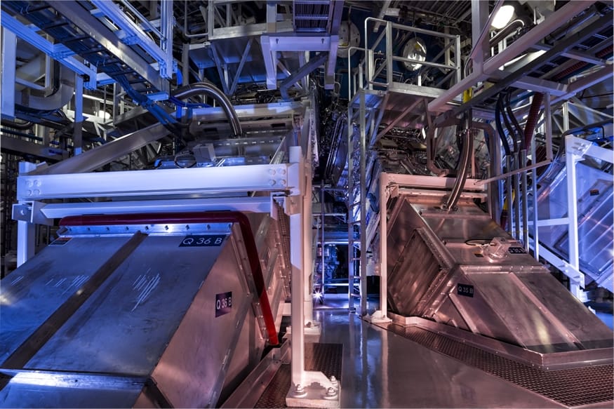
Burning question: Scientists at the $3.5bn National Ignition Facility are nearer to achieving ignition – the point at which fusion reactions generate at least as much energy delivered by the laser system (Courtesy: NIF)
To Omar Hurricane, Annie Kritcher, Alex Zylstra, Debbie Callahan and colleagues at the National Ignition Facility (NIF) in California, US, for taking a step closer to their ultimate goal of realizing “ignition”. Since NIF was turned on over a decade ago, its long-term goal has been to show it can achieve ignition – the point at which fusion reactions generate at least as much energy as its lasers put in. This involves self-sustaining reactions, in which the alpha particles that are also emitted during fusion emit heat to initiate further fusion. NIF, which is operated by the Lawrence Livermore National Laboratory, trains 192 pulsed laser beams on to the inner surface of a centimetre-long hollow metal cylinder known as a hohlraum. Inside is a fuel capsule, which is a roughly 2 mm-diameter hollow sphere containing a thin deuterium-tritium layer. Experiments between 2009 and 2012 fell well short of reaching ignition and so researchers went back to the drawing board to make improvements. That paid off spectacularly on 8 August when researchers achieved an energy yield of more than 1.3 MJ – about 70% of the energy that the laser pulse delivered to the sample. Although still short of break-even, the figure far exceeded previous markers of around 0.1 MJ and some experts have described the result as the most significant advance in inertial fusion since it began in 1972.
To researchers from the Antihydrogen Laser Physics Apparatus (ALPHA) and the Baryon Antibaryon Symmetry Experiment (BASE) collaborations at CERN, for two separate studies presenting new ways to cool particles and antiparticles. The techniques could pave the way for precision studies examining the matter–antimatter asymmetry in the universe. The ALPHA collaboration demonstrated laser-cooling of antihydrogen atoms for the first time. To achieve this, the physicists developed a new type of laser, which produces 121.6 nm laser pulses, to cool the antiatoms. They then measured a key electronic transition in antihydrogen with unprecedented precision, a breakthrough that could lead to improved tests of other key properties of antimatter. The BASE researchers, meanwhile, showed how to extract heat from a single proton via a superconducting circuit connected to a cloud of laser-cooled ions several centimetres away – a technique, they say, that could easily be applied to antiprotons.
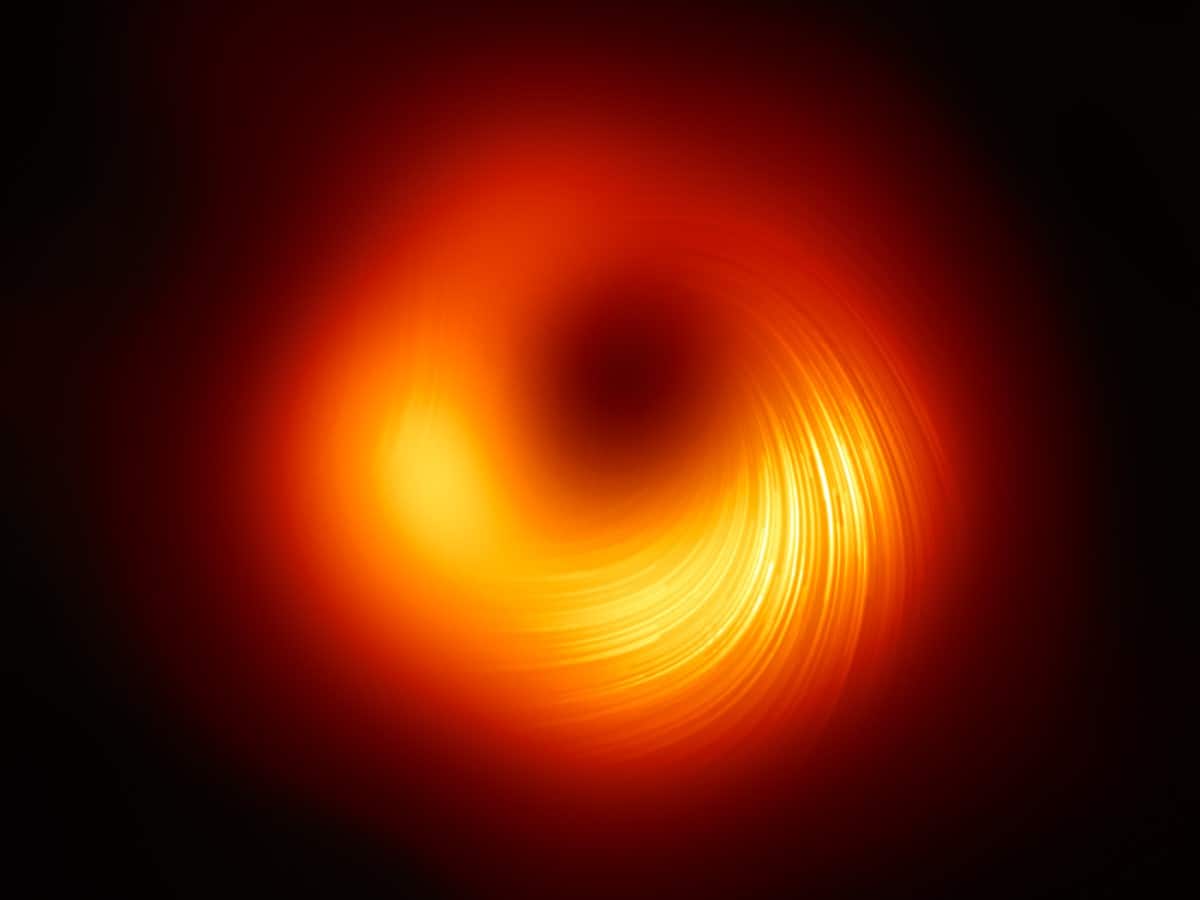
Magnetic swirl: A view of the M87* supermassive black hole in polarized light. The lines mark the orientation of the polarization, which is related to the magnetic field around the shadow of the black hole. (Courtesy: EHT Collaboration)
To the Event Horizon Telescope Collaboration (EHT) for creating the first image showing the polarization of light in the region surrounding a supermassive black hole. The polarization reveals the presence of strong magnetic fields in an area where matter is accelerating into M87*, a black hole more than six billion times the mass of the Sun. Further study of this polarization could provide important insights into how some black holes create huge jets that eject matter and radiation into surrounding space. In 2019 the EHT made history by capturing the first image of the shadow of a black hole, and the collaboration was awarded the Physics World 2019 Breakthrough of the Year for that work.
To Jörg Evers and colleagues at the Max Planck Institute for Nuclear Physics in Heidelberg and the Deutsches Elektronen-Synchrotron – both in Germany – and the European Synchrotron Radiation Facility in France, for being the first to achieve the coherent quantum control of nuclear excitations. The team used X-ray light from a synchrotron that was delivered to the nuclei in two ultrashort pulses. By adjusting the phase of the pulses, the team could toggle iron nuclei between coherent enhanced excitation and coherent enhanced emission. As well as providing a better understanding of quantum matter, the work could hasten the development of new technologies such as ultra-precise nuclear clocks and batteries that can store huge amounts of energy.
To Christian Sanner and colleagues at JILA in the US; Amita Deb and Niels Kjærgaard at the University of Otago; and Wolfgang Ketterle and colleagues at the Massachusetts Institute of Technology in the US, for independently observing Pauli blocking in ultracold gases of fermionic atoms. Pauli blocking occurs in such gases because the constituent atoms fill nearly all available low-energy quantum states, which prevents atoms from making small transitions to neighbouring states. This affects how light scatters from atoms in the gas, and all three teams observed that Pauli blocking increased the transparency of their gases as they were cooled. The effect could someday be used to improve technologies based on ultracold atoms such as optical clocks and quantum repeaters.
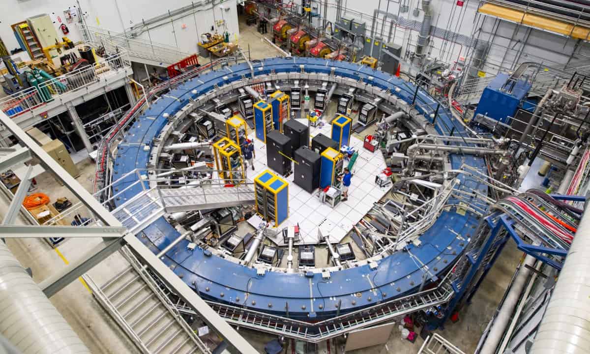
New home: The Muon g-2 ring sits in its detector hall at Fermilab, where it studies precession of muons. (Courtesy: Reidar Hahn/Fermilab)
To the Muon g–2 collaboration for providing further evidence that the measured value of the muon’s magnetic moment disagrees with theoretical predictions. The international team circulated a beam of magnetically polarized muons in a storage ring at Fermilab in the US. The magnetic moments of the muons were rotated by a magnetic field and the rotation rate gave the size of the muon’s magnetic moment. The discrepancy between theory and experiment was first revealed two decades ago at Brookhaven National Laboratory. Now the combined Fermilab/Brookhaven results put the difference between experiment and theory at 4.2σ, which is less than the 5σ required for a discovery. If the discrepancy stands the test of future experiments, it could point to new physics beyond the Standard Model.
Physics World‘s Breakthrough of the Year coverage is supported by Bluefors, a leading supplier of cryogen-free dilution refrigerator measurement systems with a strong focus on the quantum computing and information community. Our aim is to deliver the most reliable and easy to operate systems on the market which are of the highest possible quality.
Two leading quantum companies – Honeywell Quantum Solutions and Cambridge Quantum Computing (CQC) – have merged to form a new business called Quantinuum. The new company, which combines Honeywell’s expertise in quantum hardware with CQC’s software capabilities, has almost 400 employees. Quantinuum claims it is the world’s largest integrated quantum computing company, and has headquarters in both Cambridge, UK, and Colorado, US.
Honeywell’s hardware is based on trapped-ion qubits and the firm had achieved what it said was the highest-ever “quantum volume”, which indicates how many useful calculations a processor can do before breaking down due to decoherence. CQC, meanwhile, has focused on software that can be used in quantum chemistry to, for example, search for new drugs or develop novel carbon-capture materials. CQC’s software is compatible with all types of quantum computers – not only those using trapped ions – and Quantinuum will be similarly “platform agnostic”.
As well as having a majority stake of 54% in Quantinuum, Honeywell has additionally invested almost $300m in the new company. Quantinuum is due to launch a quantum cryptography product this month, and a software package in 2022 aimed at corporate clients in various industries from pharmaceuticals to agrochemicals.
Ilyas Khan, who founded CQC and is now chief executive of the merged business believes that the new firm will help “to bring real, quantum computing products and solutions to large, high-growth markets as quantum computers scale in capacity and quality”. Meanwhile, Tony Uttley, who was head of Honeywell Quantum Solutions, becomes president and chief operating officer of Quantinuum. “I am thrilled to help lead our new company, which will positively change the world through the application of quantum computing,” he said.
This merger is a promising sign for the industry, according to James McKenzie, who was vice president for business at the Institute of Physics from 2016-2020. “It shows commercial relevance and fuels further investment,” he says. “Honeywell is a serious international business with a long-running and strong track record in commercialization of technology, so an acquisition or investment by them is a very positive statement about the UK quantum sector.”
This news also marks a big milestone in quantum computing for Najwa Sidqi, who is knowledge transfer manager for quantum technologies at KTN, a UK-based organization that aims to drive innovation by promoting networks between hi-tech firms and other stakeholders. “This merger will speed up the deployment of the technology and perhaps also access to quantum computing for end users,” she says. “It brings together strong talent and skillsets from both Honeywell and CQC, and I look forward to seeing where it goes in future.”
What do the heart and the vocal cords have in common? They are constantly vibrating, albeit at different frequencies. While biological tissue is tough enough to withstand these vibrations, designing synthetic tissue that can survive such a dynamic environment remains a major challenge in regenerative medicine.
Scientists from McGill University have developed a new type of injectable hydrogel that is resistant to prolonged, high-frequency stimulation. Describing their findings in Advanced Science, the researchers believe that the hydrogel’s unique combination of properties, including mechanical integrity, high porosity and cell compatibility, could one day enable the repair of muscular tissues such as the heart and vocal cords.
“Our work highlights the synergy of materials science, mechanical engineering and bioengineering in creating novel biomaterials with unprecedented performance,” says Jianyu Li, who led the team alongside colleague Luc Mongeau.
“We hope that one day the new hydrogel will be used as an implant to restore the voice of people with damaged vocal cords, for example laryngeal cancer survivors,” adds co-first author Guangyu Bao.
The researchers (from left to right): Guangyu Bao, Luc Mongeau, Jianyu Li and Sareh Taheri. (Credit: Guangyu Bao)
Putting the hydrogel through its paces
Our vocal cords vibrate to produce sounds at a frequency of 100 to 300 Hz. Many hydrogels break down under these conditions due to their porous structure. Pores are an essential feature of implantable biomaterials because they allow oxygen and nutrients to perfuse through the implant, which is crucial for cell survival. Unfortunately, porosity often comes at the expense of mechanical strength, especially under dynamic loads.
To combat this, the researchers developed a specific type of hydrogel called a porous double-network (PDN) hydrogel. The two polymer networks used to form the PDN, chitosan (a sugar found in the outer skeleton of shellfish) and glycol–chitosan, are difficult to separate from one another. This results in a stretchy material that resists fracture. In fact, PDNs can resist millions of cycles of mechanical loading compared to their porous single-network (PSN) counterparts.
“Most synthetic materials developed so far for vocal cord repair are based on single-network hydrogels,” Bao tells Physics World. “Currently, patients may need periodic injections because those hydrogels do not last long. Our hydrogel offers a five to 40-fold increase in toughness, which could potentially improve the retention of the hydrogel after injection.”
Using a perfusable vocal cord bioreactor, the team subjected the PDN and a chitosan-based PSN to over six million cycles of 120 Hz vibrations – that’s the equivalent of talking for two hours every day for a week. Not only did the PDN remain intact after loading, but cells encapsulated within the hydrogel were viable throughout the seven-day period. On the other hand, the PSN fractured into pieces.
Accelerated wound repair
What sets the PDN apart from other injectable double-network hydrogels is its ability to form cell-sized pores upon injection. Injectable hydrogels are desirable in many areas of regenerative medicine, from drug delivery to lab-on-a-chip disease models. Most injectable hydrogels form nanosized pores, meaning that they suffer from low permeability. The team found that cells could not proliferate inside a nanoporous glycol–chitosan hydrogel, but could inside the PDN.
“We utilize the phase separation behaviour of chitosan to create pores on the microscale,” explains Bao. “This feature can facilitate cell recruitment from native tissues to help accelerate wound repair.”
By varying the concentration of glycol–chitosan, the researchers could further control key parameters like stiffness and porosity. They also found that their fabrication technique was compatible with other types of polymers, such as gelatin.
Future steps
The team believes that this study could form the basis of a new class of tough, injectable hydrogels. After investigating the biocompatibility, biodegradability and theurapeutic performance of the PDN in animal models, the researchers hope to apply for permission to perform clinical trials.
While their study focused on frequencies relating to vocal cords, they emphasize that the hydrogel is tough enough to repair other soft tissues under dynamic loading.
“People recovering from heart damage often face a long and tricky journey. Healing is challenging because of the constant movement tissues must withstand as the heart beats,” says Bao. “We are curious to know [the hydrogel’s] efficacy in cardiac tissue engineering and welcome collaboration with scientists in those fields.”