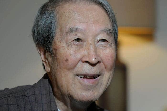
The Japanese Nobel-prize-winning physicist Toshihide Maskawa died on 23 July at the age of 81. Maskawa shared half the 2008 Nobel prize with the Japanese physicist Makoto Kobayashi for their work on the mechanism of “broken symmetry” that led to the prediction of a new family of quarks. The other half was awarded to the Japanese–American particle physicist Yoichiro Nambu for applying spontaneous symmetry breaking to particle physics.
Born in in Nagoya, Japan, Maskawa studied physics at Nagoya University and earned a PhD in particle physics from the university in 1967. In 1970 he then moved to Kyoto Univeristy before heading to Kyoto Sangyo Univeristy from 2003 to 2009. From 2010, Maskawa became the first director of the Kobayashi-Maskawa Institute for the Origin of Particles and the Universe at Nagoya University, a position he held until 2018.
Broken symmetries
Symmetry breaking seeks to explain the subtle differences in physics that enables matter to tip the balance with antimatter in the universe. Charge (C) symmetry involves particles behaving like their oppositely charged antiparticles, while “parity” (P) symmetry means events should be the same when the three spatial co-ordinates x, y and z are flipped.
Physicists were sure that all these symmetries held for elementary particles but in the 1950s, physicists discovered that charge symmetry breaks in the weak interaction, which governs radioactive beta decay. In 1956 the theorists Tsung Dao Lee and Chen Ning Yang suggested that P symmetry might break in the weak interaction, which governs radioactive beta decay. Soon after, a famous experiment by Chien-Shiung Wu and colleagues at Columbia University showed that, during beta decay, cobalt atoms emit electrons in a preferential direction. The result validated Lee and Yang’s beliefs and bagged them a Nobel in 1957.
Despite the proof that symmetries could at least be broken individually, most assumed that combined parity and charge symmetry, or so-called CP symmetry, would hold. But in 1964 tests on the radioactive decay of particles known as kaons showed that even CP symmetry could break, a result that won physicists James Cronin and Val Fitch the Nobel prize in 1980. It is the theory that explains this broken symmetry that has handed the 2008 prize to Maskawa.
In 1972, using calculations based on quantum mechanics, Maskawa and Kobayashi formulated the 3 × 3 matrix that describes how the strange quark and down quark inside a kaon can switch to and fro into their antiparticles and, in doing so, occasionally break CP symmetry. Moreover, the mixing in the matrix implied the existence of new quarks – the charm, bottom and top – all of which were discovered over the following decades. The matrix became known as the “CKM” matrix (the “C” being named after the Italian physicist Nicola Cabibbo who proposed the concept of “quark mixing”).
In 2008 Maskawa and Kobayashi shared half of the Nobel prize “for the discovery of the origin of the broken symmetry which predicts the existence of at least three families of quarks in nature.” The other half was awarded to Nambu from the University of Chicago, “for the discovery of the mechanism of spontaneous broken symmetry in subatomic physics”.
Maskawa was not widely known outside of Japan, and much of his work is in his native language. Indeed, it is thought that he had never travelled abroad and only got a passport in order to attend the Nobel prize ceremony in Stockholm in 2008. Yet according to Tim Gershon of the University of Warwick in the UK, Maskawa had an “enormous influence” in his home country. “This helped to make Japan the powerhouse of fundamental science that it is today,” adds Gershon. “Maskawa leaves an important legacy.”
Radiation therapy plays a vital role in the treatment of more than 50% of all cancer cases. But there’s a massive disparity in access to radiotherapy equipment as a function of a country’s income. In low- and middle-income countries, much of the population lacks access to radiation therapy. And in low-income countries, only 10% of patients have access to radiotherapy.
In a dedicated session at the AAPM Annual Meeting, speakers described some of the projects aiming to reduce the overall cost of delivering radiotherapy and improve access to effective cancer care for patients across the world.
Low-energy options
One potential way to lower the cost of radiotherapy could be to replace the expensive and bulky linear accelerators (linacs) used to deliver treatment with low-cost, lower-energy kilovoltage (kV) X-ray tubes, of the type used in diagnostic imaging systems.
Michael Weil from Sirius Medicine and Precision RT described just such a device: the “linear converging radiotherapy system”, or LCRS. “Why go to the effort of developing low-energy beams to treat cancer? The answer comes down to price, pictures and process,” he told the AAPM delegates.
While kV beams are historically considered unsuitable for radiotherapy, “low-energy beams can be manipulated in potentially useful ways for therapy,” said Weil. He described initial studies using high-Z contrast agents to enhance the dose to the target. Intra-lesion injection of such contrast under CT guidance, followed by delivery of kV X-rays at a modest skin dose, could provide “exquisite conformity of the high-dose region”.
Weil and collaborators used this approach to treat advanced cancers (under a palliation protocol), in 23 human patients and 80 veterinary patients with spontaneous tumours. They observed low dose to skin and significantly higher dose to deeper tumours. “Using conventional radiosurgery dosing regimens, we were confident that this approach could safely treat beneath the skin,” he said.
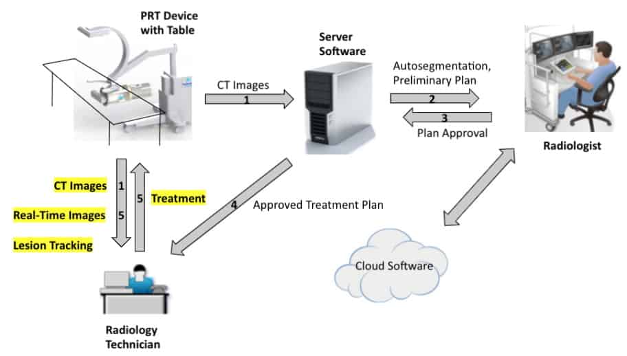
The team has now built a prototype LCRS, with the goals of cost reduction – using less expensive hardware and infrastructure – and dramatically improving the workflow. As the treatment source can also be used for real-time tracking, tighter margins can safely be used. The system also offers the possibility of performing planning and treatment in a single visit, with the ultimate aim of completing CT imaging, planning and treatment delivery in less than 1 hr.
Improving the workflow reduces the costs. The linear converging radiotherapy system aims to perform imaging, planning and treatment delivery in less than an hour. (Courtesy: Michael Weil)
The LCRS uses a magnetically swept electron beam aimed at a stationary tungsten target to create X-rays collimated in a single plane. The researchers tailored the beam geometry to spread out the entrance dose and concentrate the beam at the lesion. They also incorporated a flat-panel detector for CT, fluoroscopy and tomosynthesis. They note that the system’s footprint is similar to a standard CT system, though power and cooling requirements are greater.
To demonstrate the feasibility of treatment delivery with this kV source, the team simulated radiotherapy plans for breast and lung lesions (without contrast) using the kilovoltage arc technique (kVAT) and conventional volumetric modulated arc therapy (VMAT). For both cases, the most noticeable difference was a plateau in maximum dose for VMAT and a higher peak dose in the target with kVAT.
“We believe that this extra intra-lesion dose is beneficial in controlling hypoxic, relatively radioresistant central tumour cells,” said Weil.
Focused X-ray beams
Also looking to utilize a low-energy X-ray source, Mohammad Salehpour from the MD Anderson Cancer Center described a novel focused X-ray beam technology developed in collaboration with medical start-up company Convergent Radiotherapy and Radiosurgery (CRnR). The idea here is to combine a CT X-ray tube with an X-ray lens to create a converging 60 keV X-ray beam focused on the tumour target.
“An X-ray source such as used in a CT scanner will create a diverging beam of radiation,” Salehpour explained. “But if I had an X-ray lens, then I could create an intense beam at the focal point.”
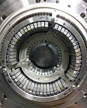
An X-ray lens made from rings of single aluminium crystals. (Courtesy: Ze’ev Harel, Convergent RnR)
CRnR has developed a proprietary focusing lens, composed of several concentric rings formed from tiles of single aluminium crystals, arranged to reflect the X-ray beam onto the target. Simulations of the 3D dose distribution showed a low-dose region slowly increasing to a high-dose focal spot in the centre before reducing again.
Salehpour notes that the depth–dose distribution resulting from a focused X-ray beam resembles the Bragg peak seen in proton therapy, with a higher intensity at depth than at the beam entrance or exit. For clinical purposes this enables delivery of high dose to the tumour and low dose to surrounding normal tissue. It’s also possible to stack these peaks to create a “photon spread-out peak”.
Salehpour and colleagues tested the X-ray lens in a water phantom containing dosimetry film, noting that the results reflected those seen in the simulations. Films irradiated by 60 keV X-rays through the lens exhibited dose circles at a depth of 20 mm, converging to a bright spot at 65–70 mm and then back to larger circles at greater depths.
“Considering the question ‘can we focus X-rays as we would focus rays of sunlight’ – the answer is yes,” said Salehpour. The team has now created a first-generation prototype with the X-ray tube and lens a housed in a robotic arm. They also added laser beams onto the robot, with the beams converging at the X-ray focal spot. As this system is based on diagnostic X-ray tubes, it requires far less shielding than other radiotherapy devices.
“Because it has a small footprint and low shielding requirements it can be made mobile and moved to different locations,” said Salehpour.
In addition to radiotherapy, where small shallow tumours are ideal treatment candidates, the technology could have many other applications. These include, for example, treating the eye condition age-related macular degeneration, combination with immunotherapy, cardiac ablation, or dose enhancement using gold nanoparticles.
Exploiting automation
Alongside the global inequality in access to radiotherapy equipment, there’s a similar gap in the availability of trained staff. To effectively expand access to radiotherapy, the introduction of lower cost radiotherapy systems must be accompanied by efforts to address of other resource issues. Tucker Netherton from the MD Anderson Cancer Center took a look at how artificial intelligence (AI) and automation could help narrow this radiotherapy gap.
Two approaches available immediately are “education” and “tele-radiotherapy”, he explained. In education, for example, the IAEA is currently developing a global curriculum for radiation oncologists, medical physicists and radiation therapists. Examples of tele-radiotherapy – defined as the use of telecoms and IT to provide radiotherapy support from a distance – include web-based treatment planning services and virtual tumour boards.
“By providing accessible treatment planning through tele-radiotherapy, automation can be used to offset task-related burdens for resource-limited clinics, saving time and increasing efficiency,” Netherton explained.
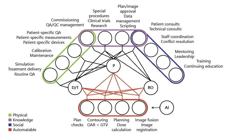
Examining which aspects of the treatment planning process could be automated, Netherton and colleagues concluded that AI can perform tasks such as plan checks, normal tissue and target volume contouring, planning and image registration. He noted that many vendors and researchers are already developing deep-learning based contouring tools with comparable performance to manual approaches.
Automated treatment planning tools, meanwhile, could address the gap in resources by providing web-based access to planning services. With this goal, MD Anderson has created the Radiation Planning Assistant (RPA), a fully-automated contouring and radiotherapy treatment planning tool. The goal of the RPA, which is currently in research phase, is to make its services available to clinics at no cost. The clinic simply uploads the CT and plan order and the RPA returns a complete treatment plan. Impressively, the RPA can complete 483 cervix plans or 258 head-and-neck plans in 24 hr.
One major caveat to tele-radiotherapy is that Internet access is needed to access such tools. But Netherton notes that the number of Internet users is increasing globally and many efforts exist to “connect the disconnected”, including those by Google, SpaceX, Microsoft and the World Bank.
“Tele-radiotherapy can increase safety, efficiency and access to radiotherapy tools to provide resources to resource constrained clinics,” Netherton concluded.
As the critical dimensions of semiconductor devices are approaching the atomic scale, continuous improvement of device performance can no longer rely on straightforward device shrinkage. The introduction of new three-dimensional structures and new materials, in addition to further reduction of device sizes, is now the main driving factor for higher device performance. The reduction of CO2 and other greenhouse gases is another growing challenge in the semiconductor manufacturing industry as the cost and energy consumption of device manufacturing continue to grow.
In this talk, after reviewing the current trend and challenges in the manufacturing processes of advanced semiconductor devices, Satoshi Hamaguchi will discuss the outlook of future technologies in this arena, especially plasma-processing technologies. It is now clear that the manufacturing processes strongly demand atomic-scale accuracy with little or no damage to the materials in device fabrication. The discussion will be focused on how the fundamental science of plasma surface interaction can contribute to the development of such technologies.
Satoshi Hamaguchi has been Professor of Engineering at the Center for Atomic and Molecular Technologies, Graduate School of Engineering, Osaka University, Osaka, Japan, since 2004. He has been working on analyses of plasma surface interaction for semiconductor processing and surface modification of biomaterials, using modelling, numerical simulations, and beam and plasma experiments. His interest also includes plasma medicine, nuclear fusion, and AI and machine learning applications in plasma science and technologies.
Prior to joining Osaka University, Dr Hamaguchi was Associate Professor of Energy Science, Graduate School of Energy Sciences, Kyoto University, Kyoto, Japan, from 1998 to 2004, Research Staff Member of Thomas J. Watson Research Center, IBM Research Division, IBM at Yorktown Heights, New York, USA, from 1990 to 1998, and Research Fellow at the Institute of Fusion Studies, University of Texas, Austin, Texas, USA, from 1988 to 1990.
Hamaguchi holds a PhD in mathematics from Courant Institute of Mathematical Sciences, Department of Mathematics, New York University, and a PhD in physics from the University of Tokyo. He is a Fellow of the American Vacuum Society and American Physical Society.
Researchers at University of Illinois Urbana-Champaign have developed a novel trimodal brain imaging system, incorporating electroencephalography (EEG), functional magnetic resonance imaging (fMRI) and a third technique, event-related optical signal (EROS) imaging, reporting their findings in Human Brain Mapping.
The development of multimodal neuroimaging
Brain function was first measured almost 100 years ago using crude electrodes placed on the human scalp. Since then, EEG has become a routine clinical and research tool, penetrating the electrophysiology of the human brain. EEG measures the change in potential at the scalp, caused by the firing of neurons within the cortex, with millisecond temporal resolution. However, this signal results from a smeared summation of signals from within the brain, due to the different conductivities of the scalp, skull and cerebral-spinal fluid. This means that EEG cannot precisely trace signals back to the region of the brain that produced them.
By 1990, functional neuroimaging reached high spatial resolution with the development of fMRI, allowing brain activity to be traced to its origin with millimetre precision. fMRI measures the blood oxygenation level-dependent (BOLD) response, which relates the change in oxygenation of cortical blood to brain activity. As this method probes the haemodynamic response of the brain, rather than the electrophysiology, there is an inherent time delay of approximately 5–8 s between activation and the measured BOLD signal.
More recently, fMRI-compatible EEG instrumentation has facilitated simultaneous EEG and fMRI, capitalizing on the temporal resolution of EEG and the spatial resolution of fMRI. However, how the signals from these two modalities relate to one another is poorly understood.
Introducing EROS
EROS uses near-infrared light sources and detectors placed on the scalp to measure changes in ion transport during neural activity. Ion transport causes neurons to swell and shrink, subsequently scattering the light. By measuring these changes in light scattering, the technique can infer the location and temporal characteristics of cortical activity. While stand-alone EROS suffers from low sensitivity, incorporating it with EEG and fMRI clarifies the links between these two different measures.
To create a proof-of-concept trimodal system, the scientists integrated EROS sensors (based on optical fibres and photomultiplier tube light detectors) into an MRI-compatible EEG–EROS cap. The cap, seen in the image above, does not produce any artefacts during the scanning.
University of Illinois Urbana-Champaign psychology professors (from left) Florin Dolcos, Gabriele Gratton and Monica Fabiani, with postdoctoral researcher Matthew Moore, developed a new method to simultaneously image the brain using three technologies. (Courtesy: L Brian Stauffer)
The team tested this novel system using an emotional oddball experiment. This required participants to identify “oddball” circles presented on a screen in a string of standard (scrambled) and distracter (emotional and neutral) images. This type of stimulus is known to induce different spatial and temporal responses, making it ideal for testing the trimodal system. The researchers note that EROS is expected to be spatially similar to the fMRI data and correspond temporally with EEG.
The EROS results identified a similar spatial response to the emotional and target stimuli in the frontal cortex as the fMRI, but provided a temporal resolution similar to EEG, whose signals originated from a different location in the brain. These findings demonstrate that introducing a third simultaneous imaging modality can expand our understanding beyond that which bimodal investigations are able to achieve.
The researchers highlight the considerable advantages of the trimodal system: the ability to overcome the limitations of each imaging modality; clarifying the links between each measure; and providing an excellent platform for future investigations into the spatial and temporal characteristics of brain mechanisms associated with healthy function and disease.
Evidence of a possible moon-forming region has been discovered surrounding a giant, newly forming exoplanet. Using the Atacama Large Millimeter/submillimeter Array (ALMA) telescope in Chile, astronomers have spotted a disc of dust surrounding the recently-discovered exoplanet PDS70c. This exoplanet orbits the young star PDS70, which is 370 light-years away. The observation was made by an international team led by Myriam Benisty, who is at the Universities of Chile and Grenoble. The discovery could lead to important insights into how moons and planets form and evolve within young star systems.
The discs of gas and dust surrounding young stars often have rings, gaps, and spiral arms that are carved out by newly forming planets. These planets can also acquire discs of their own, and astronomers believe that moons can form in these discs, carving out their own rings and other structures. So far, however, this process has never been observed directly.
In 2018 and 2019, the ESO’s Very Large Telescope (VLT) made the first observations of two exoplanets yet to fully form, through direct infrared images of the disc surrounding PDS70. Since then, the giant, Jupiter-like planets have been observed using a variety of other techniques. Among these were observations of hydrogen-alpha light emission, which only occurs in regions where hydrogen is being ionized. This suggested that the planets were still accreting material.
Cool dust grains
In their study, Benisty’s team present the latest observations of PDS70 by ALMA, which can pick up millimetre wavelengths emitted by cool dust grains. With resolutions as high as 2.3 au (Earth-Sun distances), the ALMA’s images clearly show a disc surrounding the outermost exoplanet, PDS70c, with an outer radius no larger than 1.2 au.
Depending on the size of the dust grains, the researchers reckon that the disc could contain anywhere between 0.7% and 3.1% of Earth’s mass – enough material to form up to three satellites with similar masses to Earth’s Moon. In addition, the material in PDS70c’s disc is well within the radius where it would be retained by the exoplanet – providing ideal conditions for a moon to form.
Starved of dust
The innermost exoplanet, PDS70b, did not display any clear evidence for a circumplanetary disc. According to Benisty’s team, this could mean that the planet has a far smaller radius within which orbiting material can be retained. Alternatively, it could have been starved of dust grains by PDS70c, whose orbit is better placed to access material within PDS70’s circumstellar disc.
Further observations of the system offer astronomers a unique opportunity to study the formation of planets and moons through direct images – and this could improve our understanding of how moons form around young gas-giant planets. Benisty and colleagues now look forward to exploring the system in more detail using the ESO’s upcoming Extremely Large Telescope (ELT), which is currently under construction in Chile and should switch on in 2027.
As you listen to this episode of the Physics World Weekly podcast, your phone will be struck by particles created by collisions of cosmic rays with atoms in the atmosphere. The vast majority of these particles have no effect on digital electronics, but very occasionally they can flip a bit. While this is usually harmless, it can have dire effects on critical systems such as those in aircraft and driverless cars – as Physics World’s Tushna Commissariat explains.
In 2021, 6.1% of electricity worldwide was generated by wind – and the percentage can be much higher in windy countries like Denmark and the UK. However, as the size and number of wind farms continues to increase, engineers must be mindful that the efficiency of a wind turbine can be reduced significantly by the presence of its neighbours, as Enrico Antonini of the Carnegie Institution for Science, Department of Global Ecology at Stanford University explains.
Also in this episode, we chat about the latest discovery in twistronics; that twisted trilayer graphene may be a rare type of superconductor.
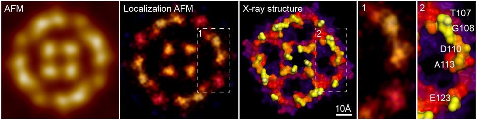
Resolution boost: localization AFM map (second from the left) of the AqpZ molecule. Also shown are a simple AFM image, the AgpZ structure derived from X-ray crystallography and a comparison of LAFM and X-ray results in a detail of the LAFM image. (Courtesy: Simon Scheuring/GR Heath et al. Nature 594 385)
The conventional resolution limit for biological samples in atomic force microscopy (AFM) has be broken without needing to improve the physical experimental setup. George Heath and his team at Weill Cornell Medicine in New York City have done this by applying concepts of localization microscopy to molecular AFM studies and showed that AFM images can be given new life. The research, led by Simon Scheuring, is described in a recent paper in Nature.
Seeing molecular structure under the right conditions
Biomolecules are complex, dynamic structures that are difficult to study because they must be observed under conditions mimicking the environment of a real biological cell. Furthermore, sufficient resolution is needed in both space and time to fully capture molecular behaviour. AFM can already be used to image molecules at physiologically relevant temperature and pressure, and it can also be used in a high-speed mode (HS-AFM), where sufficient temporal resolution can be reached to observe conformational changes of molecules in real time.
The remaining challenge had been obtaining sufficient spatial resolution at the sub-molecular scale. Until recently, AFM has been limited by the radii of AFM tips, the smallest of which are on the order of a few nanometres, and hence not sharp enough to resolve significant details on a single, corrugated, soft and flexible biomolecule in liquid. However, inspired by other super-resolution fluorescence microscopy techniques, Heath and his colleagues have now shown that resolution down to the angstrom-range (10-10 m) can be extracted from AFM images using a post-acquisition image reconstruction technique dubbed localization AFM (LAFM).
The impact of post-acquisition reconstruction
The LAFM approach is based on the idea that resolution superior to physical limitations can be achieved by determining the localization of isolated signals with high spatial precision using localization algorithms to process several images of the sample. The results are then merged to create a compiled map. Specifically for AFM, these signals are local maxima in the height traces – the locations where the force between the tip and the sample is largest. These are extracted from several images at specific locations and then merged in a peaking-probability map.
Scheuring describes the technique: “Imagine the high-resolution molecular features were marbles on a table, and you were trying to sense them by scanning a basketball over them. You most likely cannot resolve them. However, if you imagine that they, like individual ping-pong balls, stochastically bounce up, you will be able to localize them individually. Merging later all localization coordinates, you will be able to reconstruct the pattern of the marbles.”
New insight into biomolecular properties
Using LAFM, the team resolved single features in the surface protruding loops from aquaporin-Z (AqpZ) tetrameric channels. AqpZ is a protein that acts as a water channel in cell membranes. They were able to resolve details comparable to those in X-ray crystallographic structures, details that were previously undetectable in the AFM images (see figure). The team also produced videos showing pH-dependent conformational changes in the molecule CLC-ec1, which is found in many organisms and cells. This provided important information about the transport mechanism of this dimeric transporter molecule.
A LAFM map can be reconstructed either by recording many molecules in a few frames, or by observing a single molecule over a longer time. This makes it possible to observe either time- or environment-dependent conformational changes, but also acquire high-resolution information of individual molecules. Such capabilities can help to significantly advance understanding of biomolecular structure and function in the future, and Scheuring hopes that LAFM maps will become the new standard for biomolecular imaging.
Will it ever be feasible to emulate Harry Potter’s levitation charm, “Wingardium Leviosa”, and manoeuvre tiny objects by scientific means? The manipulation of nanosized materials such as biological molecules, quantum dots and other inhomogeneous nanoparticles has been achieved via several approaches, including magnetic, electric and acoustic fields. The major drawback of many existing techniques, however, is their inability to control nano-objects over a large area. Furthermore, it is almost impossible to trap the tiniest particles and manipulate them over a large scale without creating additional background interference.
To overcome these limitations, scientists at Duke University developed a hybrid technology known as acoustoelectronic nanotweezers (AENT) to dynamically manipulate sub-100 nm particles. Instead of using sound waves to directly move the nanoparticles, AENT uses sound waves to create electric fields that provide the push.
AENT combines the benefits of electric tweezers and acoustic fields to precisely pattern the nanoparticles, while also controlling their orientation before they are eventually used for downstream applications – such as designing sensors to investigate disease, building devices for nanoelectronics and understanding particle–particle interactions.
In the past decade, the focus of nanomanipulation technology has been to capture nanomaterials and investigate their properties over a small area. However, with the AENT technology, scientists can now transport the trapped nanoparticles over larger distances and perform several tests at different positions and times. Most importantly, the researchers are able to trap particles smaller than 10 nm while minimizing the interference that often occurs in such devices due to acoustic streaming. Using this technology, it is possible to study DNA, proteins, exosomes and single nanoparticles at a high resolution.
Previous studies reported that combining acoustic and electric fields to trap particles could lead to excessive disturbances due to the forces acting on the nanomaterials. However, the team devised a means of minimizing this hydrodynamic interference by deploying integrated transducers on a piezoelectric (PZE) layer. This technique generates elastic deformations that propagate as acoustic waves through the PZE-layer and establish periodic electric fields.
‘’This method enabled us to perform levitation, transformation, particle pairing and rotation at a large [centimetre] scale with minimal hydrodynamic perturbations,’’ explains Tony Jun Huang, senior author of the study, details of which are published in Nature Communications.
To test the efficiency of the AENT technology, the researchers exposed polystyrene particles (100 nm) to acoustic waves in both electrically shielded and non-shielded regions. Less than 2 s after exposure, the particles were seen to form distinct linear patterns in the non-shielded region, while particles in the shielded region were seen to be stationary after their trapping.
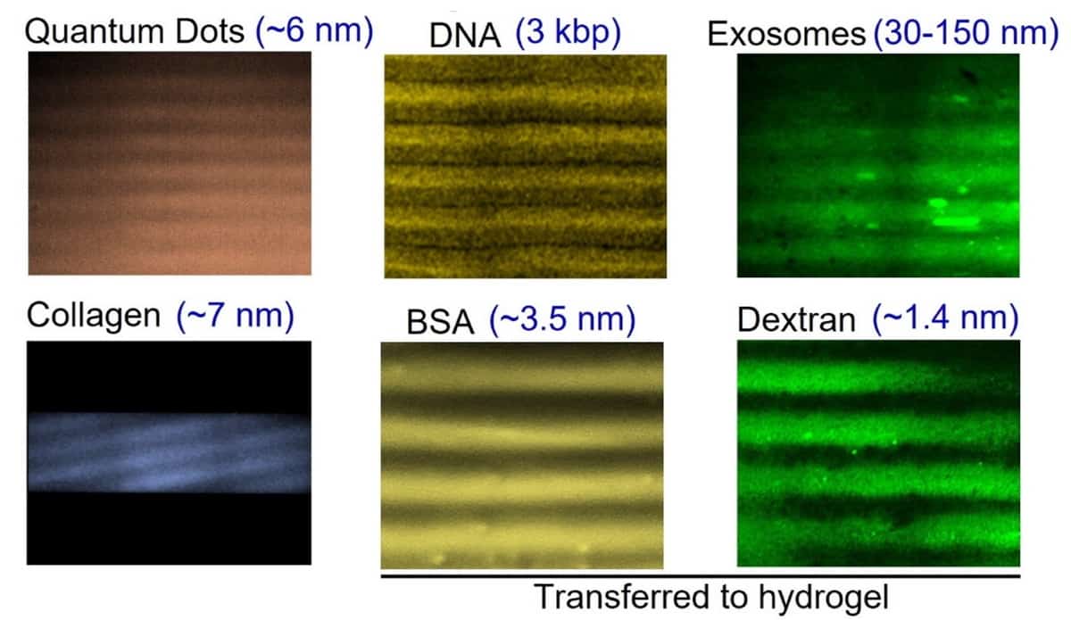
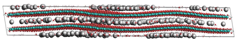
The patterns formed by nanosized biomaterials, such as quantum dots, DNA, exosomes, collagen, BSA and dextran, after nanomanipulation by AENT. (Courtesy: CC BY 4.0/Nat. Commun. 10.1038/s41467-021-24101-z)
The team also manipulated several other nanoscale objects including quantum dots (6 nm), exosomes (30–150 nm), silicon dioxide gold nanobeads (100 nm) and graphene flakes (30–40 nm), and observed similar patterns.
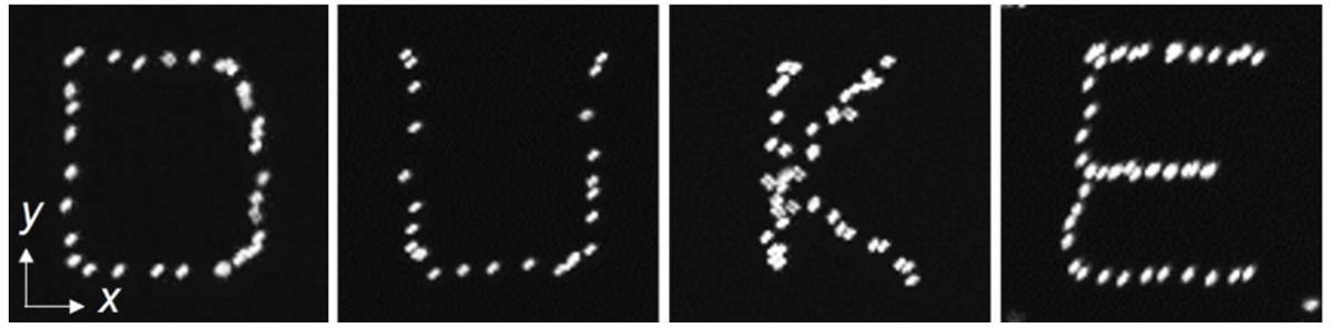
To demonstrate highly specific manipulation, the team managed to dynamically control single particles and push them around to spell out the letters D, U, K, E. The researchers believe that AENT holds great potential as a next-generation nanomanipulation technology that will find application across many disciplines, including nanofabrication, electronics, molecular physics, chemistry, metamaterials and biomedicine.
Researchers at Duke University manipulate nanoparticles in a dynamic manner to spell out the letters D, U, K and E. (Courtesy: CC BY 4.0/Nat. Commun. 10.1038/s41467-021-24101-z)
Understanding host–guest interactions of layered inorganic solids ushered in the modern era of portable electronics powered by lithium ion batteries. Future improvements in the power capability of these devices, and their potential use in emerging technologies such as water treatment, critical element recovery and ion-based electronics, depend on the development and understanding of new ion insertion hosts with fast insertion kinetics. One avenue is via tuning the interlayer environment of a layered material, since many are flexible hosts, whose interlayers can accommodate not just electrolyte ions but also solvents, organic molecules, polymers and organometallics.
This webinar reviews the Augustyn Research Group’s investigation of the mechanistic understanding of water-mediated ion intercalation in transition metal oxides. Topics covered include the role of ordered and confined water networks in transition metal oxide hydrates; design of metastable hydrated oxides via displacement of interlayer ions by water; and the role of disordered and confined water networks in electrochemical capacitor materials. The fundamental understanding of these materials, which blur the distinction between solid and liquid, paves the way for electrochemical ion insertion applications with high-power capability.
Veronica Augustyn is an Associate Professor of Materials Science and Engineering and a University Faculty Scholar at North Carolina State University (NC State), US. From 2013–2015, she was a Postdoctoral Fellow at the University of Texas at Austin, US. She received her PhD from the University of California, Los Angeles, US, in 2013 and BS at the University of Arizona, US, in 2007, both in Materials Science and Engineering. Her research group focuses on the design, synthesis and characterization of materials for electrochemical energy technologies including batteries, electrochemical capacitors, electrolyzers, and fuel cells. In particular, Augustyn is interested in the relationships between material structure, composition and morphology, and the resulting electrochemical mechanisms. She leads SciBridge, an award-winning international project at NC State that develops renewable energy research and education collaborations between universities in Africa and the United States. Her research group was recognized with a 2019 Sloan Fellowship in Chemistry, 2019 DOE Early Career Award, and 2017 NSF CAREER Award. In 2021, she was named an NC State Goodnight Early Career Innovator and received the George H Blessis Advising Award for her mentorship of undergraduate students.
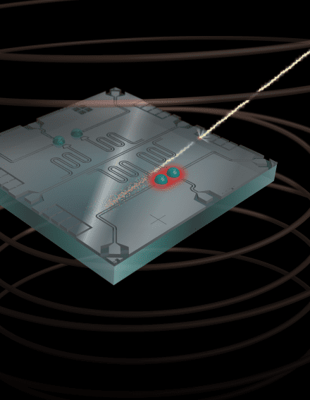
Cosmic intruders: Energetic particles from space and natural background radiation can trigger hard-to-correct errors when they collide with chips containing superconducting qubits. (Courtesy: Chris Wilen)
Quantum computers may need a redesign to protect them from background radiation, say physicists. After earlier experiments showed that cosmic rays can severely disrupt the operation of superconducting quantum bits (qubits), an international team led by Robert McDermott of the University of Wisconsin-Madison, US, has now concluded that a leading error-correction method is unlikely to fix the problem on its own. Instead, McDermott and colleagues suggest that a combination of shielding and changes in qubit chip design may be required to keep errors at a manageable level.
Cosmic rays have created headaches in classical computing for decades. When these energetic particles fly in from space and strike a silicon computer chip, one or more bits in the chip may change state, or flip, in ways that programmers never intended. If these errors go uncorrected, damaging glitches may result – including, in one case, injuries to passengers on a Qantas flight after a bit-flip error fed incorrect data to the aeroplane’s instruments.
Surface code error correction
For quantum computers, the problem is more complicated since qubit states can flip in two directions (representing the X and Z axes) rather than one. Nevertheless, a form of error correction known as a two-dimensional surface code should, in principle, be able to handle qubit flips as long as the quantum processor meets certain requirements.
Surface code error correction works by encoding information in a flat sheet of superconducting qubits, each of which is connected to its nearest neighbours. If the error rates of individual qubit operations are low enough, it should be possible to use some of these qubits to identify and correct errors in neighbouring qubits via multi-qubit operations. The other requirement is that errors cannot be correlated – in other words, an error that affects one qubit cannot affect its neighbours at the same time.
Unfortunately, McDermott’s team discovered that errors caused by cosmic rays and gamma rays from background radiation do not meet this second condition. “We basically are finding multiple mechanisms for correlated errors,” Chris Wilen, a PhD student at Wisconsin and the lead author of a new study about the research, tells Physics World.
Quasiparticle poisoning
To study these correlated errors and quantify their effects, McDermott and colleagues constructed a chip containing two pairs of qubits: one pair separated by 340 μm, the other by 640 μm. While performing quantum operations on this four-qubit system, the physicists observed numerous simultaneous jumps in the charge induced on the paired qubits. When they modelled these bursts of charge using a standard particle-physics toolkit, they determined that the correlated jumps stem from collisions between the chip and a mixture of gamma rays and cosmic rays.
The probability of correlated jumps was highest for the qubit pair with the smallest physical separation, indicating that spacing qubits further apart reduces the direct effects of energetic particles striking the chip. However, the group also encountered a thornier problem: the energy released in these strikes ultimately gets transferred to the qubit substrate in the form of phonons, which are vibrations in a material and can lead to the creation of quasiparticles. As these phonons spread, they produce other kinds of correlated errors, and these errors affect the entire millimetre-scale chip. This phenomenon is known as quasiparticle poisoning, and Wilen says it “could be really damaging for error correction” unless it can be mitigated.
Writing in Nature, the researchers suggest two possible solutions. One is to protect the quantum processor by shielding it with lead and shifting it to an underground site, as is already done for dark matter and neutrino detection experiments that are especially sensitive to radiation. Another is to reduce the sensitivity of the qubits by, for example, adding materials to the chip that can trap quasiparticles or funnel them away from the qubit substrate. “It’s a roadblock that we’re going to get over,” Wilen says, adding that the Wisconsin group plans to explore several of these mitigation strategies in the future.
Chunyang Ding contributed reporting to this article.

 Satoshi Hamaguchi has been Professor of Engineering at the Center for Atomic and Molecular Technologies, Graduate School of Engineering, Osaka University, Osaka, Japan, since 2004. He has been working on analyses of plasma surface interaction for semiconductor processing and surface modification of biomaterials, using modelling, numerical simulations, and beam and plasma experiments. His interest also includes plasma medicine, nuclear fusion, and AI and machine learning applications in plasma science and technologies.
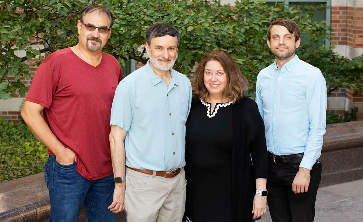
 Understanding host–guest interactions of layered inorganic solids ushered in the modern era of portable electronics powered by lithium ion batteries. Future improvements in the power capability of these devices, and their potential use in emerging technologies such as water treatment, critical element recovery and ion-based electronics, depend on the development and understanding of new ion insertion hosts with fast insertion kinetics. One avenue is via tuning the interlayer environment of a layered material, since many are flexible hosts, whose interlayers can accommodate not just electrolyte ions but also solvents, organic molecules, polymers and organometallics.
 Veronica Augustyn is an Associate Professor of Materials Science and Engineering and a University Faculty Scholar at North Carolina State University (NC State), US. From 2013–2015, she was a Postdoctoral Fellow at the University of Texas at Austin, US. She received her PhD from the University of California, Los Angeles, US, in 2013 and BS at the University of Arizona, US, in 2007, both in Materials Science and Engineering. Her research group focuses on the design, synthesis and characterization of materials for electrochemical energy technologies including batteries, electrochemical capacitors, electrolyzers, and fuel cells. In particular, Augustyn is interested in the relationships between material structure, composition and morphology, and the resulting electrochemical mechanisms. She leads SciBridge, an award-winning international project at NC State that develops renewable energy research and education collaborations between universities in Africa and the United States. Her research group was recognized with a 2019 Sloan Fellowship in Chemistry, 2019 DOE Early Career Award, and 2017 NSF CAREER Award. In 2021, she was named an NC State Goodnight Early Career Innovator and received the George H Blessis Advising Award for her mentorship of undergraduate students.