Frontotemporal dementia (FTD) is a brain disorder that most commonly affects those under the age of 60. Due to an overlap of clinical symptoms (for example, loss of enthusiasm, empathy or motivation) with other neurological disorders, as well as the gradual nature of its onset, it is not uncommon for patients with FTD to be misdiagnosed with late-life depression. Therefore, it is critical to find a distinct symptom that can be used to diagnose FTD.
Muireann Irish and her team at The University of Sydney are the first to differentiate a distinct symptom in patients with FTD: anhedonia, the inability to enjoy pleasant experiences. Their research, published in Brain, also gives insight into the neurobiological workings of anhedonia, distinguishing it from other neuropsychiatric conditions such as apathy and depression.
“We are very excited by these findings as they reveal a symptom that has not previously been documented in frontotemporal dementia, as well as the neural bases of this symptom, opening the door to potential treatments,” explains Irish.
A unique symptom of frontotemporal dementia
In a study of FTD patients, patients with Alzheimer’s disease and healthy controls, the researchers strove to characterize this multi-faceted condition through two diagnostic approaches: cognitive and neuroimaging assessments. Behavioural tests revealed a high prevalence of anhedonia in FTD syndromes as opposed to other forms of dementia. Specifically, FTD patients with anhedonia would experience a lack of interest in rewarding experiences and enjoyable hobbies, such as eating a favourite meal or spending time with friends.
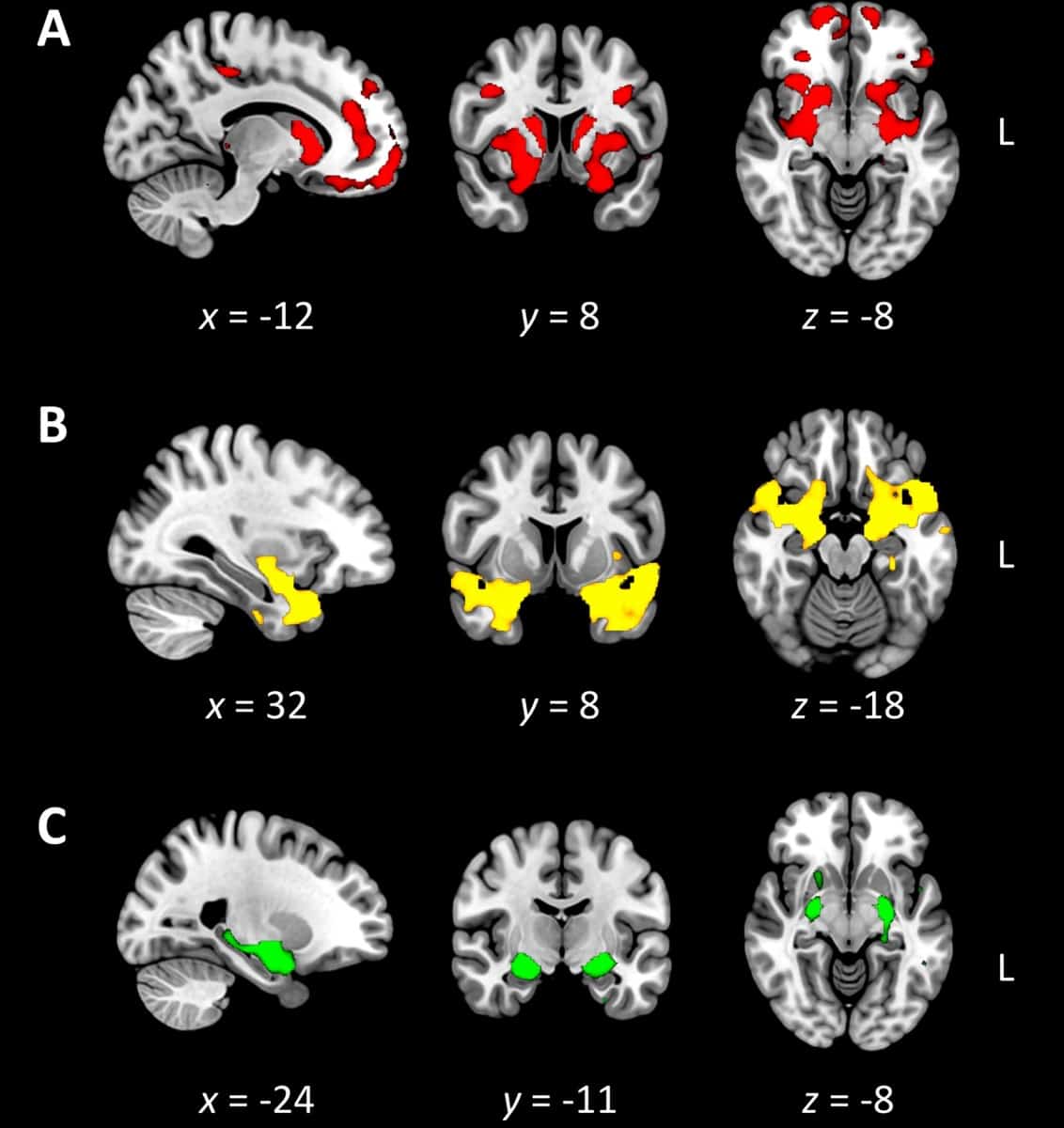
The researchers then performed voxel-based morphometry analysis of participants’ whole-brain MR images to examine voxel-by-voxel changes in grey matter signal intensities. They discovered that that the neural circuitry of anhedonia differs from that of apathy and depression. Specifically, FTD patients diagnosed with anhedonia show deterioration mostly within a frontostriatal grey matter network responsible for experiencing pleasure.
The researchers note that participants with Alzheimer’s disease, who did not show clinically significant anhedonia, exhibited different patterns of grey matter atrophy to the FTD patients.
Shedding light on anhedonia
While this work provides insight on potential treatment areas that could improve the quality of life for these patients, more research is needed to gain a comprehensive understanding of anhedonia in FTD. Future investigations would focus on the relationship between the deterioration of the brain’s reward circuit and the manifestation of anhedonia in the patient’s everyday life.
“Our findings are also important for understanding the subjective experience of the person living with dementia, for the delivery of personalized care, as well as revealing broader insights into a fundamental aspect of the human condition,” says Irish. “If we pause to consider what it might be like to lose our capacity to experience pleasure, we can appreciate the immense need for future work in this field to restore some of the simple pleasures in life to those affected by these cruel disorders.”