X-ray imaging plays a vital role within diagnostic medicine, with modalities such as radiography, fluoroscopy and computed tomography (CT) enabling visualization of the internal structure of the human body. But exposing the body to ionizing radiation comes with an associated risk, and the medical imaging industry is continually seeking new ways to reduce the required imaging dose, via sensitive, high-resolution detectors.
One emerging approach is to use lead halide perovskites to create high-sensitivity X-ray detectors. These materials contain heavy elements such as lead and iodine that have a large X-ray scattering cross section. And as they directly convert X-ray photons into an electrical signal, perovskites offer higher sensitivity and lower cost than competing scintillator-based detector technology. Integrating these perovskites into standard microelectronics, however, remains a challenge.
Now, researchers in Switzerland have demonstrated that 3D aerosol jet printing, a low-cost technique that deposits material with micron precision, can be used to build X-ray detectors from the perovskite methylammonium lead iodide (MAPbI3) on graphene. As they report in ACS Nano, the resulting devices exhibited a record sensitivity of 2.2 × 10⁸ μC/Gyair/cm2 when detecting 8 keV X-ray photons at low dose rates – four orders of magnitude higher than today’s best-in-class detectors.
“By using photovoltaic perovskites with graphene, the response to X-rays has increased tremendously,” says group leader László Forró, from EPFL’s Laboratory of Physics of Complex Matter, in a press statement. “If we would use these modules in X-ray imaging, the required X-ray dose for forming an image could be decreased by more than a thousand times, decreasing the health hazard of this high-energy ionizing radiation to humans.”
Device optimization
Halide perovskites such as MAPbI3 form elongated crystallites when dissolved in a polar aprotic solvent. During aerosol jet printing, as this solution travels through the nozzle, the crystallites grow and land on the substrate as crystalline nanowires containing little solvent. Once a droplet reaches the substrate, the solvent is mostly evaporated, reducing any spatter and creating the well-defined 3D structures needed to construct an X-ray photodetector with high spatial resolution.
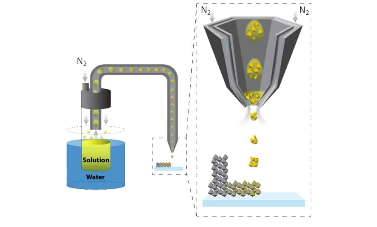
Schematic of the aerosol jet printing method. The agitated MAPbI3 perovskite solution is focused to a predefined position at the nozzle by the nitrogen. The nanocrystals formed in flight do not spread out on the graphene substrate, allowing the creation of 3D architectures. (Courtesy: Glushkova et al ACS Nano)
Using the aerosol jet printing device at CSEM in Neuchatel, the team first characterized the properties of different MAPbI3 geometries printed on microstructured silicon surfaces. A 3D-printed pillar showed an increased charge carrier collection compared with a 2D spot. Both 2D and 3D geometries enabled detection of low light intensities, down to 31.4 μW/cm2 under 650 nm illumination.
To further improve the photodetection characteristics, the researchers 3D-printed the perovskite on graphene, which amplifies the generated photocurrent. After testing several architectures and operation modes (resistor, diode and heterojunction) they found that the optimal pixel design was a heterojunction, with a MAPbI3 pillar printed on a graphene layer spanning the gold electrodes.
X-ray detection
To create the X-ray photodetector, the researchers fabricated a sensing chip containing 3D-printed MAPbI3 walls of about 600 μm in height. They investigated the X-ray detection properties of the fully integrated detector by exposing it to an 8 keV X-ray source for 10 s at various dose rates. The X-ray detection limit was reached at a dose rate of 0.12 μGy/s, below which the signal could not be distinguished from the background noise. At the highest dose rate examined, 358 μGy/s, the device exhibited high photocurrents of up to 4000 μA/cm2.
As the sensitivity of the device is dose-rate dependent, the team determined its sensitivity at 100 mV bias for three dose-rate regions. The sensitivity of a detector pixel (with an estimated surface of 0.075 mm2) was largest at dose rates below 1 μGy/s, reaching 2.2 × 10⁸ μC/Gyair/cm2. Above 1 μGy/s, the photocurrent begins to saturate, yielding sensitivities of 2.5 × 10⁷ μC/Gyair/cm2 at up to 40 μGy/s, and 2.9 × 10⁶ μC/Gyair/cm2 from 40 to 100 μGy/s.
One obstacle preventing commercialization of perovskite-based optoelectronics is their stability. To protect the active MAPbI3 layer in their device, after wire-bonding, the researchers encapsulated the detector in a PDMS polymer. They note that the assembled detector unit was stable for over nine months, with no degradation in performance.
“We are confident that this X-ray detector unit architecture is extremely promising for highly sensitive X-ray imaging,” the researchers conclude. “This approach could allow significant lowering of the radiation doses required for X-ray imaging, resulting in safer and more affordable CT imaging systems.”
For medical imaging, the detector units will need to be assembled into a large surface area X-ray detector. The team also note that as CT scans use higher X-ray energies than the 8 keV source employed in this work, measurements should be repeated for 100 keV photons to confirm the device’s suitability for medical applications.
At 2.46 p.m. local time on Friday 11 March 2011 a massive earthquake hit north-eastern Japan. Striking 130 km offshore, the complex double quake measured 9.0 on the Richter scale and lasted around three minutes. In that time, a 650 km-long section of the sea floor shifted 10–20 m horizontally, while Japan moved a few metres east and the local coastline subsided half a metre.
Initially the Fukushima Daiichi nuclear power plant – located around 225 km north-east of Tokyo – survived the earthquake. But around 40 minutes later it was engulfed by a 15 m-high tsunami. The three operating reactors had turned off automatically following the earthquake, but the immense wave disabled the emergency diesel generators that were supplying back-up power to the cooling systems. With nothing to cool the reactors, their nuclear fuel cores started to overheat and melt. Powerful hydrogen explosions caused extensive damage to the reactor buildings (shown in photo above) and released radiation and radioactive material.
The worldwide nuclear community engaged in a multi-pronged effort to improve the safety of nuclear reactors
Brent Heuser, University of Illinois
Following the accident at Fukushima, members of the nuclear industry started working on advanced fuel concepts to increase accident tolerance. Their goal is to create fuels that can tolerate any potential loss of cooling in a reactor, and other adverse events, for longer than current fuels – thereby increasing safety margins. In the US, this initiative was brought together by the Department of Energy (DOE) under its Accident Tolerant Fuel Development Program. Launched in 2012, it aims to have test fuel rods in a commercial reactor by 2022.
“The worldwide nuclear community engaged in a multi-pronged effort to improve the safety of nuclear reactors, which are already very, very safe,” explains Brent Heuser, a nuclear engineer at the University of Illinois, US. “And one of those approaches was to attack a specific problem related to the behaviour of the cladding that encapsulates the uranium-dioxide fuel.”
In most commercial reactors the fuel rods consist of uranium-dioxide fuel pellets stacked inside a long cladding tube made of zirconium alloy. Used as a barrier to protect the pellets, zirconium is very resistant to corrosion under normal operation conditions, in which the fuels rods sit in water at temperatures of around 300 °C. (Being under such high pressure, the water doesn’t boil.) “It’s kind of ironic. Zirconium is one of the most reactive metals, but it performs very, very well in reactors under normal operating conditions,” says Heuser. “Its corrosion behaviour is very good. It is radiation tolerant. It is really the first engineering barrier. And the probability of cladding failure is incredibly remote.”
At very high temperatures zirconium reacts with water to produce hydrogen, which generated the explosions at Fukushima
Jonathan Cobb, World Nuclear Association
However, if there is a temperature excursion and the water coolant starts to boil, the cladding is exposed to high-temperature steam, which zirconium easily reacts with. It converts rapidly to zirconium oxide and becomes very brittle, which can make it crack and therefore no longer serve as a protective barrier.
This cladding was responsible for the explosions seen during the Fukushima incident, explains Jonathan Cobb, senior communication manager at the World Nuclear Association. “At very high temperatures zirconium reacts with water to produce hydrogen,” he says. “And that hydrogen gas is what generated the explosions that were seen at Fukushima Daiichi.”
Improved claddings
The approach the nuclear industry has settled on for the first generation of accident-tolerant fuels is to coat the cladding, to stop the zirconium from being directly exposed to high-temperature steam during accidents. This is what Heuser’s lab does – it tests chromium coatings on traditional zirconium-based alloy cladding.
“The advantage of improving the existing cladding is that you’re not starting from scratch,” says Heuser. “You’re taking something that’s been proven to work very well under normal operating conditions and you’re putting a coating on it, which represents a very minor perturbation to the normal operating parameters of a reactor, but gives you this built-in defence should a transient occur and the cladding becomes exposed to high-temperature steam.” Heuser’s team has conducted tests both under normal operating conditions and under exposure to high-temperature steam, and the chromium coatings perform well.
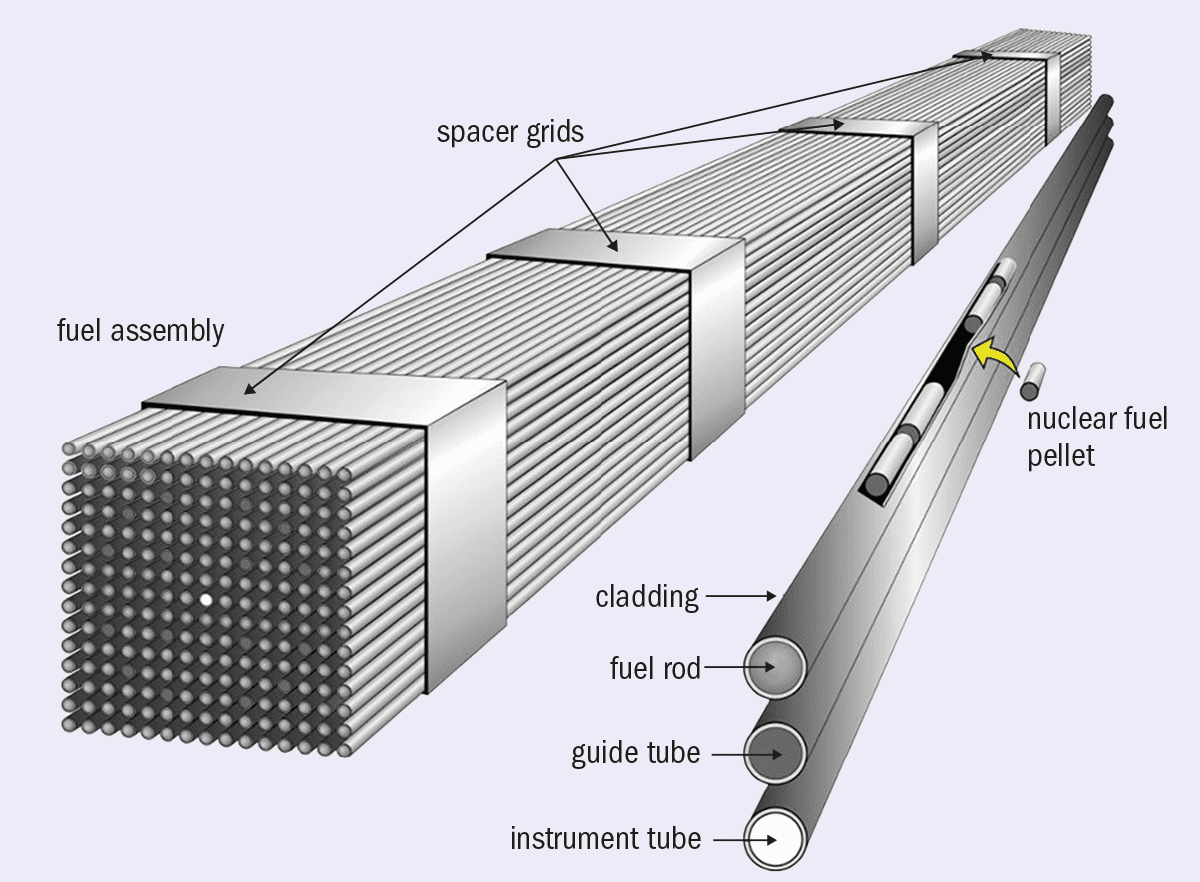
1 From pellet to power Nuclear fuel in a reactor is contained within fuel assemblies sitting in 300 °C water held at high pressure to prevent boiling. The assemblies feature a series of rods, most of which are fuel rods containing uranium-dioxide fuel pellets. The rods traditionally have zirconium alloy cladding to protect the pellets from the water, but during an accident this cladding can become damaged due to the water becoming steam. (Adapted from US Department of Energy original)
Indeed, chromium-coated claddings are now starting to appear in commercial nuclear reactors. As part of the 2012 programme, the DOE contracted three companies to develop accident-tolerant fuels: Framatome, Westinghouse and GE Research. Both Framatome (a French firm) and Westinghouse (from the US) have been developing chromium-coated zirconium-alloy fuel rods. Westinghouse installed test rods featuring their advanced cladding in the Byron Nuclear Generating Station in Illinois in 2019, and the Doel Nuclear Power Station in Belgium in 2020. Meanwhile Framatome’s chromium-coated fuel rods were loaded into a reactor at the Vogtle Electric Generating Plant in Georgia in the US in early 2019. All three reactors are currently using these upgraded rods for a few fuel cycles, after which they will be examined.
Delayed reaction
The aim of accident-tolerant fuels is to delay a reaction during an accident, not prevent it entirely. “The goal is not to say, OK this has happened, we can just let nature take its course and in three days we’re going to be fine,” Heuser says. “That was never the goal. It’s completely unrealistic. The question is: how much time can we buy?” He points out that research suggests that a chromium coating can prevent the high-temperature steam reaction with the underlying zirconium cladding for a couple of hours, which could be enough time for reactor operators to fix the problem.
If you could buy yourself a few hours to get your cooling back under control, then you might save the reactor
Dave Goddard, National Nuclear Laboratory
As Dave Goddard, the technical lead for the UK’s advanced nuclear fuel programme at the National Nuclear Laboratory (NNL), puts it: “If you could buy yourself a few hours to get your cooling back under control, then you might save the reactor.” But he is unsure if this delay period for chromium-coated rods would be long enough. “A lot of the modelling that’s been done on the chromium-coating concept is suggesting that it may actually be minutes, not hours.”
Coating and doping A fuel rod featuring chromium-coated cladding and chromia-doped fuel pellets developed by Framatome being tested at the Vogtle Electric Generating Plant in Georgia, US. (Courtesy: Framatome)
There are, however, other options, such as changing the cladding material completely. Silicon-carbide composites, a material more akin to a ceramic, are one potential solution currently being looked into. These materials have exceptionally good high-temperature performance. “You can probably reach up to about 800 °C with silicon-carbide cladding before you start seeing any sort of detrimental behaviour,” Goddard explains. But making these composites into 4 m-long tubes with a thin wall is challenging. Currently this is done using a chemical vapour deposition technique that can take weeks, creating problems for the economic viability of the technology. But researchers in the UK, supported by NNL, are working on improving this manufacturing technique.
Meanwhile, the US company GE Research has been developing two products: a new iron-based alloy cladding material known as IronClad, and ARMOR, a coating for zirconium cladding. The ARMOR coating is not chromium, Russ Fawcett – who manages the fuel programme at GE – explains, adding that it is a proprietary product and the company has not disclosed its composition.
As with other accident-tolerant fuel concepts, the aim is to provide time to stabilize a power plant during a severe accident. Fawcett says that in a blackout situation similar to Fukushima’s, research suggests that GE’s cladding products could buy between three and six hours. So far, they have been tested with prolonged exposure to high-temperature steam at GE Research and in reactors at the Idaho National Laboratory in the US, with both products exhibiting improvements in resistance to high-temperature steam compared to the standard zirconium-alloy cladding. In 2018 they were also installed in a commercial reactor at the Hatch Nuclear Power Plant in Georgia in the US, and in 2020 GE performed poolside examinations, which it says showed the prototypes work very well.
The company is now working with the US Nuclear Regulatory Commission to license its ARMOR product for use in nuclear power plants, while development of IronClad will continue. GE hopes to be selling the former by 2025 and the latter four or five years later.
New fuels
Accident tolerance could also potentially be improved by changing the uranium-dioxide pellets that sit inside the cladding tubes.
At Elizabeth Sooby Wood’s lab at the University of Texas at San Antonio, US, researchers are looking at advanced technology fuels. “That’s where we’re getting a better fuel economy or accident-tolerant fuels. And then some products kind of overlap both,” Sooby Wood explains.
When it comes to accident-tolerant fuel, one of the big factors Sooby Wood and her colleagues are looking at is thermal conductivity – that is how efficiently the fuel can dissipate its heat so that it doesn’t reach really high temperatures during adverse incidents. In a reactor, all fuel has to be above a critical temperature. Fuels that are less thermally conductive do not heat up evenly, leading to spots where the fuel is much hotter than the critical temperature.
There are accident scenarios where uranium silicide does perform much better
Elizabeth Soddy Wood, University of Texas at San Antonio
In contrast, fuels with a higher thermal conductivity can run at a much more even temperature in the reactors, reducing the overall temperature of the fuel and meaning it has further to go from operating temperature to melting point in an accident. The cooler fuel gives you more of a safety margin. “It’s about improving different material families so that they tolerate these abnormal scenarios better,” Sooby Wood says.
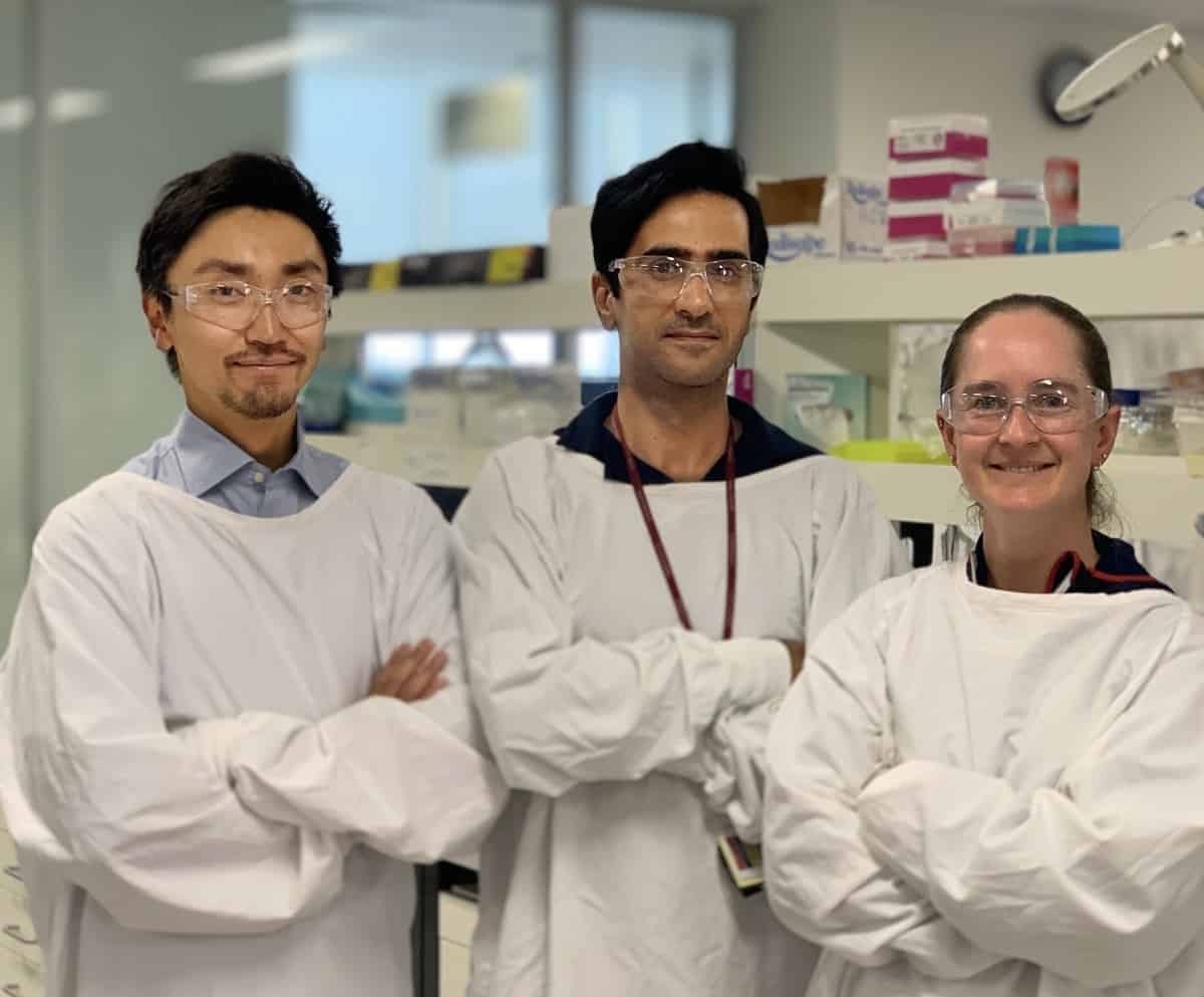
Extreme testing Elizabeth Sooby Wood and doctoral student Geronimo Robles operating arc melting furnace in her lab at the University of Texas at San Antonio. The apparatus is used to test uranium compounds for new accident-tolerant fuels. (Courtesy: The University of Texas at San Antonio)
Currently her team is looking at uranium silicide as an alternative to the traditional uranium dioxide. On its own, uranium silicide is actually not very accident tolerant – if it is exposed to the reactor coolant it has a worse reaction than uranium dioxide. “But it does have a much higher thermal conductivity,” adds Sooby Wood. “So there are accident scenarios where it does perform much better. We are seeing if we can engineer the uranium silicide to withstand exposure to the coolant.”
Just as materials are added to iron to create stainless steel – a much less corrosive material than plain iron – so the researchers are adding metals to make uranium-silicide alloys. So far, they have been adding both aluminium and chromium. The resulting alloys are tested in a furnace that can go up to 1600 °C, and steam is flowed through to simulate coolant exposure of the fuel elements and the uranium compounds. Ideally there will be no reaction: the uranium-silicide alloys will not degrade or ignite. To date, the researchers have run these steam oxidization tests at temperatures from 200 °C to 1000 °C and the results have been promising, with delayed onset of reaction compared with plain uranium silicide. Now Sooby Wood’s team and others are looking at different ways to incorporate the metals to get further delays.
At this stage, the work is just fundamental research and development, questioning whether alloying works and if it can improve the behaviour of the fuels. But if successful, Sooby Wood says these new fuels could also improve fuel economy as they have a higher uranium density: more uranium per unit volume. “So, for the exact same footprint you have more fissile material. More stuff making power.” This means higher power output from reactors, many of which are currently running at peak capacity. It also leads to longer fuel cycle times.
The higher uranium density could also enable the use of more advanced cladding materials. Sooby Wood explains that some of the cladding materials being looked at have a higher neutron-capture rate than zirconium. This means that they soak up more of the neutrons that are produced and used in fission chain reactions. However, higher-density fuels produce more neutrons in the first place, making this loss less of an issue.
Added benefits
The advanced cladding materials could also improve fuel performance and provide economic benefits. For example, the chromium coatings make the cladding more durable, reducing fuel rod failures and unplanned outages, Goddard says. And GE claims that its new claddings also cut corrosion. Power plants could therefore keep the fuel rods in the reactors for longer, extending fuel cycle lengths and making them more profitable. GE hopes that the improved fuel performance, combined with a slight increase in fuel enrichment, will reduce the number of fuel assemblies a power plant needs by around 10–20%.
Evolving fuel GE Research testing its ARMOR and IronClad accident-tolerant fuel rods at the Edwin I Hatch Nuclear Plant in Georgia, US. (Courtesy: Southern Nuclear)
Ultimately this could also reduce fuel waste. “If your fuel can be in the reactor for longer, if you have reloads every 18 months instead of every 12 months,” explains Cobb, “then that is going to reduce the already low volumes of used fuel that comes out of the reactors.”
Some experts argue, however, that the pursuit of improved performance and economic benefits is shifting accident-tolerant fuels away from their original aims. Edwin Lyman, director of Nuclear Power Safety at the Union of Concerned Scientists in the US, says that while the idea of accident-tolerant fuels sounds good, he has been disappointed with the results so far. He adds that in the US, industry is moving away from any pretence of improved safety. “Instead, they’re focusing on trying to reduce their operating costs by increasing the burnup and the enrichment of fuels.”
Lyman says extending the coping times by a couple of hours does not make a significant difference to safety – studies suggest that these time frames have a fairly small impact on the probability of a core melt. “I’m not saying it’s of no benefit,” he adds, “but it’s a minimal benefit.”
The US nuclear industry is focusing on trying to reduce their operating costs by increasing the burnup and the enrichment of fuels
Edwin Lyman, Union of Concerned Scientists
This becomes a real issue when you try to extract more energy from the fuel. By increasing the burnup and keeping the fuel in the reactors for longer you increase the risk of fuel fragmentation and related issues, which could reduce the safety margins in a loss-of-cooling accident, like that at Fukushima. “If you just substituted one of these chromium-coated fuels and didn’t try to increase the burnup, then you might have an additional safety margin,” Lyman says. “But if you push the burnup to the limits of what the fuel can withstand, then you’re not realizing that safety benefit.”
John Allen of GE Research says that while the company will seek to recognize the other benefits of these advanced fuels, “the margin to safety will increase”. His colleague, engineer Evan Dolley, adds: “Our primary objective is to increase fuel reliability and safety, and provide more coping time. We’ve never veered from that.”
Meanwhile, Heuser believes the research and nuclear industry has found a viable solution for accident-tolerant nuclear fuels. “I think the future is bright for nuclear power. And I think it needs to be part of a balanced energy portfolio along with renewables,” he says. “We need to move away from fossil fuels because global climate change is a real problem to address and nuclear power can be and should be part of that solution.”
A positron (β+) deposits energy along its track through the formation of “spurs” and a terminal “blob”, before positronium formation and annihilation to generate two 0.511 MeV photons. (Courtesy: CC BY 4.0/Sci Rep 10.1038/s41598-021-81910-4)
Positron-emitting radionuclides have long been employed for diagnostic imaging, with PET scans using fluorine-18 (18F)-labelled fluorodeoxyglucose (FDG) playing an essential role in cancer diagnosis. But positrons could also be used to destroy cancer cells. Perhaps due to their prevalence within diagnostics, this therapeutic potential has to date been largely overlooked. A research team in Australia aims to address this oversight.
“We refer to it as positron emission radionuclide therapy, or PERT,” says senior author Dale Bailey.
Cell studies
When the radionuclide 18F undergoes decay it emits a positron (a beta-plus particle emitted from a proton-rich nucleus). The positron will ultimately annihilate with an electron, leading to the emission of two 0.511 MeV photons. And it is these photons that are detected to create PET images.
But before this final annihilation process, the positrons lose kinetic energy in discrete quantities (roughly 100 eV) via multiple interactions along their track, creating positron “spurs” – nano-sized spheres of electron/positive-ion pairs – and a terminal positron “blob”. These spurs and blobs are all sources of highly reactive species and deliver a relatively large radiation dose when they interact with biomolecules such as DNA.
To investigate the potential of positrons in cancer medicine, the researchers examined the survival of prostate cancer cells exposed to sodium fluoride (18F-NaF) solution for 18 h. They found that a dose of 20 Gy 18F positrons killed over 90% of the cells, while a 10 Gy dose caused 70% cell kill.
To quantify the relative biological effectiveness (RBE) of 18F positrons, the researchers compared their results with high-dose rate X-ray irradiation. They assessed cell survival at various absorbed doses for positrons and for X-rays from a small-animal radiation research platform (SARRP). By comparing the mean absorbed doses required for 50% cell survival, they calculated a mean RBE of 0.42 for 18F positrons relative to SARRP irradiation. This is three times higher than the RBE of radionuclides that emit beta-minus particles (electrons emitted from a neutron-rich nucleus), such as 90Y and 177Lu.
Takanori Hioki (left) and co-authors Yaser Gholami (centre) and Kelly McKelvey.
“Clinically speaking, the dose rate and linear energy transfer (LET) of positron emitters is expected to be higher than that of most beta-minus emitters that are currently used in radionuclide therapy, predominantly due to the relatively shorter half-life and more ionizing radiation of many positron emitters,” explains first author Takanori Hioki. “Furthermore, radionuclide therapy targets metastatic lesions, while external-beam radiotherapy is generally used for larger sites or primary lesions.”
Damage simulation
Hioki and colleagues also performed a Monte Carlo simulation of a linear DNA model to determine the frequency of DNA single strand breaks (SSBs) and double strand breaks (DSBs) caused by positron or beta-minus irradiation at kinetic energies from 250 eV to 1.5 keV. They observed that the lower energies produced larger numbers of SSBs and DSBs.
The simulation revealed that positron tracks induce 1.5- and 2.2-fold more SSBs and DSBs, respectively, than beta-minus tracks. The greatest difference occurred at 400 eV, where positrons caused 55% increase in SSBs and 117% increase in DSBs compared with beta-minus particles.
These results imply that the direct interaction of a single positron with DNA should create more lethal damage than caused by a single beta-minus. “As each positron that is emitted loses energy as it interacts, an accumulation of the simulated interactions causes the total damage that we observe in the in vitro experiment,” Hioki notes.
Plotting the LET (a measure of how much energy an ionizing particle deposits per unit path length) revealed that maximum SSB and DSB production should occur at 250 eV (the kinetic energy near the end of its track) for both positrons and electrons. At this energy, a positron has roughly 7% higher LET than a beta-minus.
The spur model suggests that beta-minuses and positrons initially have similar radiation tracks, but behave differently at the lowest energies. For a sub-keV beta-minus, the mean separation between spurs is 20 times larger than the diameter of the DNA helix, while a sub-keV positron continuously forms spurs and builds up a blob along and at the end of its track. Thus, at sub-keV energies, a positron has a higher LET than a beta-minus. Additional ionization at the terminal annihilation event further increases the total number of ionizations per positron track.
“The biggest contribution to the higher DNA damage from positrons in comparison to beta-minuses is the higher LET of the particles at sub-keV energies, as well as contributions from the difference in charge,” says Hioki.
Dual role
The researchers point out that, in addition to an untapped therapeutic potential, positron-emitting radionuclides could also play a role in emerging theranostic strategies, for use as a combined therapeutic and diagnostic agent. For clinical use, however, the highly penetrating, low-ionizing nature of the emitted annihilation photons will require careful safety considerations when administering therapeutic doses of the radioactive compound.
“As this study demonstrated the therapeutic potential of positrons, we are currently working on the next step – to optimize the administered activities to maximize its treatment efficacy,” Hioki tells Physics World. “We are also performing biological assays to demonstrate the impact that positrons have on the cellular mechanisms that lead to the results we observed in the cell survival assays.”
A new isotope of darmstadtium and a new excited state of copernicium-282 have been found in the decay chains of the superheavy element flerovium by an international team of researchers. The discoveries made by Anton Såmark-Roth of Lund University in Sweden and colleagues provide important clues to nuclear physicists trying to make long-lasting superheavy elements that lie within islands of stability.
First synthesized in 2002, oganesson has an atomic number of 118 and is currently the heaviest known chemical element. Since its discovery, researchers have continued their search for even heavier elements but face a major barrier. As elements become heavier, growing imbalances between proton and neutron numbers tend to make them increasingly unstable, making it increasingly difficult for researchers to synthesize them in the lab.
This trend is reversed somewhat when nuclei contain “magic” numbers of protons or neutrons. This creates “islands of stability” in an otherwise turbulent part of the chart of nuclides – which plots nuclei in terms of their proton and neutron content. These islands could provide crucial steppingstones for researchers aiming to synthesize elements heavier than oganesson. One such island is believed to occur at around flerovium-298, which is predicted to be “doubly magic” for protons and neutrons. This isotope cannot currently be produced in the lab but is a target of great interest to nuclear physicists.
Alpha decay
In their experiment, Såmark-Roth’s and colleagues studied the decays of two lighter versions of that element: flerovium-288 and 286. These isotopes were created by firing an intense beam of calcium ions into plutonium, using the TASCA facility at the GSI Helmholtz Centre for Heavy Ion Research in Darmstadt Germany. After its formation, the flerovium decays rapidly by emitting alpha particles (helium-4 nuclei), producing unstable nuclei that go on to decay themselves.
The team studied the decay chains using high-resolution nuclear spectroscopy, which involves measuring the different types of radiation emitted by decaying nuclei. Although dozens of chains were detected, two were of particular interest. One is flerovium-288 decaying to copernicium-284, which itself decays to darmstadtium-280 – an isotope that had not been observed before. In the second decay chain, flerovium-286 decays to an excited state of copernicium-282, which contains even numbers of both protons and neutrons. Again, this had never been seen before in an excited superheavy nucleus.
The observation of these decay chains and the existence of the excited state of copernicium-282 provide important information for physicists developing theoretical models of flerovium-298 and could also point physicists in the right direction to discover islands of stability.
A full-scale prototype muon tomograph that can peer inside cargo containers has been created by researchers in Italy and the US. The team, led by Francesco Riggi at the University of Catania, combined layers of muon detectors with a specialized reconstruction algorithm to deliver high-resolution 3D images of a small lead block inside a large sensing area. The technology could make it far easier for cargo authorities to stop dangerous nuclear materials from being transported illegally.
Cargo containers are widely used to transport high volumes of goods and because of their robust steel construction and large size, small objects can easily be concealed inside them. As a result, there is a growing concern that containers could be used to transport illicit nuclear materials, including plutonium and uranium.
There is a need, therefore, for an efficient way of screening containers without disrupting global trade. Among the most promising emerging techniques for doing so is muon tomography, which uses the natural showers of muons created when high-energy cosmic rays collide with molecules in the upper atmosphere. When muons interact with a dense material such as uranium, they are scattered and absorbed in characteristic ways, depending on the atomic number of the material.
Egyptian pyramid
Muon showers strike all parts of the Earth and have been studied by physicists for nearly 90 years. As a result, scientists have a very good understanding of the energies, fluxes, and angular distributions of muons at moderate altitudes. By comparing these values before and after muons have passed through hidden volumes, researchers can determine both the compositions and positions of concealed materials. The technique is now being used in a growing number of research areas, and even led to the discovery of a large chamber inside an ancient Egyptian pyramid in 2017.
Muon tomography is particularly desirable because cosmic muons are uniformly available across the Earth’s surface. Furthermore, muons can penetrate far deeper into dense materials than conventional imaging methods, including X-rays. On the downside, however, because muons fluxes are relatively low, long scanning times are needed with current detector technologies.
Now, Riggi and colleagues have combined several techniques previously developed to overcome the flux problem to create a full-scale muon tomograph. Their setup features multiple layers of scintillator-based muon detectors positioned above and below the sensing area. These detectors track changes in the paths of muons as they are scattered by dense materials. Then, an algorithm analyses the muon trajectories to estimate the point of closest approach between the muons and heavy atomic nuclei. From this information, the team can create high-resolution 3D images of dense materials inside the sensing area.
The technique enabled Riggi’s team to precisely determine the 3D position of a small lead block about 20 cm across within a sensing cross-sectional area of 18 m2 – which is large enough to accommodate a standard cargo container. The team says that their successful demonstration paves the way for devices that can efficiently detect concealed nuclear materials. With further improvements to reduce scanning times, the inspection devices may one day become an integral part of cargo-handling facilities worldwide.
The Integral Quality Monitor (IQM) is a simple solution that offers advanced real time QA for every segment and beam. The novel design introduces a single ion chamber that provides exceptional reproducibility and accuracy for every treatment technique, from small-field SRS to large conventional treatments. All this is combined with workflows that are fully automated and require no extra steps in planning or treatment delivery.
This webinar, presented by Juergen Oellig and Marlies Pasler, will cover the following topics:
The IQM development project
The IQM technology
– Reference calculation
– Signal measurement
– Signal reproducibility
– Beam attenuation
Clinical experience with IQM at Lake Constance Radiation Oncology Center, Friedrichshafen, Germany
– Error detection sensitivity
– Clinical experience with IQM treatment monitoring for all fractions
– Testing IQM for QA purposes (beam flatness and symmetry)
Juergen Oellig is the managing director of iRT Systems. He began his career in radiation therapy in 1995 and has been responsible for the sale and service of treatment planning systems as well as dosimetry and quality assurance equipment.
In 2013 he founded iRT Systems with the sole focus on developing, certifying and marketing the Integral Quality Monitor (IQM) System. A unique aspect of the IQM development project was the dynamic programme of research co-operation with radiation therapy departments all over the world. Groups on four continents contributed their investigative work before and after the product release, addressing various aspects of using IQM for online Fraction QA, for Plan QA for complex treatment techniques, as well as for linear accelerator quality assurance.
Today, his focus is on showing clinicians how they can realize clinical benefits and achieve more patient safety with IQM.
Marlies Pasler is a medical physicist at the Lake Constance Radiation Oncology Centre, Friedrichshafen, Germany. As a physicist, her areas of expertise are versatile, ranging from routine tasks such as treatment planning, plan verification, dosimetry and linac constancy checks, to more projecting tasks such as the implementation of new techniques, hardware and software beta-testing. Her fields of research include VMAT treatment planning, QA for rotational techniques with a special focus on log files and dosimetry and multi-institutional auditing concepts.
Her work is published in several journals and she presented her projects at various conferences including DEGRO, ÖGRO and ESTRO.
The Greek philosopher Plato once imagined a city that provides full justice to its citizens. Setting out his ideas in the Republic almost 2500 years ago, Plato did not, however, think that such a city could ever be realized. Radical (and surely unachievable) transformations in education, culture and government would be required to establish and sustain it. “Ridiculous,” Plato concluded.
In a similar vein, the US cultural anthropologist Vincent Ialenti envisions a fictional city whose citizens have been trained to think so that humans don’t need to flee the planet to survive. So utopian is the picture that Ialenti – writing in his new book Deep Time Reckoning – calls it “absurd”. Yet that notion is no less absurd, he continues, than the way humans are now acting, “careening toward an Anthropocene cliff”.
Based at George Washington University, Ialenti developed this picture by drawing on three years of fieldwork in Finland, where he’d studied experts who were evaluating the risks of a permanent repository for nuclear waste. The experts had been asked to develop methods to convince Finnish regulators that the facility, being cut out of granite bedrock underneath the island of Olkiluoto, would not expose future populations – thousands and even hundreds of thousands of years from now – to radiation above the country’s legal limits.
Long-term predictions face enormous scepticism and deeply embedded resistance from the public and government officials, especially when they concern global matters
The Finnish experts developed various strategies to envision “deep time”. For example, they implemented unusual computer modelling methods to integrate a variety of datasets, scenarios, maps and reports over an unprecedented range of issues, including climate change, geological events, shorelines, human demographics, vegetation growth and ecosystems. For clues on the long-term evolution of materials and planetary landscapes, they studied everything from ancient Roman nails and 2100-year-old Chinese cadavers to cannons from a sunken 17th-century Swedish warship and traces of a crater in Finland caused by a meteor 73 million years ago.
Playing the long game
Ialenti is fully aware of the deficiencies and partialities of the Finnish project and of his own study. Sample size was one concern, based as it was on just two dozen experts from a single firm associated with the nuclear-power industry. Another was the fact that Finland is the most sparsely populated country in the European Union. Finland also has a much higher public trust of scientists and technical experts – certainly higher than other large nations such as mine, where tens of millions of citizens can vote for leaders who think that consulting scientific advice is for losers.
Furthermore, contemporary humans are soaked in a world of tools, technologies and institutions that reinforce short-term – rather than long-term – thinking. Decisions about buying and selling stocks must be made in seconds, apps can be updated every few weeks, and start-up companies must rethink their plans every year or so. Long-term predictions face enormous scepticism and deeply embedded resistance from the public and government officials, especially when they concern global matters.
Somewhere in deep time looms a catastrophe that we don’t yet have the imagination to envision, nor the will to confront
Climate-change predictions, even for 2050, seem hopelessly far in the future, and tainted by politics, guesswork and subjectivity. Thinking about the present seems more do-able, while thinking about tens or hundreds of thousands of years in the future appears starry-eyed and abstract. But Ialenti believes the exact opposite is true. What’s abstract (in the sense of detached from reality) is what Ialenti calls “a manic fixation on the present”, and not being able to think about humanity thousands of years hence.
Ialenti is less interested in the conclusions reached by the Finnish experts than by their audacious aims, which are to develop methods to break free from what he calls our “shallow time discipline”. He then tries to devise ways to retrain our habits to encourage humans to think long-term; for him, Deep Time Reckoning is not a stale academic treatise but more of a “practical toolkit”.
This toolkit includes high-school civics classes devoted to teaching long-term developments: of the universe since the Big Bang 13.8 billion years ago; of Earth since 4.5 billion years ago; of Earth’s life, dinosaurs and humans; and of the evolution of languages and technologies. It envisions school pupils reading about futuristic visions by Ray Kurzweil and Marxist descriptions of world utopias.
Ialenti even asks his university students to examine the tools that the insurance industry uses to protect companies against future calamities, and the methods that the Catholic Church uses to maintain institutional continuity. Practised over generations, Ialenti thinks, such an education would eventually make deep time thinking “less wacky and aloof”, and more second-nature.
Ialenti proposes a range of details in his deep-time-thinking world that are more New Atlantis than Republic. His deep-time-thinking world has monuments, but they are to volcanoes, glaciers, asteroid collisions and other testimonials to deep time. Internet competitions and TV gameshows are devoted to preparations for and speculations about the future. Even the screensavers of his utopian world remind computer users of deep time. The United Nations, meanwhile, might support institutions with names like “Global Deep Time Reckoning Association”, precursors to which, he thinks, already exist in organizations like the Planetary Society and the Aspen Institute, a global non-profit think tank.
The critical point
Plato meant the Republic to be a beacon for humans to think about justice in the present, not as the blueprint for an actual city to be realized in the future. After all, if you head straight towards a lighthouse, you usually end up on the rocks.
Somewhere in deep time looms a catastrophe that we don’t yet have the imagination to envision, nor the will to confront. Ialenti thinks he finds in the Finnish nuclear-risk experts glimmerings of what it might take to cultivate the human behaviour needed to do so. Humanity’s long-range hope, Ialenti suggests, hangs on what we might call the Finlandization of the planet.
Air purifiers in lifts (elevators) and other confined spaces could actually increase the spread of airborne virus droplets, according to new calculations by two researchers in Cyprus. This surprising result means that health and safety authorities should revise current guidelines accordingly by considering how these droplets circulate and disperse, they say.
When people infected with the Cov-SARS-2 virus, responsible for the current COVID-19 pandemic, sneeze, talk or simply breathe, they expel thousands of contaminated saliva droplets. These droplets are now recognized as being the main way in which the virus spreads to other individuals and people are being advised to wear masks to curb contamination. Regularly airing indoor spaces to reduce the concentration of viral particles is also important.
Since the quality of air in a small space can quickly deteriorate even when there is just one person in that space (mainly because of the increased CO2 levels produced as we breathe out), lift manufacturers routinely include ventilator systems. These can, however, increase the rate at which air – possibly laden with bacteria and viruses – circulates. While adding an air purifier to the lift in addition to the ventilator should help in theory, the way in which the purifier affects air circulation, and thus transmission of airborne viruses is unclear.
Computational study
Air purifiers work by sucking in stale air and exhausting cleansed air, but this adds to the overall circulation of air – an aspect that previous studies, or indeed air purifier manufacturers themselves, have not considered.
To investigate this issue, Dimitris Drikakis and Talib Dbouk of the University of Nicosia analysed how air circulates in a three-dimensional volume of 27.28 m3 (x=1.24; y=1; z=2.2 m), which is equivalent to the space in a lift designed for five people. They equipped their virtual lift with a standard ventilation system and then installed an air purifier at a height of 1.9 m (z=1.9 m) in the lift. The purifier has an air intake (outlet) and an air exhaust (inlet) and circulates air at roughly 60 m3/h.
Using a (Eulerian–Lagrangian) multiphase model that they recently developed, the researchers simulated what happens when someone in the lift coughs lightly. They did this by ejecting 1000 “contaminated” saliva droplets (each with a mass of 20 mg) at a speed of 1 m/s from the person’s mouth, which they placed at two different positions (x=0; y= 0.41; z=1.6) and (x=0; y=0.67; z=1.6) in the lift. They then modelled the spatial distribution of saliva droplets inside the lift. They performed their computations at 20 °C, 50% relative humidity and atmospheric pressure.
The researchers found that the air purifier actually increases the spread of saliva droplets in the cabin. This is because the air intake integrated inside the purifier equipment induces flow circulation that can add to the transport of these droplets, explains Drikakis. They also found that the risk of airborne virus transmission is lowest for low ventilation rates. This is due to reduced flow mixing inside the lift, says Dbouk.
Revise guidelines?
Unsurprisingly, the risk of contamination increases with the number of infected individuals in the lift, add the researchers. Restricting the number of people allowed in such a small place could therefore minimize virus spread as would designing better air purifier and ventilation systems that reduce the flow of circulating air.
The results of the study, which is detailed in Physics of Fluids, could be applied to any confined space, they explain. These include small rooms, underground car parks, shops, aircraft cabins, submarines and spacecraft.
“The broader implications of the present findings are that health and safety authorities must revise guidelines accordingly by considering the flow circulation and droplet dispersion effects arising from air purifiers and sanitizers in confined spaces,” say Drikakis and Dbouk.
“For our part, we now plan to investigate the airborne transmission mechanisms for COVID-19 and other diseases,” they tell Physics World. “Indeed, a recent study we published regarding fluid mechanics and epidemiology shows how temperature, humidity and wind effects can predict the second wave of the pandemic.”
It’s one of the fundamental challenges facing radiotherapy equipment manufacturers: how to balance the relentless clinical demand for automation and enhanced workflow efficiency versus the desire of care providers to customize treatment planning and radiation delivery to meet the personalized needs of every cancer patient. Back in May last year, Elekta, a specialist equipment provider in precision radiation medicine, went some way towards squaring this circle with the market introduction of Leksell Gamma Knife Lightning, an automated treatment planning system that integrates with Leksell Gamma Knife, the manufacturer’s flagship stereotactic radiosurgery (SRS) platform for the treatment of benign and malignant intracranial tumours as well as vascular and functional disorders in the brain.
For context, the Gamma Knife SRS system exploits multiple narrow beams from different directions to deliver conformal, high-dose radiation to the disease target in one or a few fractions while minimizing collateral damage to surrounding healthy tissue and organs at risk (OARs). Despite widespread deployment in cancer centres worldwide, it’s fair to say that the precision targeting inherent to SRS remains a non-trivial dose optimization challenge for the medical physics team – and not least when it comes to focusing “high-payload” radiation onto metastatic small lesions (as small as 2 mm3 in extent) and having it fall off as quickly as possible.
If that’s the problem, Leksell Gamma Knife Lightning is shaping up a lot like the solution. This next-generation treatment planning system enables medical physicists, radiation oncologists and neurosurgeons to automatically create multiple SRS treatment plans in less than a minute, with optimization based on dose constraints to one or more lesions and OARs. The result: fast-track comparison of treatment plan options followed by selection of the optimal plan for each patient (in terms of conformality and tissue sparing). In some cases, that can include a reduction in “beam-on” time by as much as 50% compared with manual forward planning.
The view from the clinic
As part of the network of alpha test sites for Lightning, Prague Na Homolce Hospital (Czech Republic) was ideally positioned last summer to become one of the first Elekta customers to deploy the software clinically for SRS patient treatments. The department of stereotactic and radiation neurosurgery at Na Homolce handles around 1200 patients annually in its Leksell Gamma Knife centre, with Lightning now used to plan all but the most straightforward single-shot SRS treatments on its Icon device. (Worth noting, though, that Lightning is also fully compatible with Perfexion, Elekta’s other commercial Gamma Knife system.)
Josef Novotný “Lightning streamlines the overall treatment planning process for SRS.” (Courtesy: Na Homolce Hospital)
“The benefits of Lightning are especially evident in complex cases when very high dose conformity and protection of critical structures like the optic nerve are required,” explains Josef Novotný, head of medical physics at Na Homolce. Novotný should know: he has notched up more than 20 years of clinical experience with Leksell Gamma Knife, having participated in the development of the Perfexion and Icon systems while also supporting clinical roll-out at over 150 Leksell Gamma Knife centres around the world.
Equally instructive, says Novotný, is the impact of Lightning on Na Homolce’s SRS clinical workflow and patient throughput. “Lightning streamlines the overall treatment planning process for SRS, such that the typical calculation time per plan is less than one minute,” he adds. “We also see a reduction in beam-on time by almost 15% owing to efficient planning and use of a very high number of isocentres – akin to ‘dose painting’ of the tumour targets.”
An initial study by the Na Homolce team, the results of which are to be formally presented at the Third European Congress on Medical Physics (ECMP) in Turin this summer, provides quantitative evidence of Lightning’s clinical benefits. Novotný and colleagues report that Lightning improves both selectivity (i.e. dose reduction outside the target) and Shaw conformity index (a measure of targeting accuracy) by about 9%, while reducing the 12 Gy volume (which is often used as a predictor for complication rate) by about 5%. In selected cases, the researchers also observe enhanced sparing of critical structures by about 12% (optic nerve), 9% (cochlea) and 5% (pituitary).
“It can be generally said that Lightning is superior to previous methods used for treatment planning on Leksell Gamma Knife,” notes Novotný. “That means better treatment plans versus multiple metrics – whether in terms of dosimetry characteristics, lower doses to critical structures, as well as reduced beam-on time.”
Collective progression
Another enthusiastic early-adopter of Lightning is Ian Paddick, chief medical physicist at the Queen Square Radiosurgery Centre in London and immediate past-president of the International Stereotactic Radiosurgery Society (ISRS). For the past two decades, Paddick has provided training, mentorship and consultancy to more than 100 Gamma Knife centres around the world. It’s within this wider frame of reference that he sees Lightning as a catalyst for community-wide progression and innovation in SRS treatment planning. Put another way: a “rising tide lifts all boats” scenario that will ultimately promote convergence towards clinical best practice and enhanced patient outcomes across the Gamma Knife user base.
Ian Paddick “If you want to master the art of manual SRS planning, you really need to pit yourself against Lightning.” (Courtesy: Medical Physics Limited)
The upsides are already evident at a more granular level. For starters, claims Paddick, Lightning will do a better job on the majority of tumour targets than an expert treatment planner – in terms of enhanced conformality, selectivity and dose gradient. What’s more, Lightning is also incredibly fast. “I could not believe it when I first used the software,” he adds. “In fact, I’ve just used Lightning for a case with six complex metastases and it took about 35 seconds to produce an optimized treatment plan. Generating the same plan manually took me around 40 minutes.”
Of course, the treatment plans generated by Lightning still need to be reviewed and signed off, with the opportunity to manually adjust dose distribution ahead of treatment delivery (allowing further optimization, for example, if the dose is spilling out of the target in the vicinity of a cranial nerve). “In this way,” adds Paddick, “Lightning can be used to do most of the heavy-lifting, with the final ‘sculpting’ and optimization of the dose distribution being hand-crafted by the medical physicist, neurosurgeon or radiation oncologist.”
Herein lies another opportunity: the use of Lightning to fast-track learning and development among inexperienced SRS planners at new Gamma Knife centres. “If you want to master the art of manual SRS planning, you really need to pit yourself against Lightning,” Paddick argues. “Used in the right way for performance benchmarking, Lightning will always show you what can be achieved in terms of dose optimization and plan quality.”
Novotný, for his part, believes that Lightning will simultaneously drive improvements within established Gamma Knife centres while helping new SRS users to achieve consistent, high-quality treatment planning sooner – i.e. without the lengthy learning curve and accumulated experience from planning many stereotactic cases. “It’s clear that Lightning lowers the barriers to entry in SRS,” he concludes. “Once the target volume and critical anatomical volumes are defined, the operator just has to specify prescription dose to the target volume and dose constraints to any adjacent structures. The rest is handled automatically by Lightning.”
A polymer-based gel made by researchers in the US and inspired by the Venus flytrap plant can snap, jump and “reset” itself autonomously. The new self-propelled material might have applications in micron-sized robots and other devices that operate without batteries or motors.
“Many plants and animals, especially small ones, use special parts that act like springs and latches to help them move really fast, much faster than animals with muscles alone,” explains team leader Alfred Crosby, a professor of polymer science and engineering in the College of Natural Sciences at UMass Amherst. “The Venus flytraps are good examples of this kind of movement, as are grasshoppers and trap-jaw ants in the animal world.”
Snapping instabilities
The Venus flytrap plant works by regulating the way its turgor pressure – that is, the swelling produced as stored water pushes against a plant cell wall – is distributed through its leaves. Beyond a certain point, this swelling leads to a condition known as a snapping instability, where the tiny additional pressure of a fly’s footsteps is enough to cause the plant to snap shut. The plant then automatically regenerates its internal structures in readiness for its next meal.
Researchers have previously used these swelling-induced snapping instabilities to amplify the power of artificial motors, as they are good at converting the motor’s output into motion (think of pulling a rubber band and letting go). The problem is that so far, artificial gel-based devices have only been able to snap once, as they lack the internal self-regenerating mechanisms that makes repeated snaps possible in living structures like the Venus flytrap.
Transient deswelling
Crosby’s team got around this problem by harnessing an often-overlooked process called transient deswelling. They discovered the mechanism for this process by chance, when they observed a long, elastic strip of polymer gel moving as its internal liquid evaporated. Although most such movements were slow, every so often the researchers saw them speed up. “These faster movements were snap instabilities that continued to occur as the liquid evaporated further,” they explain. “Additional studies revealed that the shape of the material mattered and that the strips could reset themselves to continue their movements.”
To investigate the mechanisms behind this phenomenon, the researchers made a strip of material 0.2 mm thick, 5 mm wide and 60 mm long from poly(dimethylsiloxane), PDMS, and placed it in a solvent of n-hexane. They chose n-hexane because it has a high affinity for PDMS, allowing the material to fully swell up. Once the strip swelled, they placed it on a sheet of black poly(tetrafluoroethylene), PTFE. This polymer minimizes the effect of friction on the strip’s motion and prevents solvent from absorbing into the substrate.
Abrupt snaps
The researchers observed that during the first 90 seconds of deswelling, the strip buckled and transformed into different shapes in a seemingly random fashion. These complex buckling transitions included both smooth, simple bends and abrupt snaps.
To analyse how the snapping affected the strip’s movement, the researchers measured the change in the position of its apparent centre of mass and one endpoint (tip). They found that the centre of mass moved, on average, at 0.007 m/s, with a maximum speed or around 0.2 m/s. In contrast, the average speed of the tip was roughly 0.018 m/s, with a maximum of 1.1 m/s. This “extraordinarily” fast speed, which occurs at distinct moments and makes up only a few of the observed movements, must come from snapping, Crosby and colleagues say, since deformations caused by swelling or deswelling would only produce speeds of about 0.006 m/s or less.
Snapping structures perform complex tasks
The researchers, who detail their work in Nature Materials, also showed that the snapping motion could be used to perform tasks. “We used our understanding of the physics of the strip and made spherical shells from these structures,” Crosby tells Physics World. “These shells harnessed the snap instability to jump up a sloped substrate on their own as well as autonomously climb a ladder.”
The results demonstrate how materials can generate powerful movement by making use of interactions with their environment, such as through evaporation, he adds. “They are important for designing new robots, especially at small sizes where it’s difficult to have motors, batteries, or other energy sources.”
The UMass Amherst team says it’s now planning to extend its work on snapping strips and shells to create microscale bundles that act like synthetic muscles while generating power. “We will be able to more directly engineer these structures into microscale robots and other moving structures at small size scales,” says Crosby.


 Juergen Oellig is the managing director of iRT Systems. He began his career in radiation therapy in 1995 and has been responsible for the sale and service of treatment planning systems as well as dosimetry and quality assurance equipment.
 Marlies Pasler is a medical physicist at the Lake Constance Radiation Oncology Centre, Friedrichshafen, Germany. As a physicist, her areas of expertise are versatile, ranging from routine tasks such as treatment planning, plan verification, dosimetry and linac constancy checks, to more projecting tasks such as the implementation of new techniques, hardware and software beta-testing. Her fields of research include VMAT treatment planning, QA for rotational techniques with a special focus on log files and dosimetry and multi-institutional auditing concepts.