3D foams could control light

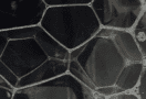
Scientists at Princeton University in the US have discovered that a material known as a Weaire-Phelan foam can act as an optical filter. As well as adding to our understanding of such foams, which have been studied for more than 130 years, the discovery might also spur the development of novel optical telecommunications devices.

Foams have a multitude of practical applications and are common ingredients in products ranging from chemical filters to heat exchangers. In mathematical terms, they are known for forming structures that minimize the surface area of the geometrical shapes, or cells, that make them up. It was this property that attracted the attention of the 19th-century Scottish physicist Lord Kelvin. In 1887, Kelvin proposed that the “luminiferous aether” thought to permeate all of space might have a foam-like structure. He then attempted to find the most efficient way of filling a 3D space by sub-dividing it into interlocking cells of equal volume and minimizing the surface area between cells. The resulting bubble-like structure came to be known as a Kelvin foam.

Extra efficient space-fillers

More than a century later, the Irish physicist Denis Weaire and his student Robert Phelan improved on Kelvin’s conjecture by putting forward an alternative arrangement that requires even less surface area. Although a Weaire-Phelan (WP) foam looks superficially similar to the disordered froth of soap bubbles or the head on a glass of beer, it is in fact a precisely structured arrangement containing two types of cells (as opposed to just one in Kelvin’s original proposal) of equal volumes. Twenty-five years after its discovery, a WP foam remains the most efficient space-filling ordered bubble foam known to exist.

3D photonic networks of foam edges

Much research has been done on WP foams and many of their physical properties are well-understood. The Princeton team of Michael Klatt, Paul Steinhardt and Salvatore Torquato, however, took a different approach, studying the optical properties of 3D photonic networks made from the edges of a WP foam, a Kelvin foam and another type of dry crystalline foam known as C15. Foams such as these contain very little liquid, and their edge structures are characterized by a set of relationships known as Plateau’s laws. These laws dictate that the borders of individual cells within the foam meet in sets of four, with tetrahedral bond angles equal to around 109° at each vertex. In principle, it would be possible to turn such edge structures into photonic networks by solidifying the foam and coating it with a dielectric material.

A photonic band gap

The team simulated Maxwell’s electromagnetic wave equations for these structures to determine how they behave when light passes through them. These calculations were executed by Klatt on the supercomputing facilities of the Princeton Institute for Computational Science and Engineering, and were computationally intensive, requiring a detailed set of calculations based on analysing the foams using a software tool called Surface Evolver, which optimizes shapes according to their surface properties. The results showed that all three foams have refractive indices that vary on the length scale of electromagnetic waves such as visible light – a phenomenon known as a photonic band gap. The presence of such gaps affects how light or other waves propagate through the material, allowing some wavelengths to pass through while completely reflecting others.

Band gaps are typically measured in percentages that indicate the size of the frequency gap relative to the gap’s central frequency. According to the researchers’ calculations, the Kelvin foam has a band gap of 7.7%, while that of the C15 foam is 13%. The WP foam has the largest photonic band gap of the three, at 16.9%. These figures are comparable to or greater than the band gaps found in self-organizing photonic crystals such as synthetic opals. The band gaps of all three foams are also highly isotropic, meaning they do not have strongly directional properties. This could be useful for designing photonic waveguides and other optical circuits.

The rise of “phoamtonics”

The researchers say that their calculations open up a host of possibilities for future work on WP foams and similar materials, in a field they dub “phoamtonics” (from “foam” plus “photonics”). One possibility would be to use these foams to transport and manipulate light, for example in telecommunications applications. At present, much of the data travelling across the Internet is carried by glass optical fibres, but when it reaches its destination, the photonic signal is converted into an electrical one, with an associated loss of speed and precision. Torquato suggests that photonic bandgap materials could guide the light much more precisely than conventional fibre optic cables, and might even serve as optical transistors that perform computations using light.

The finding also expands the range of 3D heterostructures available for photonic applications beyond photonics crystals, quasicrystals and amorphous networks, Torquato adds. “While the WP foam does have a smaller band gap than other well-known materials like ordered diamond networks (31.6%), it might offer some advantages thanks to its multifunctional properties,” he tells Physics World.

The researchers report their work in the Proceedings of the National Academy of Sciences of the United States of America (PNAS).
Alumina glass bends rather than breaking

Flexible glass that does not shatter on impact could soon be made using insights from a study of a glass-like material made from aluminium oxide. Erkka Frankberg at Tampere University in Finland and colleagues have come to this conclusion after studying the molecular mechanisms the prevent cracks forming in the material.

Glass has lots of very useful properties including optical transparency, durability and low electrical conductivity. However, the inherent brittleness of the material has prevented it from finding a wider range of applications.

Glass is brittle because it no way to effectively dissipate mechanical energy when it is deformed by external forces. Instead, the energy accumulates around microscopic defects. This leads to localized concentrations of stress and eventually to the propagation of sharp cracks and shattering.

Blunter cracks

This weakness is particularly pronounced in traditional glasses made of silicates (silicon oxides), which form rigid tetrahedral structures that encourage the propagation of sharp cracks. In principle, brittleness in glass could be overcome by blunting the tips of cracks as they propagate – something that occurs in ductile materials. If achieved, this would give glass far higher mechanical strength, and make it less likely to fail because of defects.

Frankberg’s team created a new type of glass from aluminium oxide (alumina) using a technique called pulsed laser deposition. This was a significant challenge in because the material normally occurs in a crystalline form, rather than in an amorphous glassy state.  In contrast to silicates, amorphous alumina can deform irreversibly at room temperatures. Through a combination of transmission electron microscopy with molecular dynamics simulations, they explored the mechanisms by which this deformation occurs.

The team discovered that molecular bonds in amorphous alumina are up to 25 times more likely to break and reform when distorted – compared to bonds in silicate glass. This allows mechanical stresses in the materials to relax. Furthermore, localized strain events within the material can accumulate into ductile flows instead of concentrated stresses, allowing blunter cracks to form. The team then showed that this “viscous creep” mechanism allows amorphous alumina to endure far higher strains without fracturing. Indeed, they were able to the materials elongate by up to 100% in the most extreme scenarios.

Despite the excitement surrounding their findings, Frankberg’s team acknowledges that they looked at idealized samples of amorphous alumina that were free from defects. This means that commercialized products incorporating the material are currently an unrealistic prospect. However, the researchers say that their results provide important guidelines for developing generalized strategies for tailoring the mechanical properties of oxide glasses.

The research is described in Science.

Super slippery toilet is self-cleaning, the physics of logjams


“A robust bio-inspired, liquid, sludge and bacteria-repellent coating that can essentially make a toilet self-cleaning,” is how Tak-Sing Wong describes a recent invention of his research group at Penn State University.

The coating is sprayed onto the toilet surface in two steps – the first puts down a layer of hair-like molecules and the second makes those hairs extremely slippery. The team then tested its efficacy using artificial poo – yes, it is available – as well as bacteria commonly found in toilets. Neither were able to stick to the surface. As well as boosting hygiene and reducing odours, the team says that the coating could reduce the water used to flush toilets by 50%.

You can read more in “New, slippery toilet coating provides cleaner flushing, saves water”.

Moving on from toilets to logjams, which aren’t actually jammed according to geoscientists Nakul Deshpande and Benjamin Crosby at Idaho State University in the US. The duo studied a logjam in Idaho’s Big Creek using a number of techniques including time-lapse photography. They charted the motion of the logs in May and June 2016, as the river crested its annual peak. The jam had been formed two years earlier when a snow avalanche pushed dead trees into the river.

Deshpande and Crosby found that the logjam exhibited creep and clogging behaviours that are also seen in some disordered materials. This, they conclude, could provide insights into how to mitigate hazards associated with logjams. You can read more in “Logjams are not jammed: measurements of log motions in Big Creek, Idaho”.

Gold plasmons guide light in new photonic switch

A highly compact, low-energy device capable of switching the paths taken by light within photonic systems has been unveiled by physicists in the US and Switzerland. The new switch could provide a basis for artificial-intelligence (AI) systems that mimic human decisions, allowing for a diverse range of applications.

“All-optical computers” use light instead of electronic signals to process information. In principle, they could be faster and much more energy efficient than conventional computers. However, it is proving very difficult to create compact and energy efficient photonic devices that can switch and process optical signals at high speeds.

The new device was created by Chris Haffner and colleagues at the National Institute of Standards and Technology (NIST), ETH Zurich and the University of Maryland.

Light enters the switch via a linear silicon waveguide that is adjacent to a racetrack-shaped cavity that is etched onto a silicon disc. If the wavelength of the incoming light resonates with the racetrack cavity, some of the light will enter the racetrack and circulate around it many times.

Gold membrane

The device also incorporates an extremely thin, circular gold membrane, suspended just a few nanometres above the disc. This membrane is connected to the disc by doped silicon and gold bridges, enabling Haffner and colleagues to apply a varying voltage between the two structures. This allows them to bend the membrane either up or down, creating a variable gap between it and the waveguide.

As some of the light travelling around the cavity escapes, it strikes the membrane, creating collective oscillations of electrons (called plasmons) on the gold’s surface. These plasmons vibrate at the same frequency as the light, but with much shorter wavelengths. This means that the plasmons can be converted back into light with a high degree of control.

When the membrane is bent upwards, the rest of the light in the waveguide passes through unaffected. When positioned downwards, however, the membrane plasmons oscillations allow light to leak from the waveguide into the cavity. This light can then interfere destructively with light in the waveguide, essentially turning off the transmission of light. Furthermore, light can be transferred to a second waveguide positioned close to the membrane – rerouting the path of the light.

The compactness and low energy requirements of the device allowed the team to integrate it onto a single computer chip.  They envisage a wealth of AI-related applications for their device, including self-driving cars which can rapidly redirect light beams to scan for other cars and pedestrians. More generally, the technology could bring about advanced circuits that form the backbones of neural networks – capable of recognizing patterns and making human-like decisions about complex tasks.

The new switch is described in Science.

Artificial intelligence in medical physics, quantum computing in silicon and a return to physics in film

This week’s episode focuses on the interface between physics and computing, with deep dives into how artificial intelligence (AI) is contributing to medical physics and how silicon could form the basis of a future quantum computer.

First, we hear from Tami Freeman, Physics World’s resident expert on medical physics, about a new positron emission tomography (PET) scanner that can image a patient’s whole body much more quickly (or at higher resolutions) than is possible with current commercial scanners. We then stick with the medical theme to discuss three recent examples of how AI is being used in medicine: firstly to diagnose skin conditions (but, disturbingly, only if the patient’s skin is white); secondly to help radiologists detect lung tumours in X-rays; and thirdly to develop better radiotherapy treatment plans.

The second part of our podcast switches from classical computing to the quantum world. There are several ways of constructing the qubits, or quantum bits, that make up a quantum computer, and this week we hear from a trio of researchers – Fernando Gonzalez-Zalba, Alessandro Rossi and Tsung-Yeh Yang – who have been developing silicon-based qubits. Their work is part of a Europe-wide collaboration between universities, government laboratories and companies called MOS-Quito, and you can read more about it in their article for the Physics World Focus on Computing.

And finally, if you’ve been dying to hear the answers to last week’s parlour game, be sure to listen to the end of the podcast and groan along with our editors at some truly terrible amazing physics-in-film wordplay.

MRI-compatible electrodes line up for use in neural probes

Deep brain stimulation (DBS) – in which electrodes implanted in the brain send electrical signals to areas that control movement – is increasingly employed to treat symptoms of movement disorders such as Parkinson’s disease, essential tremor or dystonia. It is also used in epilepsy and is under investigation as a potential treatment for traumatic brain injury, addiction, dementia, depression and several other conditions.

Patients with implanted electrodes often undergo brain MRI, for example to guide electrode placement, investigate DBS outcomes or evaluate implantation-related abnormalities. DBS electrodes are generally made from thin-film platinum or iridium oxide. However, such metal-based electrodes are affected by the magnetic fields of the MR scanner, and can cause image artefacts, move or vibrate, or even generate heat.

To tackle these problems, San Diego State University (SDSU) engineers have created a glassy carbon microelectrode for use instead of the metal version. Working in collaboration with researchers at Karlsruhe Institute of Technology, they have now shown that the new electrode does not react to MRI scanning, making it a safer option for DBS (Microsyst. Nanoeng. 10.1038/s41378-019-0106-x).

“Our lab testing shows that, unlike the metal electrode, the glassy carbon electrode does not get magnetized by the MRI, so it won’t irritate the patient’s brain,” explains first author Surabhi Nimbalkar.

The glassy carbon electrodes, first developed in 2017 at SDSU, are designed to last longer in the brain without deterioration. The researchers previously demonstrated that while metal electrodes degrade after 100 million electrical impulse cycles, the glassy carbon material survived 3.5 billion cycles. Another benefit is that glassy carbon electrodes can read both chemical and electrical signals from the brain.

“It’s supposed to be embedded for a lifetime, but the issue is that metal electrodes degrade, so we’ve been looking at how to make it last a lifetime,” says senior author Sam Kassegne. “Inherently, the carbon thin-film material is homogenous so it has very few defective surfaces. Platinum has grains of metal, which become the weak spots vulnerable to corrosion.”

In their latest study, Kassegne and colleagues fabricated probes made from glassy carbon and thin-film platinum microelectrodes supported on a polymer substrate. They placed the probes in a brain-tissue-mimicking agarose phantom and imaged them in a 3 T MRI scanner using clinical MRI sequences.

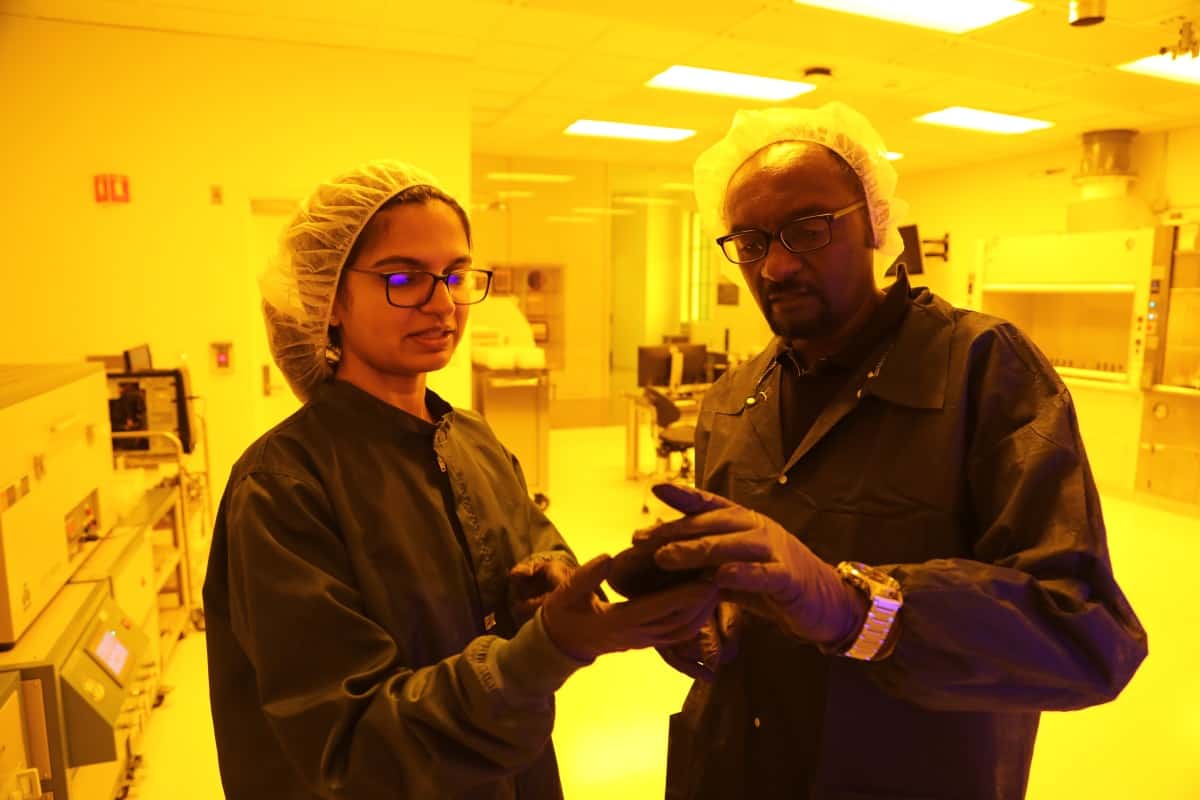
Sam Kassegne and Surabhi Nimbalkar

The researchers found that, because of their low magnetic susceptibility and lower conductivity, the glassy carbon microelectrodes caused almost no susceptibility shift artefacts and no eddy-current-induced artefacts compared with the platinum microelectrodes. Tests in a high-field (11.7 T) magnet exhibited similar findings.

The team also used a novel instrument developed at KIT to precisely measure gradient-induced vibrations in the electrodes during 1.5 T MRI. Both the platinum and glassy carbon microelectrode samples had vibration amplitudes below the limit of detection (indistinguishable from that of non-conductive PMMA plates).

Theoretical analysis, however, revealed that while the platinum microelectrode was at the limit of detection, the glassy carbon microelectrode had an approximately 40-fold weaker response. The team also note that gradient-induced vibration scales to the power of four with implant radius, so for larger electrodes, the smaller conductance of glassy carbon will be advantageous.

Finally, to examined induced currents in the two microelectrode types, the researchers fabricated glassy carbon and platinum ring electrodes supported on a silicon wafer. Induced currents measured with a 1 Ω resistor indicated that induced current in glassy carbon was at least a factor of 10 less than in the platinum sample.

The researchers conclude that glassy carbon microelectrodes demonstrated superior MR compatibility to standard thin-film platinum microelectrodes, experiencing no considerable vibration amplitudes, minimally induced currents and generating almost no image artefacts. While they did not examine RF-induced heating in this study, the lack of RF-induced eddy currents (a large source of heating) in glassy carbon microelectrodes suggests that they will also be superior to platinum in this aspect.

With lab testing completed, Kassegne’s clinical collaborators will now test the glassy carbon electrode in patients, while Nimbalkar and Kassegne plan to test different forms of carbon for use in future electrodes.

Light forks one way in a Bose–Einstein condensate

Researchers at the universities of Bonn and Cologne in Germany have developed a new way of splitting photon wavepackets that involves cooling them down to a Bose–Einstein condensate (BEC) in a double-ridge microresonator structure. This thermodynamic method differs from the usual optical beam-splitting techniques because it is irreversible, meaning that the original beam cannot be reconstructed. The BEC-based process might be extended in the future to make new optical sources for entangled and correlated light states for applications in quantum computing.

The research team was led by Martin Weitz from the Institute of Applied Physics at Bonn University, who made headlines in 2010 when he and his colleagues created the first BEC from photons – 15 years after the first BEC was made in 1995 from a cloud of rubidium atoms cooled to a fraction of a degree above absolute zero. BECs form when bosons (particles with integer quantum spin) are cooled until they are all in the same quantum state. At this point, a BEC made up of tens of thousands of particles will behave as if it were in fact just a single quantum particle.

First photon BEC

Weitz and his colleagues made their photon BEC, or “super-photon”, by firing a laser beam into a microresonator cavity made of two concave mirrors separated by about a micron. This separation defines (to within an integer multiple) the maximum wavelength, and thus the minimum energy, of a photon trapped longitudinally within the cavity. The cavity is filled with a dye solution that is held at room temperature, meaning that its thermal energy is only about 1% of the energy of the photons.

Because the dye has so much less energy than the photons, it is highly unlikely that additional photons will emerge from the dye, or that the dye will completely absorb a photon. What happens instead, Weitz explains, is that when the photons collide with the dye molecules, they are briefly “swallowed” and then spat out again. After repeated absorption and re-emission cycles, the photons acquire the temperature of the dye solution and are thus cooled down to room temperature without being lost.

By increasing the laser intensity irradiating the dye solution, the researchers increase the number of photons in the cavity until it reaches about 60,000. This strong concentration of the light particles, combined with simultaneous cooling, causes the individual photons to coalesce into a photon BEC – much like a liquid drop condensing in a gas.

Photons pass into lower-energy states

In their new work, Weitz’s team used the same experimental set-up as before. This time, however, one of the two cavity mirrors was not completely flat. Instead, it contained two small valley-like optical ridges, or potential energy minima. When the laser light beam enters one of these ridges, the distance it travels, and therefore its wavelength, increases slightly. As in previous work, the photons in this beam are cooled by the dye solution before passing into a lower-energy state in the ridges.

The study’s lead author, Christian Kurtscheid, compares the photons in their system to marbles rolling over a sheet of corrugated metal. Whereas marbles with a high velocity would simply skip over the sheet’s surface, slower marbles will settle in the valleys of the corrugations. Similarly, the cooled photons in their experiment “roll into the valleys of the ‘corrugated sheet’ and remain there,” Kurtscheid says.

Tunnel coupling

In the experiment, the two ridges are close enough together to allow for a phenomenon called tunnel coupling, he says. This means it is no longer possible to determine which photons are in which ridge – only that they are in the lowest energy state in the cavity. “This state is the symmetric linear combination of photons localized in the two potential minima,” Weitz explains. “We observe that the photons condense into a BEC into this low-energy, spatially bifurcated state.”

This process irreversibly splits the wavepackets of light as if they were passing through an intersection at the end of a one-way street while the tunnelling between the ridges leads to one of the hybridized wavefunctions of the photons coupling.

Irreversible, coherent light splitting

The researchers observed the light wavepacket splitting by using a camera to monitor the optical radiation transmitted through one of the cavity mirrors. They also monitored the coherence of the beamsplitting by recombining the light beam paths. In this way, they were able to observe interference fringes of the beams.

“Our technique is a new, energetically-driven way to prepare optical quantum states,” Weitz tells Physics World. “We have demonstrated how to irreversibly create coherently split light.”

The method, which is detailed in Science, might be extended in the future to realize new optical sources for entangled and correlated states of light, he adds.

The researchers now plan to prepare photons in a periodic lattice potential and enhance effective photon interactions. In such a set-up, highly entangled many-body states can become the lowest energy state in the cavity. “We could directly populate these quantum states by cooling using our method and these states could be used in applications such as quantum information and communication,” Weitz says.

How well can biological cells sense their environment?

Biological cells adapt to chemical changes in their environment by sensing certain molecules as they bind to specific receptors on the cell surface. Previous work to measure this sensing ability typically assumed that concentrations of these binding molecules, known as ligands, remain constant or change at steady rates over time. Now, researchers in France and the US have developed a mathematical model for a cell’s sensitivity that better reflects real-world conditions. Their work could shed light on dynamic processes within biological systems, such as rapid cell growth in an embryo or the motion of bacteria in response to chemical stimuli.

The new model was developed by Thierry Mora of the ENS in Paris and Ilya Nemenman of Emory University in Georgia, and it describes the rapid shifts in a cell’s chemical environment with a non-linear randomly changing numerical field. This formulation allows the researchers to apply techniques from stochastic field theory that are routinely employed to solve problems in quantum and statistical physics.

Calculating the smallest fluctuations

Cells sense chemical concentrations by binding external ligands to specific receptors on their surface. Mora and Nemenman’s model derives the probability of a ligand binding to a cell within a given time period to calculate the smallest fractional fluctuations of concentrations that the cell can detect.

They found that the cell can sense smaller fractional fluctuations as the overall concentration of a biochemical increases, or as a receptor’s binding rate increases. In previous calculations that assumed a constant, non-fluctuating, environment, the cell’s sensitivity – expressed as an error in the concentration, c – was related to the biochemical concentration and the receptor binding rate by a ½ power law known as the Berg and Purcell bound.

In the new model this sensitivity changes more slowly with the concentration and binding rate and obeys a ¼ power law. Indeed, it scales as δc/c ∼ (Dacτ) -1/4. In this equation, D is ligand diffusivity, a is the linear size of the receptor, and τ is the ligand fluctuation time scale.

Model works for a real-world situation

The researchers, who have reported their work in Physical Review Letters, say they have already applied their model to a real-world situation in which an external chemical drives a network of signals inside the cell after it binds with the cell’s receptors. Their computer simulations showed that under these circumstances, the cell can detect the molecule within the fundamental limit they derived.

Robert Endres of Imperial College London, who was not involved in this research, says that the problem of sensing a fluctuating ligand concentration by a receptor is certainly relevant for biology. He adds that deriving the sensing limit via a field-theoretic approach, as Mora and Nemenman did, is “very elegant”. However, he also downplays the degree to which their findings differ from earlier work.

“Although a great result, I would not say that it is a radically different limit from the usual Berg and Purcell limit,” Endres says. The Berg and Purcell limit, he explains, is a lower limit, a kind of “noise floor”, while Mora and Nemenman’s new limit is higher due to a fluctuating ligand concentration. Second, as the researchers explain themselves, the new limit can be reconciled with the Berg and Purcell limit using an optimal averaging time – that is, by making the measuring time interval as long as possible to better average the results obtained, and short enough for the concentration of a ligand to vary less.

Neutron dripline extended to fluorine and neon isotopes

The maximum number of neutrons that can be packed into fluorine and neon isotopes have been determined by nuclear physicists working on an experiment in Japan. These are the first new measurements of the neutron dripline in 20 years and could provide physicists with important information about how to model the atomic nucleus. The same experiment failed to determine the dripline for sodium, which is the next element in the periodic table beyond neon.

The neutron dripline refers to the maximum number of neutrons that can be packed into an atomic nucleus before it becomes unbound. Until this latest work, physicists had measured the driplines of the eight lightest elements (hydrogen up to oxygen). In general, the maximum number of neutrons in a nucleus increases with the atomic number. However, there appears to be an exception to this rule with the dripline isotopes carbon-22, nitrogen-23 and oxygen-24 – which all have 16 neutrons. This is called the “oxygen anomaly” and suggests that 16 may be a magic number for neutrons, signifying the completion of a stable shell of neutrons.

Now, Deuk Soon Ahn and colleagues working on the BigRIPS experiment at the RIKEN Radioactive Isotope Beam Factory have looked at the next three elements in the periodic table: fluorine, neon and sodium.

Fragmenting nuclei

To look for the most neutron-rich isotopes of these elements, the team fired a high-energy beam of calcium-48 ions at a beryllium target. The calcium nuclei undergo fragmentation to create smaller nuclei, which were studied by the team. This was done using BigRIPS, which sorts nuclei according to their mass and charge.

Before the study was done, the heaviest known isotopes of these elements were fluorine-31, neon-34 and sodium-37. However, it was not known if heavier isotopes existed. The team was unable to detect fluorine-32, fluorine-33, neon-35 and neon-36 – providing strong evidence that fluorine-31 (with 22 neutrons) and neon-34 (with 24 neutrons) are dripline isotopes.

The team also looked for sodium-38 and sodium-39 and although they saw no evidence for sodium-38, they did spot one sodium-39 nucleus – which has 28 neutrons. As a result, they conclude that the neutron dripline must be at or beyond 28 neutrons for sodium.

These observations do not fully agree with state-of-the-art calculations of the dripline for these elements – which suggest that both fluorine and neon should have a maximum of 24 neutrons. The model of the nucleus used in these calculations will therefore have to be revised.

Looking to the future, the Facility for Rare Isotope Beams (FRIB) at Michigan State University in the US will open in 2022 with beams that are significantly more intense than those at RIKEN. This should make it possible for physicists to resolve the dripline for sodium and begin to study magnesium, which is the next element in the periodic table.

The research is described in Physical Review Letters.

Twinkle, twinkle little star, how I wonder where the trabecular meshworks are

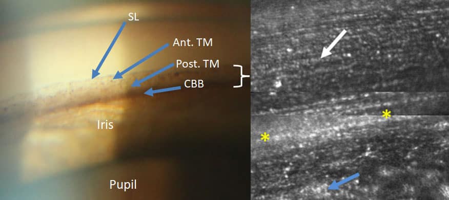
Astronomers use adaptive optics to see past the Earth’s atmosphere, which distorts celestial objects and makes them appear to twinkle. A new technique called adaptive optics gonioscopy (AOG) applies similar principles to image the human eye.

Researchers at Indiana University published the results of their proof-of-concept study in Translational Vision Science & Technology (10.1167/tvst.8.5.5).

Glaucoma: origins and impact

The clinical motivation for developing this new imaging technique is glaucoma, an eye disease currently affecting 76 million people worldwide. Interventional procedures, such as laser therapies or surgery, have mixed outcomes, and the physiology of the disease is poorly understood.

In an eye that functions normally, clear fluid circulates throughout, supplying nutrition and maintaining the eye’s shape. The eye’s trabecular meshwork – a series of sequentially smaller pores – acts like a sieve, allowing fluid to drain properly and regulating pressure.

The trabecular meshwork is altered in glaucoma. Fluid no longer drains properly and intraocular pressure, a risk factor for glaucoma and its progression, rises.

“As a result, the trabecular meshwork is an important structure to study, but direct imaging has been difficult,” says Brett King, lead author on the study. “AOG allows us to overcome the natural, near total internal reflection in this region and the differences in index of refraction between the eye’s cornea and the air.”

To see or not to see with AOG

King’s team successfully used AOG to visualize the trabecular meshwork at near cellular-level resolution in a proof-of-concept study with nine individuals (seven healthy volunteers and two with pigment dispersion syndrome, thought to be a precursor to glaucoma). This high-resolution visualization is a vast improvement over current clinical imaging methods, which only allow clinicians to determine the relative level of pigmentation of the trabecular meshwork and evaluate an individual’s risk of developing certain types of glaucoma.

The trabecular meshwork

The AOG technique requires an existing adaptive optics system for imaging the retina, which includes a wavefront sensor and deformable mirror. The wavefront sensor measures optical aberrations while the deformable mirror corrects for them. The researchers added cameras to assist clinicians in placing the modified gonioscopy lens on a patient’s eye and added a head mount to reduce patient motion.

Shooting for the stars

One challenge that the researchers faced was knowing what it was they were looking at. “As AOG imaging of the trabecular meshwork had not been performed before, we had difficulty at first realizing where we were and what structures we were seeing,” says King.

The researchers compared their images to pathology images that were often distorted from surgical or post-mortem artefacts. They also tested the technique with a model eye used to train surgeons.

King and colleagues are now building a new device to image even deeper within the eye and account for anatomical differences between individuals. They hope that this work will improve our understanding of age-related and pathological changes that occur in the human eye, as well as responses to pharmacological and surgical interventions.

Copyright © 2026 by IOP Publishing Ltd and individual contributors