Call made for Germany’s Helmholtz Association to boost diversity

The Helmholtz Association, Germany’s largest research organization, needs to increase the diversity of its workforce, including the number of senior female researchers. That is according to an evaluation of the organization conducted by around 600 external scientists from 27 countries. The review will now be used for a major evaluation of the organization next year that will set the association’s research priorities until 2027.

We really need to increase diversity to bring in other ideas and make progress

Sören Wiesenfeldt

With an annual budget of more than €4.5bn, the Helmholtz Association employs around 40,000 people and brings together 18 scientific research centres – including the DESY accelerator centre near Hamburg and the Research Centre Jülich. While the Helmholtz Association has previously conducted regular assessments of its individual centres, Sören Wiesenfeldt, head of research at the Helmholtz Association, told Physics World that this is the first time it has assessed its overall scientific performance. As well as looking at individual centres, the assessment examined the performance of research programmes that often involve multiple research centres.

Need to improve

The report finds that the Helmholtz Association conducts high-level, interdisciplinary research with the 18 centres well positioned in their respective research fields and that they collaborate well together. The association does, however, face challenges. The organization needs to strengthen its data science research and use tools such as artificial intelligence to better manage the huge volumes of data that are generated at Helmholtz centres. In particular, there is a need to improve data sharing among centres so that results can be used across different organizations and research programmes.

According to Wiesenfeldt, the reviewers found that the Helmholtz centres are “very German” – around 6000 workers are foreign – and that the organization’s “culture for diversity has to improve”. The evaluation discovered that while there is almost gender parity at PhD level, only 19% of full professorships are held by women. Wiesenfeldt adds that the association was aware of the gender gap and it is funding several initiatives to increase female researchers at all levels with an overall aim to boost the proportion of female professors to 24% by 2020. “We really need to increase diversity to bring in other ideas and make progress,” says Wiesenfeldt.

Bioengineered bile ducts mimic the real thing

Diseases of the bile duct can lead to serious medical conditions such as liver cirrhosis and eventually liver failure. But research into these diseases, and possible treatments for them, has been frustrated by a lack of realistic models for the functional cells that line the bile duct, known as cholangiocytes.

Researchers in the Netherlands have now devised a two-step technique that exploits stem cells taken from mouse livers to create cholangiocyte-like cells (CLCs). The CLCs have the same biological characteristics as in vivo cholangiocytes, and combining the cells with hollow fibre membranes also enabled the team to construct life-like bile ducts that could transport typical bile acids (Biofabrication 10 034103).

The main function of the bile duct, and in particular the cholangiocytes that line its surface, is to carry away waste products from the liver, such as excess cholesterol, bilirubin and hormones, as well as drugs and toxins. Any damage to the lining of the bile duct can usually be repaired by the cholangiocytes multiplying themselves, but various biliary diseases can emerge when this natural mechanism breaks down. Left untreated, these cholangiopathies have the potential to get progressively worse and cause severe liver disease.

This new system offers us an unprecedented new way to analyse and modulate cholangiocyte function in vitro

Chen Chen of Utrecht University, the Hubrecht Institute-KNAW and the University Medical Center Utrecht

Although researchers have been able to culture primary cholangiocytes from different species, these cultures typically only survive for about three or four weeks. More importantly, the in vitro models that have been developed so far do not have the same physiology as natural cholangiocytes.

“To our knowledge, a source of sustainable and genetically stable stem cells that can differentiate into functional cholangiocytes has not been reported,” says study lead author Chen Chen of Utrecht University, the Hubrecht Institute-KNAW and the University Medical Center Utrecht. “Although cholangiocytes have been generated in vitro from induced pluripotent stem cells, this technique is inefficient, time-consuming and produces cells that are genetically compromised.”

According to Chen, researchers have recently found that many organs, including the stomach, intestine, liver and pancreas, contain adult stem cells that can differentiate into the cell types of the respective organ. In the liver, for example, cells marked with a specific receptor (Lgr5) can be isolated from small pieces of liver tissue. These cells can then be made to form organoids – 3D structures with the same biological properties as the organ – by creating conditions that mimic the physiological environment in which the tissue self-renews or repairs.

Building on this previous research, Chen and colleagues have now developed a two-step differentiation process to generate CLCs in vitro from mouse liver organoids. The first step exploits a hydrogel composed of collagen and Matrigel, a culture medium that contains various growth factors plus extracellular matrix components of the basement membrane. “In this first step, the liver organoids quickly proliferate, providing us with an ‘unlimited’ cell source,” explains Chen. “In the second step, we supplemented the cells with two more compounds to inhibit proliferation and enhance cell function.”

The CLCs are genetically stable and have the same morphology and biological function as primary cholangiocytes. What’s more, integrating the cells with collagen-coated hollow fibre membranes allowed the researchers to bioengineer structures that looked like native bile ducts.

This new system offers an exciting new way to analyse and modulate cholangiocyte function in vitro, Chen told Physics World. “It could help us investigate, for example, how cholangiocytes protect against bile acids and how they regulate bile acid homeostasis,” he says. “These studies will help us develop treatments for diseases of the bile duct system.”

Spurred on by their work on mouse tissue, the researchers say they will now attempt to engineer human bile ducts. “We will also be testing different tubing materials that could better mimic the liver microenvironment,” adds Chen.

  • Read our special collection “Frontiers in biofabrication” to learn more about the latest advances in tissue engineering. This article is one of a series of reports highlighting high-impact research published in Biofabrication.

Beefing up X-rays at the European Synchrotron Radiation Facility

Synchrotrons push the boundaries of science at every frontier. On the one hand, synchrotrons such as the Large Hadron Collider at CERN in Geneva – accelerators designed to smash particles together at high energies – test the very foundations on which physics is based. On the other hand, synchrotrons can also be used to explore the less grandiose, but just as important “science of the everyday”. No particle-smashing takes place at synchrotron facilities; instead the goal is to harness the light – typically X-rays – generated when charged particles are accelerated continuously in a ring. These X-rays can be focused on pretty much any material – biological tissue, protein crystals, cosmetics, engineering alloys, superconductors and dinosaur fossils to name just a few – in order to determine how they behave, and understand why.

Initiated back in 1988, the European Synchrotron Radiation Facility (ESRF) in Grenoble, France, was the first “third generation” synchrotron. This meant that the charged particles it accelerated, electrons, were forced to wiggle inside special magnets to generate X-rays of extremely high brilliance. Over the years, the facility has been integral to several momentous discoveries, such as the structure of the ribosome – the subject of the 2009 Nobel Prize for Chemistry. But now, 30 years and 30 000 publications into its life, the ESRF is on the verge of a massive upgrade. The Extremely Brilliant Source (EBS) – as its new electron storage-ring will be known when it launches in 2020 – will multiply the brightness and coherence of X-rays by 100, illuminating the science of the everyday with unprecedented clarity. Though not the very first fourth-generation synchrotron, it will be the first operating at such a high energy – which is why four key areas of instrumentation must also be up to scratch.

Optics in focus

It probably comes as no surprise that incredibly bright and coherent X-rays demand incredibly high-quality optics. Optics transform a raw X-ray beam into a form best suited for a particular experiment, by tuning characteristics such as shape, energy, divergence and polarization. At a major facility such as the ESRF–EBS, the electron storage ring is surrounded by dozens of beamlines through which the X-rays are emitted at tangents, and every beamline is geared towards a specific type of experiment. Get its optics wrong, and a beamline could lack precision and stability, and its characteristics could even degrade over time.

That is why the ESRF–EBS project has a special development programme to address optics. The essential goal is to minimize the spatial and temporal perturbation of X-ray wave-fronts, and transfer X-ray flux to the samples at a beamline as efficiently as possible. Getting it right involves computer modelling, high-precision engineering, the development of new fabrication techniques, and testing outside and inside beamlines. The types of optics under scrutiny are crystal analysers (which measure the energy of X-rays in spectroscopy), multilayers (which focus X-rays, or select X-rays of a certain energy range), refractive lenses, mirrors, and measurement devices.

The ESRF has a strong pedigree in optics. Only last year, its ID16A beamline set a record for the highest resolution obtainable with high-energy X-rays – less than 13 nm – thanks to the installation of a new multilayer mirror. Besides nano-focusing, techniques such as imaging and coherent scattering (a special type of imaging in which the X-rays do not transfer energy to a sample) rely strongly on the quality of beam manipulation. But the impact of the optics programme will be far-reaching, giving a lift to the ESRF–EBS’s entire beamline portfolio.

Data collection

Few aspects of instrumentation capture the attention of users so much as detectors. Changes to detectors can alter the amount of data generated in an experiment by orders of magnitude. The ESRF is always improving its detectors, and in the next year or two several ongoing projects will come to fruition. One of these is the next-generation Frelon camera – a fast-readout detector used on a quarter of all ESRF beamlines, and at many other synchrotrons the world over. For the EBS, however, a dedicated detector development plan (DDP) aims to go further. Aside from developing advanced new detector systems, the DDP involves collaborating with external laboratories to access other detectors already in development, as well as improving related technologies – such as scintillators, which convert X-rays to visible light – that will help maintain the quality and productivity of ESRF–EBS beamlines.

The impact of the optics programme will be far-reaching, giving a lift to the ESRF–EBS’s entire beamline portfolio

The detectors themselves are being developed along two lines. The first, SMARTPIX, will be a photon-counting pixel detector with a 55 μm distance between pixels, based on the MEDIPIX3RX readout circuit developed by the international Medipix3 Collaboration, of which the ESRF’s Detector and Electronics Group is a partner. SMARTPIX will be faster and more flexible than previous versions, and it will also have the potential for greater temporal resolution. The second line of development is a series of large-format detectors for high-energy X-ray diffraction. These include fast detectors able to simultaneously detect both very intense and very weak X-ray signals, and detectors whose output current is proportional to the incident X-rays.

That’s not all. To supplement detector development, the ESRF is advancing three strategic technologies: X-ray sensors, which convert X-ray photons into a measurable signal; control and data-acquisition, which manages the throughput of data; and energy-dispersive detection technology, which involves the use of silicon drift diodes and germanium detectors to record the number and energy of X-rays emitted from a sample.

Robotic controls

With the launch of the EBS, many of the ESRF’s users will see data-acquisition times drop from milliseconds down to microseconds, and lower. That poses an issue for control systems, which will need to be more responsive to keep up, while maintaining synchronization with the EBS storage ring.

It isn’t purely about timing, however. As more and more experimenters look towards the nanoscale, and want to perform their experiments more efficiently, control systems also need to become both more precise and more automated. This is where robots – mechatronics – can help. The ESRF already has plenty of expertise here, for example with the almost fully automated MASSIF beamlines at ID30, which can be set up to collect millions of diffraction images of protein crystals every month, with practically zero human intervention – a truly massive time saver for drug discovery. For the EBS, a mechatronics task force has been created to work between groups within the ESRF’s Instrumentation Services and Development Division, and to provide a platform to share expertise in modelling, mechanics, electronics and software. A mechatronics laboratory is also forthcoming.

Various areas will see the benefits. Currently in development, a new double-crystal monochromator – the optical component that filters bundles of X-rays down to a single wavelength – will incorporate mechatronic concepts to improve precision and repeatability. Similarly, the Nano-positioning and Active Stabilization Stage, or NASS, project – part of a collaboration with engineering physicist Christophe Collette at the University of Brussels in Belgium and colleagues – is working on a prototype for a new generation of highly integrated end-stations in which sensors, measurement systems and mechatronics are all tightly integrated.

Managing data deluges

Faster experiments at higher resolution can only mean one thing: more data. So much data, in fact, that making sense of it will be a challenge in itself. To adapt their measurement strategies in real-time, users will need new, sophisticated ways of visualizing data. They will also need new ways of processing it on-site or online, as many of them will lack the appropriate computing facilities at their home institutions.

Fortunately, solutions are in the pipeline. The foundations for a new beamline control system, BLISS (which stands for BeamLine Instrumentation Support Software), have been laid, and it is already being deployed on the ESRF beamlines ID15 and ID31, as well as those of the Structural Biology Group. Meanwhile, software engineers have been busily developing SILX, a library of common data-analysis routines, generic applications and software. Now in its sixth release, SILX is being used in an increasing number of areas, and has been demonstrated to cut data-analysis times dramatically.

Myriad other changes behind the scenes will help funnel the data deluge. For example, a new ESRF data policy requiring metadata to be systematically recorded is being implemented; it is present on 11 beamlines already and will become the standard on a further 10 beamlines every year, until they are all covered. In the future, the policy could extend to acquiring and storing data in the flexible “HDF5” format, and standardizing its management, archiving and publication.


Climate strategy needs tailoring to poorest

An effective climate strategy to protect everyone on Earth, and the natural world as well, is what the planet needs. But Austrian-based scientists have now confirmed something all climate scientists have suspected for more than a decade: there can be no simple, one-size-fits-all solution to the twin challenges of climate change and human poverty.

That catastrophic climate change driven by “business as usual” fossil fuel energy reliance will by 2100 impose devastating costs worldwide, and drive millions from their homes and even homelands,  has been repeatedly established.

So has the need to shift from fossil fuels to renewable resources, almost certainly by imposing some kind of “carbon tax” worldwide.

But a new study from the International Institute for Applied Systems Analysiswarns that if agriculture is included in stringent climate mitigation schemes, there will be higher costs in the short term.

If humans don’t act, then climate change driven by global warming will create conditions that will put an extra 24 million people, or perhaps 50 million extra, at risk of hunger and malnutrition.

Crop yields could fall by 17%, and market prices could rise by 20% by 2050.

And if they do act with a global carbon tax or its equivalent, then by 2050 an extra 78 million – or perhaps 170 million, many of them in sub-Saharan Africa and India – could be priced out of the food market.

So for many of the poorest people on the planet, the cure could be worse than the disease.

“The findings are important to help realise that agriculture should receive a very specific treatment when it comes to climate change policies,” said Tomoko Hasegawa, a systems engineer and researcher at IIASA, and of Japan’s National Institute for Environment Studies.

“Carbon pricing schemes will not bring any viable options for developing countries where there are highly vulnerable populations. Mitigation in agriculture should instead be integrated with development policies.”

Studies such as these should not be understood as excuses for doing nothing: they are precautionary exercises in foresight. All human acts impose some kind of environmental and social costs. Rich nations can absorb the price of climate mitigation. The poorest communities, ironically the ones most at risk from climate change, cannot.

Hasegawa and her co-authors report in the journal Nature Climate Change that they looked at eight global agricultural models to analyse a range of outcomes for 2050.

Their scenarios contemplated socioeconomic development options. These included the one in which the world actually pursued the sustainable programme implicitly agreed in 2015 in Paris, when 195 nations vowed to contain warming to “well below” 2 °C by 2100.

They also included one in which the world followed current development trends, along with various levels of global warming, and various mitigation policies.

And the researchers concluded that, instead of simply focusing on reducing emissions, policy-makers would have to look at the big picture.

Carbon taxes will in various forms raise the prices of food, in some models by 110%. But the same study offers potential solutions. Right now, grazing animals in the developing world produce three-quarters of the world’s ruminant greenhouse gases, but only half its milk and beef. So techniques used in the developed world could if introduced at the same time reduce greenhouse gas emissions, promote economic growth, reduce poverty and improve health in the poorest nations.

There are other options: money raised from carbon taxes could be used for food aid programmes to help those areas hardest hit. The point the researchers make is that when it comes to mitigation policies, governments and international organizations need to think carefully.

“Although climate change is a global phenomenon, its specific impacts and efforts to mitigate its impacts will be realized at national and local levels,” the scientists conclude. “As such, future research will be required to assess the unique local and national challenges to adapting to and mitigating climate change while also reducing food insecurity.”

Easy-to-image microbeads continuously monitor disease biomarkers

BPM

A patient’s disease state can be revealed via detection of biomarkers (molecules that are measurable indicators of specific diseases) in their bloodstream. Biomarker detection normally requires a blood sample to be drawn from the patient and sent to a specialized laboratory.  A primary goal of cutting-edge biomarker-detection research is to bring the lab to the patient instead, using devices that can continuously monitor a patient’s blood and provide real-time information on the presence and concentration of biomarkers.

As such, many researchers are investigating new strategies for continuous biomarker detection based on simple, durable and compact sensors with high sensitivity. For a team of researchers led by Menno Prins at Eindhoven University of Technology, the solution to this problem is something you may carry around in your pocket: the camera.

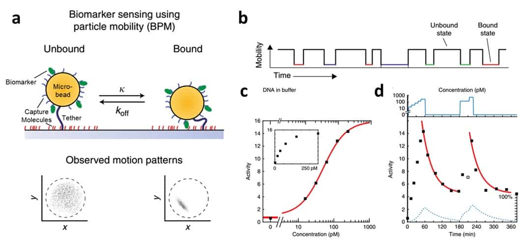
The challenge in using conventional cameras to detect biomarkers is figuring out how to create a detectable signal from molecules that are too small to photograph. In their recent paper, the team report a clever trick to detect biomarkers by monitoring the mobility of specially engineered microbeads that are large enough to photograph using a simple magnifying lens (Nature Communications 9 2541). They call their technique “biomarker sensing using particle mobility” (BPM).

In BPM, each microbead is attached to a glass surface via a long molecular tether and, under normal conditions, floats around in an area that is defined by the length of the tether. The microbead is coated with a “capture molecule” that binds to a biomarker of interest, and the glass surface is coated with another capture molecule that binds to a different side of the same biomarker. As a single biomarker molecule sticks to both the microbead and the surface simultaneously the particle is pinned down and its motion is significantly restricted.

After exposing the microbeads to a patient’s blood, a conventional camera can be used to simultaneously monitor hundreds of microbeads as they transition from mobile to immobile. A short five-minute video can then be processed to translate information about the average particle mobility to information about the amount of biomarker present in the blood sample. Specifically, the team defined “activity”, which is related to the frequency at which particles transition between high-mobility and low-mobility state, as a metric to quantify biomarker concentration.

The researchers demonstrated that BPM could detect dilute concentrations (nanomolar to picomolar) of DNA- and protein-based biomarkers in conventional buffers and even in human blood plasma, establishing BPM as a sensitive and robust technique.  As they flowed a DNA biomarker with concentrations in the picomolar range over the sensor surface, the measured activity increased.

A fundamental requirement for any continuous bloodstream biomarker sensing system is reversibility – the sensor needs to recover to baseline signal once the biomarker concentration is reduced to zero. To test the reversibility of their technique, the team added a biomarker, flushed it out, and then added the biomarker again. They indeed found that particle activity returned to near-baseline after biomarker removal and then increased again upon re-addition of biomarker.

Moving forward, the researchers hope to expand BPM’s capabilities to include the detection of more diverse biomarkers (proteins and small molecules, for example), and to implement BPM in a miniaturized, portable device.

Physics communication, a MedPhys Slam and the future of high-energy physics

The effective communication of physics is the theme of this episode of the Physics World Weekly podcast, which kicks off with an interview with the particle physicist Helen Heath. Passionate about teaching, Heath speaks about her involvement with the Bristol Institute for Learning and Teaching at the University of Bristol. She also talks about how to get more young people – and particularly young women – interested in physics and gives her take on the future of particle physics.

Next up are Physics World’s Tami Freeman and James Dacey who chat about the MedPhys Slam – an event that Freeman attended at the recent meeting of the American Association of Physicists in Medicine in Nashville, Tennessee. The competition invites PhD students, medical physics residents and postdoctoral researchers to describe their research project in a compelling and coherent manner – and do it in just three minutes. The goal is to help medical physicists develop strategies for communicating complex ideas to patients, their relatives as well as to other healthcare professionals.

European quantum-technologies roadmap is updated by leading physicists

What is covered in the quantum technologies roadmap?

The roadmap is a 24-page peer-reviewed document that includes an introduction followed by six sections that focus on important aspects of quantum technology. These are quantum communication; quantum computation; quantum simulation; quantum metrology, sensing and imaging; quantum control; and quantum software and theory. It is the work of 18 experts in quantum technology from across Europe. The roadmap provides the authors’ opinions – based on extensive discussions and feedback from the larger European quantum technologies community – of the status and main challenges of quantum-technology research as well as the advances they foresee in the future. It also serves as an updated summary of the 150-page Quantum Technologies Roadmap, which forms the basis of the European Commission’s Future and Emerging Technologies Flagship on Quantum Technologies

Why is it important for European researchers to develop a roadmap for quantum technologies?

The quantum technologies community has been maintaining a roadmap for the last 20 years and it has been an important instrument in both creating a coherent vision for the community as well as providing a benchmark for the progress in the field. This has been a key factor in demonstrating the readiness of the European community for the type of investment that the Quantum Flagship brings, as well as providing industry with a clear idea of where and when the best opportunities are for investing.

How would you characterize the state of quantum research in Europe when compared to other regions?

Research in quantum is rapidly advancing all around the world and Europe continues to play a leading role across all areas of interest. To date, the field has managed to balance being competitive and collaborative, so it doesn’t make too much sense to say any one region is “winning” in a particular area; this is a long race and there will be plenty of prizes for everyone.

What are the key challenges facing the European quantum community?

The field is still relatively young, but already garnering significant interest from industry. The challenge in Europe will be to develop the instruments and infrastructure that will allow not only this industry uptake, but continue to advance the basic science that underpins all of these efforts and enable the development of a sustainable innovation landscape. This is not so different to the challenges faced globally. It is important for everyone that in the rush to commercialize these quantum technologies we don’t forget to invest in the basic science that will ensure its long-term success. The roadmap can play an important role in this.

The quantum technologies roadmap: a European community view” is published in the New Journal of Physics.

Hotter temperatures reduce food and traffic regulation

Government workers such as police officers and food safety inspectors are less active when temperatures are hotter, precisely the times when food and public safety are at most risk. That’s according to a team in the US who looked at data on police stops, food inspections, fatal vehicle crashes and food safety violations.

“Climate and weather impact all humans — including those in charge of assisting their fellow citizens,” says Nick Obradovich of MIT, US. “Adapting to climate change will require assessing the vulnerability of not only the public but also public sector workers to temperature related stressors and introducing adaptations where possible. Without adaptation, climate change might amplify the marginal gap between citizen need and government assistance.”

Cold and hot temperatures boosted the risks of a fatal crash and the incidence of food safety violations, the researchers found, but decreased police stops and food safety inspections. Rainfall, meanwhile, increased fatal crash risk but saw a drop in police stops.

“The regulatory activity of food safety inspectors and police officers declines due to the same temperatures that amplify the risks these regulators are tasked with overseeing,” says Obradovich. “These environmental factors could hinder the ability of governments to meet citizen needs at the time those needs are most amplified by the very same environmental factors.”

Obradovich and colleagues looked at historical data from more than 70 million regulatory police stops in the US from 2000 to 2017, half a million fatal vehicle crashes from 2001 to 2015, and nearly 13 million food safety violations discovered during over 4 million inspections from 2012-2016. The team also analysed temperature and precipitation records.

Examining annual temperature projections for 2050 and 2099 indicated that on average police officer stops will decrease in the south of the US and increase in the north, while fatal crash risk will increase most in the south. The trends were similar for food safety inspections and violations.

Over the course of the year, the team believes, future warming may boost regulatory oversight during cooler seasons, but diminish it during hotter seasons at the same time as amplifying hazards.

Obradovich has a background in political science; his previous research showed how temperature may alter human physical activity, sleep, and sentiment. “This work is an extension of those findings to examine their implications in the political arena,” he says. “Climate factors alter our well-being, and climatic changes are likely to dramatically alter our well-being in ways foreseen and unforeseen. This study adds one more area of potential concern to the list.”

Now the plan is to obtain similar data from other countries to investigate if the effects found in the US exist in other political and economic contexts. Obradovich is reluctant to extrapolate his results too far but believes that “to [the] extent these temperature-oversight effects exist in other countries, those countries with lower levels of political institutionalization, greater bureaucratic discretion, and lower levels of economic development (for climate adaptation purposes) might observe larger effects”.

The team reported the findings in PNAS.

Ion-beam CT improves particle therapy planning

Ion-beam CT

Treatment planning for particle therapy is currently performed using X-ray CT images of the patient, with the CT Hounsfield units (HU) converted into relative stopping power (RSP). This conversion process leads to uncertainties, however, and an error margin of around 3% is generally employed to mitigate any potential errors.

A better option may be to use protons or other ion beams to create patient images for particle therapy planning. At the recent AAPM Annual Meeting, Sebastian Meyer discussed the potential benefits of ion-beam CT, which he is investigating in the framework of his PhD research at LMU Munich, in collaboration with the LMU University Hospital, Yale School of Medicine and Heidelberg Ion Beam Therapy Centre.

“Ion-CT provides a direct estimation of relative stopping power and eliminates conversion uncertainties,” he told the delegates. “It can also eliminate metal artefacts.”

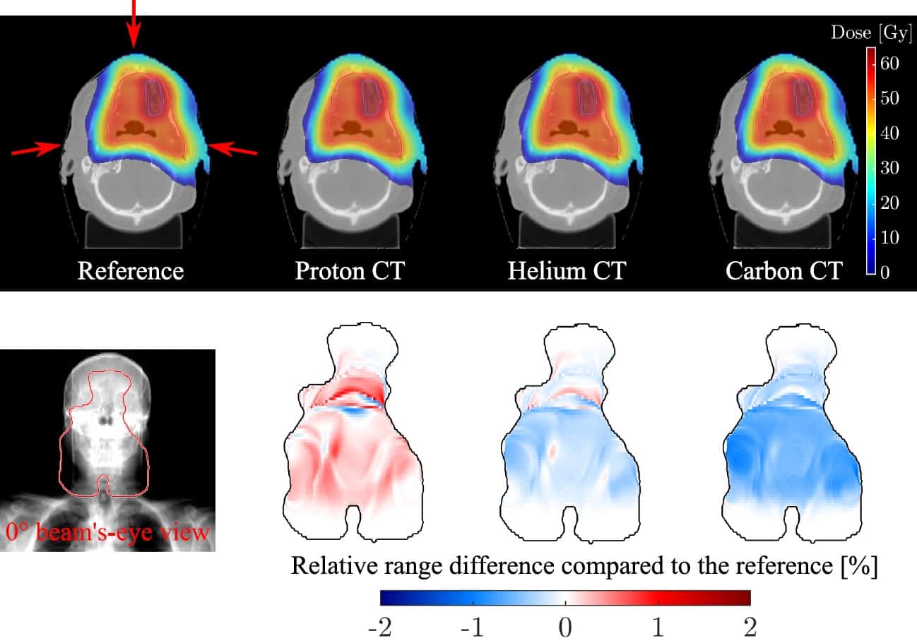
Meyer presented results from FLUKA Monte Carlo simulations comparing proton, helium-ion and carbon-ion CT for proton therapy planning. He described the simulation of ion-CT images at 2 mGy physical dose using an ideal single-particle tracking detector, for two head-and-neck cases treated with scanned proton pencil beams.

After reconstructing the images, Meyer and colleagues imported them into the RayStation treatment planning software. Proton therapy plans were then optimized on the reference anatomy and recalculated on the various ion-CT scans.

The reconstructed proton-CT, helium-CT and carbon-CT images were similar in appearance, although helium-CT and carbon-CT were slightly noisier, and all displayed comparable dose calculation performance.

The researchers quantified image quality by calculating the RSP relative error. Meyer noted that for heterogenous image slices, proton-CT exhibited the highest error, but that this reduced for more homogeneous slices. The average RSP relative errors over all image slices were 1.4%, 1.1% and 1.4%, for proton-CT, helium-CT and carbon-CT, respectively.

Looking at the range differences, helium-CT and carbon-CT generally underestimated the range, while proton-CT overestimated range by up to 1%. The average range accuracies were +0.1%, -0.1% and -0.4%, for proton-CT, helium-CT and carbon-CT, respectively.

“We can achieve way better accuracy than the 3% in HU conversion,” said Meyer.

Radiobiological implications

Ion beams have an increased relative biological effectiveness (RBE) compared with photons – so could the imaging dose from ion beams be problematic? To answer this, Meyer and colleagues used Monte Carlo simulations in combination with the repair-misrepair fixation model to estimate cell survival and double-strand break (DSB) yield for the various ion beams.

With respect to diagnostic 130 kVp X-ray radiation, the mean RBE for cell survival was 0.82-0.85, 0.85-0.89 and 0.97-1.03 for proton-CT, helium-CT and carbon-CT, respectively. RBEDSB (the RBE for DSB induction) was 0.82 for protons, 0.84 for helium ions and 0.95 for carbon ions.

“The RBEDSB relative to X-ray CT spectra is below one,” Meyer pointed out. “These preliminary studies on biological dose show a reduced biological implication, but more studies are required.”

He concluded that helium-CT exhibited the lowest overall RSP error, while proton-CT and carbon-CT had the highest accuracy for soft tissue and bone, respectively. All modalities showed sub-1% range accuracy for treatment planning and good dosimetric accuracy compared with the ground truth.

Activating retinal stem cells restores vision in mice

Generation of rod photoreceptors

An international research collaboration has successfully restored vision in mice by activating retinal stem cells, a feat that has never been achieved before. The researchers note that their study could transform treatment for patients with retinal degenerative diseases, which currently have no cure (Nature 10.1038/s41586-018-0425-3).

“This study opens a new pathway for potentially treating blinding diseases by manipulating our own regenerative capability to self-repair,” explains lead investigator Bo Chen from Icahn School of Medicine at Mount Sinai. “This is the first step to finding promising cures for patients that involve self-repair as opposed to medicine or invasive procedures.”

In cold-blooded vertebrates such as zebrafish, Müller glia cells (MG) act as retinal stem cells that can replenish damaged retinal neurons and restore vision. In mammals, however, MGs do not have regenerative capability after photoreceptors are lost, and therefore retina damage cannot fix itself. As a result, diseases like macular degeneration or retinitis pigmentosa that kill retinal cells are often irreversible. Chen and colleagues hypothesized that if they could somehow reactivate the MGs, they could bring back vision.

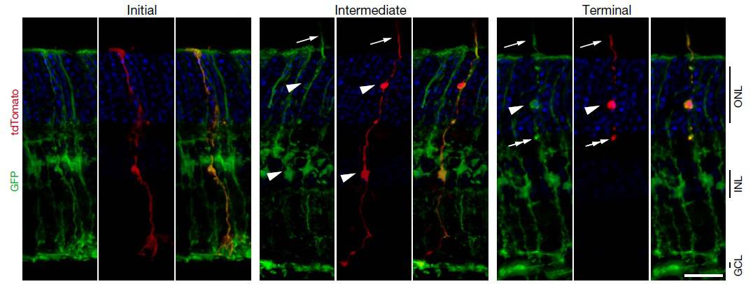
To achieve this, the team performed a two-step gene transfer to reprogram MGs into blind mice. First, they used a gene transfer process to activate dormant stem cells to turn them into active stem cells. The second step involved another gene transfer to help these stem cells develop into rod photoreceptor cells, the most abundant cell type in the retina. Rod photoreceptor cells are the first step to sensing light in the retina; they then transmit to other cells in the retina, which send signals to the brain to enable sight.

After this two-step reprogramming, new rod photoreceptors were generated and integrated into the existing retinal structure. The investigators saw no difference between these new cells and real rod photoreceptor cells: the new cells sensed light, which then triggered information to be sent to the visual cortex and restored function of the visual pathway.

Between four and six weeks after the reprogramming, the blind mice were able to sense light and regained their vision. While vision was restored to some degree, the researchers could not measure the degree of improvement, and must perform further tests to find this out.

“This could lead to extraordinary opportunities in the future where we can potentially use the same strategy to reactivate these stem cells in the diseased human eye,” said Chen. “If this works, this could transform the way we treat patients with retinal disease and possibly learn how to cure other types of eye disease like glaucoma.”

Copyright © 2026 by IOP Publishing Ltd and individual contributors