‘Oumuamua, a mysterious cigar-shaped object travelling through the solar system having arrived from interstellar space, is being propelled by outgassing as it is heated by the Sun. That is the conclusion of Marco Micheli of the European Space Agency and colleagues, who have looked at ground- and space-based observations of the motion of the object.
First spotted on 19 October 2017 by the Haleakala Observatory in Hawaii, ‘Oumuamua is 230 m long and is the first object to be identified as entering the solar system from interstellar space. ‘Oumuamua means “scout” in Hawaiian to reflect its long voyage from a distant planetary system.
The object’s extremely eccentric orbit and shiny surface initially led the International Astronomical Union (IAU) to classify ‘Oumuamua as a comet. However, that status was quickly changed to an asteroid when astronomers could not find a “coma” of gas and dust surrounding the object, something that is seen around comets. Then in November 2017, ‘Oumuamua was again reclassified by the IAU as the first ever “interstellar object” – a new classification created in light of the object’s discovery.
Extra acceleration
In this latest study, Micheli and colleagues tracked ‘Oumuamua as it moved through the solar system and found that its trajectory cannot be explained solely by the gravitational attraction of the Sun, planets and large asteroids.
Calculations done by the team rule out pressure from solar radiation and magnetic interactions with the solar wind as causes of the additional acceleration. Instead, they report evidence of a non-gravitational acceleration that is directed away from the Sun. This phenomenon is also seen in comets, which can be propelled by the gas they release as a result of being heated by the Sun.
While ‘Oumuamua appears to be comet-like, Micheli and colleagues point out that it appears to have chemical and dust properties that are unlike most known comets. They speculate that the observed lack of dust surrounding the object could mean that ‘Oumuamua is devoid of small grains of solid material – or indeed contains a very small amount of dust in general. Another possibility is that ‘Oumuamua began its long journey as a typical comet, but its surface was somehow modified along the way.
Writing in Nature, the astronomers conclude, “although ‘Oumuamua looks familiar there are differences that relate to its birth in a solar system far from our own”.
Renewables are booming across Asia, but there are variations in pace and rival options also play a role. An interesting paper by Indian academic Nandakumar Janardhanan looks at competition in renewables in developing countries in Asia, focusing on India and China.
Janardhana notes that “India and China, being major developing economies and having huge energy appetite, focused heavily on strengthening their respective alternative energy sector” so as to reduce their over-reliance on conventional fossil fuels. He adds that “India depends on external oil supplies to meet two thirds of its oil demand, one third of oil demand in China is met by imports”. As a result, the renewable energy sector has gained great momentum in these two countries and “as innovation and development began to lead the growth of alternative energy sector, opportunities for expansion within their respective borders as well as outside emerged as promising avenues for the industry from both countries”.
China has done especially well: “the competition in the domestic energy sector in China led to the emergence of cheaper and better technologies which will enjoy a natural competitive edge over its counterparts from other countries” – solar PV is an obvious example – and the Chinese government “has been keen in enhancing its ties with the developing Asian economies in all possible ways”. Basically, “China sees that capturing the developing Asian market is important to its economy”, and believes that “spreading influence among the economies in the region is in its greater strategic interest”.
By contrast, although it too has some regional involvement via ASEAN (Association of South East Asian Nations) cooperation agreements and initiatives, India has done less well on most counts, e.g. it has around five times lower national renewables output. And on trade, China is clearly winning so far: “a critical advantage China enjoys in the region is its sheer size of economy”, and it is “able to present itself as an alternative power which can offer financial support and business opportunities”. But the contest continues, with India trying hard to catch up, while China is having to cope with the problems of being in front, including the issue of curtailment. See my earlier post. With programme costs also rising, it has recently decided to throttle back on its very rapid PV solar growth, so that only 29 GW extra may be added this year, instead of the 48 GW expected.
Both India and China have nuclear programmes, which inevitably siphon off some support from renewables, but less so in the case of China – the nation is now getting around ten times more output from its rapidly expanding renewables programme than from its nuclear plants: the output from its huge wind programme – 170 GW and rising – overtook that from nuclear a while back. That’s also now the case for the smaller wind programme in India (33 GW so far), but the output from India’s other renewables is still quite low, although growing. It aims to have 175 GW of renewables by 2022, with PV accelerating.
The other major contenders in the region are also well behind China. Japan clearly has the industrial capacity but, still recovering from the shock to its energy plans represented by the Fukushima nuclear disaster, it’s dragging its feet on renewables, PV apart.
In theory, with most of its nuclear plants closed, Japan is aiming to move away from nuclear and expand its use of renewable sources so that they supply 22-24% of its power by 2030. However, even given this relatively low target (less than the UK gets now), progress is relatively slow, although some of the big PV projects are quite spectacular. But Japan is still trying to upgrade and restart more nuclear plants, which is expensive, and it is faced with the huge Fukushima clean-up bill, which may be vastly more than the official estimate, perhaps £150 bn.
South Korea might do better, having decided to abandon its nuclear programme, but like Japan, it still has a long way to go on renewables – it’s aiming to get just 20% of its power from 58.5 GW of renewables by 2030. What’s more, although the government seems resolute, there are still those who want to rescind the nuclear decision – and one plant build has been restarted, although an old reactor is to be closed ahead of its 2022 retirement schedule.
Similar issues are being fought out in Taiwan, which, with massive popular opposition to nuclear, is aiming to phase it out by 2025 and wants to get 20% of its power from renewables by then. However, there have been signs of a back-slide .
Backing off from nuclear is clearly hard work. Japan is having to import a lot of gas, at massive cost and with significant emissions being produced. In effect, and embarrassingly, it has had to abandon its Kyoto climate pledges. And the direct impacts of the 2011 Fukushima disaster are also far from over. More than 130,000 people left or were evacuated from the region. Some are being asked to return and some want to, and some already have. But a recent survey by Greenpeace Japan in the towns of Iitate and Namie in Fukushima prefecture, including the exclusion zone, found radiation levels in some locations up to 100 times higher than the international limit for public exposure. The organisation claims that the high radiation levels in these areas pose a significant risk to returning evacuees until at least the 2050s and well into next century.
This view will be unpopular with the government, since it is looking to the upcoming Olympics for an economic boost. So the last thing it wants is bad news about radiation risks, which might deter visitors. The issues can perhaps be portrayed as being far away and of no consequence as far as the Olympics in Tokyo are concerned. But it may not in fact be just a local issue, with contamination being found up to 60 miles away. Or a trivial one, with hot particles still turning up
It’s hard to see how it can all be decontaminated. There is a harrowing report and video, which looks at the issues and shows the clean-up so far, with the vast areas of water tanks and top soil in bags. It all adds up to a horrible warning about what can happen when nuclear goes wrong, with some saying that the impacts on local people amount to a human rights violation. Let’s hope that China and India, Taiwan or South Korea don’t have to face this at some point.
However, some clearly see it all differently and argue that, albeit with the benefit of hindsight, as it turned out, the costly and disruptive Fukushima evacuation was a mistake. The problem was that, at the time, no one knew what would happen next – very major radiation releases were possible. And the long-term health impacts of what did happen are still debated. Statistically, as an Financial Times article has noted, the Fukushima evacuation may have, in effect, extended the life of those moved from the worst zones by a few months from what they would have been if they had been left exposed. But statistical analysis and on-the-ground reality may differ and certainly may be perceived differently. Even now, South Korea is still refusing to import some sea foods from the Fukushima area. That may be seen as over cautious, but in the final analysis, it is the uncertainty about nuclear risks and impacts that is the killer in public policy and public reaction terms, and that makes the alternative energy options look far more appealing, the uncertainties adding to the case against nuclear, especially near population centres.
The nuclear issue is of course only one aspect of Asian – and global – energy policy. Its significance may well diminish as renewables continue to expand. In my next post, I look at the debate on how fast that may happen and at some possible constraints.
Colorectal cancer is one of the most deadly malignancies, but is preventable and curable if diagnosed at a precancerous or early stage. Optical colonoscopy is a key component of any colorectal cancer screening programme. However, many patients are reluctant to undergo colonoscopy due to its invasive nature and requirements for bowel preparation, sedation and pain medication.
To increase screening uptake, a more patient-friendly test is needed. With this aim, an international research team has developed an ingestible X-ray imaging capsule that requires neither colon cleansing nor sedation. The capsule – which is being commercialized by Israeli diagnostics company Check-Cap – scans the colon as it travels through the gastrointestinal tract and sends data to a dermal patch receiver. The technology could reduce colon cancer mortality by screening for precancerous polyps and lesions before they become malignant.
In preliminary studies, the researchers confirmed the safety of the X-ray capsule in 138 volunteers (Abdom. Radiol. 42 1291). Now, they have published results from the first prospective trial examining its efficacy for polyp detection (Gut 10.1136/gutjnl-2018-316127).
Capsule design
The capsule incorporates a short-lived 191Os radioisotope, which emits three rotating beams of low-dose 65-75 keV X-ray photons, plus photon-counting X-ray detectors. The patient swallows the capsule, along with small quantities of an iodine-based contrast. A scanning control algorithm (SCA) then uses real-time positional data to determine whether the capsule has moved along the colon and, if so, transmits a command to perform a scan.
“Ideally, since the scan width is about 5 mm, the SCA should scan every 5 mm for 100% coverage of the colon surface,” says Yoav Kimchy, founder and CTO of Check-Cap. “In reality, since our focus is on polyps of 10 mm and above, we estimate that a scan every 10 mm, which gives 50% coverage, may still catch most of these polyps.”
The colon wall is imaged based on two processes: generation of secondary X-ray fluorescence (XRF) photons at 27 keV as the emitted photons interact with iodine atoms; and Compton scattering as the photons interact with atoms in the colon wall and lumen, creating scattered photons with energies of 52-60 keV.
“Measuring both XRF and Compton photons brings a number of advantages,” Kimchy explains. “Since both the distance from the capsule [to the colon wall] and the contrast concentration at each imaging location are unknown, having two measurements allows us to solve for distance and concentration simultaneously.”
The system combines the XRF and Compton data to estimate these parameters and reconstruct the colon surface. Kimchy notes that the two techniques behave differently when encountering a gas bubble in the colon, enabling readers to distinguish between a polyp and a bubble. False readings are also reduced by requiring both data sets to confirm the presence of a polyp.
Feasibility study
To test the capsule’s ability to detect polyps, the team analysed imaging data from 45 patients with and without polyps. Patients underwent the capsule procedure and a faecal immunochemical test (FIT, an indirect colorectal cancer detection test), followed by a reference colonoscopy. The researchers calculated the sensitivity and specificity of the capsule findings as a function of the scan imaging density.
The capsule’s overall sensitivity for polyp detection was 44%, compared with 37% for FIT. In a subgroup of 19 patients with scan imaging density above 50%, this increased to 78%, while for 12 patients with imaging density above 70%, sensitivity was 100%. Specificity was high in all cases, at between 86 and 90%.
The average total transit time of ingested capsules was 52±32 hours. There were no device-related serious adverse events and the procedure was well-tolerated by the participants. The average total X-ray dose was 0.051 mSv.
After the trial, the researchers developed an improved SCA and retrospectively implemented it on the study data. This increased the number of subjects with scan imaging density above 50% from 21 of the 45 patients to 41. “The new SCA showed an improvement in the filtering of body movements, providing more homogeneous spread of scans along the colon. This amounts to a better scan imaging density in the retrospective analysis, which we hope will translate to improved sensitivity for polyp detection,” Kimchy explains. “We are collecting new clinical data with this algorithm to show statistics on sensitivity.”
Following CE Mark approval in January 2018, Check-Cap plans for commercial sales of the capsule in Europe, with additional regulatory clearances to market in Israel and the Far East. They are also developing improved software and algorithms, performing a multicentre clinical trial, and working with the FDA to start clinical trials in the USA. In parallel, the company is preparing for high-volume manufacturing, with GE Healthcare as a partner.
As a child I was fascinated by biology and aspects of physics such as electricity and magnetism. I was very lucky that my physics lessons at school were taught by a very enthusiastic teacher, who stimulated my interest strongly. I was especially interested in nuclear physics.
What area was your physics degree in, and did you ever consider a permanent academic career?
As I preferred applied physics over theoretical physics, I chose to study the former at the Delft University of Technology. I did my MSc in radiation physics at the Reactor Institute Delft, which is a research institute, not a power plant. I worked at the institute as a research scientist for a few years after finishing my degree. After a while, I switched to working as a medical physicist – first at the Radiotherapeutic Institute RISO, and later in the radiology and nuclear-medicine departments at the Deventer Hospital in the Netherlands – for more than 30 years of my career. In that time, I published some papers and completed a PhD at the University of Utrecht, on treatment of prostatic cancer using I-125 seeds.
How did your interest in photography emerge?
In the late 1990s, while teaching radiation physics and radiation safety to radiographers and physicians as part of the hospital’s programme, I found that even very thin objects (such as flowers) can be imaged when using very low energy X-rays. After a few years, I started to colour some of these X-ray images, and people found them interesting. I got my own licensed X-ray studio in 2007 and, after retiring from the hospital in 2012, I have been working full-time creating “bioramas” – nature scenes involving flowers, plants and animals. I was inspired by the unbelievable beauty of nature, and became aware of its wonderful complexity.
How do you create your portraits?
I set up a natural scene, and then X-ray it in one session as a whole – the images are not stacked or layered digitally. The animals I use are dead, as I dont think I could justify exposing living animals to X-rays for my art. I source the animals in different ways – I find traffic-victims along the road side, or birds who have flown into windows. The fish I buy at a market, while my cat catches the occasional mouse or mole. I also have a friend who breeds reptiles, who gives me their carcasses. I use animals as I find them – especially in the case of traffic victims, the anatomy mostly is mutilated, so you will see animal injuries in a lot of images.
And how do you do this in practice?
Once the scene is set, I position an analogue silver bromide X-ray film (in a light-tight envelope) with the biorama on it, and place it on the floor. The X-ray tube is about 100 cm above the film, which is a fine-grain (high-resolution) film, with a steep gradient (high contrast). But it has relatively low sensitivity, and so a high dose of radiation is required for sufficient optical density. First, I take a low energy 2.5-minute exposure to image the thinner parts (such as the petals or leaves) of the biorama, immediately followed by a higher-energy exposure of 3.5 minutes to image the thicker parts. The film needs a maximum total dose of about 300 milliGray, resulting in a maximum optical density of about 3. After processing the exposed film in a dark room, I judge each analogue image and measure its optical density. I digitize the X-ray image using a scanner, and edit the grey levels with Photoshop. I pick and colour some areas of the image and, often, it is inverted.
What are some of your current projects?
This year, I exhibited my 3D X-ray images at the Natural History Museum in Rotterdam – you observe the portraits through a View-Master. My photographs were also included in a recent Dutch children’s book titled Binnenstebinnen, published by Gottmer, Haarlem. I hope it gets a sequel.
How has your physics background helped?
It’s not easy creating these X-ray images where there are huge differences in thickness – from the very thin petals of a flower or the feathers of a bird, to the relatively much thicker bodies of animals. I think that my medical background in X-ray physics was of great value, and allows me to now take the perfect X-ray.
Any advice for today’s students?
You are studying during a wonderful period of great discoveries and interesting discussions in physics – enjoy it!
Hospital-acquired infections (HAIs) are one of the biggest challenges in modern healthcare. Within this wider problem, urinary-tract infections associated with catheter use are a particular concern: in 2001 a US study found more than half a million cases each year, accounting for approximately 40% of all HAIs (Int. J. Antimicrob. Agents17 299). Such infections often stem from biofilms that form when a catheter is inserted into a patient’s urethra. These biofilms – which are made up of micro-organisms and their extracellular detritus – act as refuges for bacteria, making infections persistent and difficult to treat. Developing novel catheter materials that resist biofilm attachment is thus a promising strategy for reducing the number of HAIs.
Modified materials
The most common material for urinary catheters, silicone rubber, is a polymer with a backbone consisting of silicon and oxygen. This backbone makes it possible to tailor the material’s texture and elasticity to different applications, and it also makes it highly chemically inert – all excellent characteristics for a medical device. Silicone on its own is, however, hydrophobic, so it is relatively easy for hydrophobic biofilms to adhere to it. This is why BioModics, a Danish medical-supplies company where two of us (MA and PT) are senior managers, has been developing ways of modifying silicone to make it more resistant to biofilm attachment.
At BioModics our approach is to transform silicone by treating it with carbon dioxide at high temperature and pressure. Under these conditions, the carbon dioxide becomes a supercritical fluid – it can penetrate materials like a gas, while also acting as a solvent for the silicone. This process causes the silicone to expand, such that it can be impregnated with hydrophilic molecules that then polymerize inside the silicone network. The result is an interpenetrating polymer network (IPN) of silicone and hydrophilic gel, or “hydrogel”; the hydrophilic nature of this system makes it more difficult for biofilms to adhere to it (Eur. J. Pharm. Biopharm.94 305).
Making catheters from this hydrophilic hydrogel-silicone could significantly reduce the risk of patients developing catheter-related infections, but BioModics’ patented technology also has potential applications in drug delivery. Whereas silicone on its own repels water, the hydrogel in BioModics’ IPN material serves as a reservoir for liquids in which small hydrophilic molecules – such as antibiotics to destroy any bacteria that do manage to colonize the catheter – could be suspended. This might make it possible to deliver active pharmaceutical ingredients locally instead of systematically, limiting their side effects. Again, the change from a hydrophobic to a hydrophilic material is key.
To improve BioModics’ technology and optimize the material processing conditions for different applications, we wanted to understand precisely how the silicone and hydrogel are distributed through the IPN structure. For example, understanding and controlling properties such as the connectivity and pore sizes of the hydrogel network is critical for ensuring that the network is suited for transporting specific drugs. However, studying the hydrogel structure is tricky because the two polymer networks (silicone and hydrogel) are integrated on the nanoscale, and inspecting them with a light or electron microscope reveals little contrast between the two materials.
To overcome this problem, BioModics turned to a project called LINX, which stands for Linking Industry to Neutrons and X-rays. This initiative was designed to bridge the gap between academic research and industrial R&D in the field of neutron and X-ray scattering, and it facilitates collaborations between three Danish universities (each with its own particular expertise) and companies from a wide range of industries (see Physics World Focus on Neutron Science October 2017 pp17–18). The project’s long-term goal is to help Danish industry make the most of nearby large-scale research facilities such as the European Spallation Source, the MAX IV synchrotron and the European X-Ray Free Electron Laser.
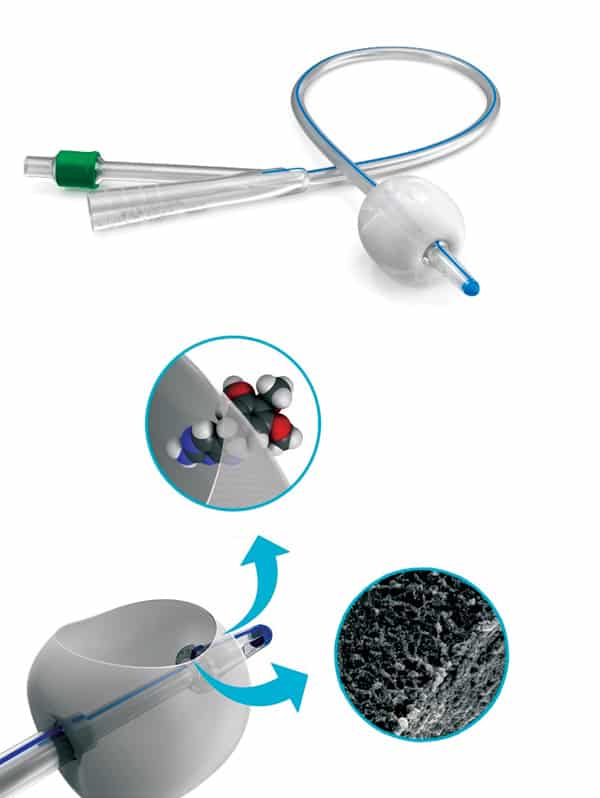
Fine details: A BioModics catheter, with the inset diagrams showing enlarged views of the catheter wall (middle) and its nanoscale structure (bottom). (Image courtesy: BioModics)
BioModics’ first port of call was the LINX team at the University of Copenhagen, which specializes in small-angle scattering techniques and where one of us (EB) works as a physicist. In both small-angle X-ray scattering (SAXS) and small-angle neutron scattering (SANS), a beam of radiation (X-rays or neutrons) impinges on a sample and scatters off it. Detectors record the scattered radiation as a function of the scattering angle, which is typically less than 5°. The resulting pattern is related to the Fourier transform of the sample’s structure, so by comparing the scattering data to geometric models, researchers can extract information about the shape and size of the sample’s structure.
Both SAXS and SANS are great for investigating structures at length scales between 1 and 100 nm (and sometimes larger, depending on the specific experimental setup), so they are a good fit for BioModics’ nanoscale silicone–hydrogel IPN. There are, however, some key differences between SAXS and SANS, and these had important consequences for BioModics’ investigations. Whereas X-rays interact with the electrons around the atomic nucleus when they scatter, neutrons interact with the nuclei themselves. This means that neutrons can “see” light elements such as hydrogen, which are practically invisible to X-rays. A subtler but equally important fact is that neutron scattering is isotope-sensitive: if the water in a sample is replaced with heavy water (D2O), the SANS results will look different.
Different tools, different results
Researchers at the University of Copenhagen began by investigating BioModics’ IPN material with SAXS, hoping that these measurements would reveal the hydrogel’s structure. Unfortunately, the electron densities of silicone and hydrogel are nearly the same, so it was not possible to distinguish the two materials using this method. In fact, the observed X-ray scattering profile turned out to be dominated by scattering from the silica (SiO2) particles used as filler to make the silicone more mechanically stable.
After this setback, the team turned to SANS, for which the contrast between silicone and hydrogel is better, especially if the hydrogel is loaded with D2O. Using SANS did, however, present some initial hurdles. While SAXS experiments can be performed using a conventional X-ray tube available in many laboratories, SANS requires a nuclear reactor or a spallation source based on a particle accelerator. These large-scale experimental facilities are usually reserved for academic research, and although industry-access programmes exist, the usual rate for proprietary beam time is thousands of euros per day.
To overcome this financial barrier, BioModics applied for beam time via a project called SINE2020, which offers short periods of free beam time to companies that want to find out whether neutron scattering is a feasible technique for studying their materials. BioModics was awarded beam time at the Institut Laue-Langevin (ILL), a major neutron-scattering facility in Grenoble, France, where one of us (CB) works as an industrial liaison officer. After BioModics shipped their materials from Denmark to France, scientists at the ILL performed measurements on samples of pure silicone, dry IPN and on IPN samples that had been soaked in D2O for a week.
Making catheters from hydrophilic hydrogel-silicone could significantly
reduce the risk of catheter-related infections
The SANS data from pure silicone and dry IPN were strikingly similar, which unfortunately means that the strongest scattering signal was still coming from the filler material. However, SANS data on the samples soaked in D2O looked quite different. This made it possible to differentiate the signal of the hydrogel from that of the silicone, and thus to learn about the IPN structure. By applying fractal network models to the data, we succeeded in deriving a characteristic correlation length related to the pore size of the hydrogel, as well as a parameter describing the roughness of the interfaces between hydrogel and silicone. After doing this for two samples with a different silicone base but the same amount of hydrogel, we found that the samples had different correlation lengths (58.6 nm and 32.5 nm), while the interface between silicone and hydrogel was also markedly different. In one sample the interface was quite smooth, whereas in the other it was rough or jagged.
Drug delivery
This result suggests that an IPN made from the material with the smoother interface may be the better of the two for transporting drug molecules. Although this finding would, of course, need to be confirmed by more direct measurements of drug transport through the material, it was intriguing enough for BioModics to purchase additional beam time at the FRM-II research reactor in Garching, Germany. In a later series of experiments, we mapped out how the IPN’s structure changed as a function of hydrogel content, in order to determine how to produce a structure that is optimized for drug delivery. These data, taken in summer 2017, are being analysed by researchers in the LINX team at Copenhagen, but so far the results are promising. Ultimately, we expect that the project will help BioModics select the optimal silicone type, hydrogel content, and possibly other parameters, in order to achieve the best possible properties for our devices – including an optimal hydrogel structure for drug delivery and optimal mechanical properties, such as softness and flexibility, for the catheter material. In the fight against catheter-related infections, these are useful weapons.
The ability to make decisions distinguishes robots from basic machines. Most robots are wired with electronic circuits for decision making, a convention that has developed into a sophisticated art capable of fast data-dense operations. However as robotics broaches applications such as surgery and disaster response, the demands for soft robots increase, raising compatibility issues with the hard world of silicon electronics. By demonstrating binary logic operations using origami, researchers at the Wright-Patterson Air Force Base in the US demonstrate a type of mechanologic that may provide a useful complement to electronics.
Credit: CC-BY 2.0 Josey
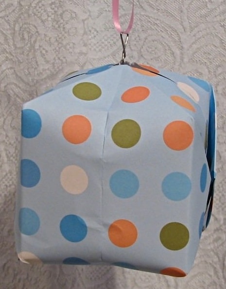
Benjamin Treml, Andrew Gillman, Philip Buskohl, and Richard Vaia investigated the possible logic operations of a waterbomb-base origami pattern, where inward and outward pointing configurations can indicate 0 and 1 states in binary logic. In general origami systems have localized folding with flat planes that can accommodate hard electronics to complement the mechanologic. However, the water bomb structure in particular has additional advantages as the apex can point inwards or outwards without affecting the mount or valley nature of surrounding folds. “We believe these properties allow the mechanical bit to switch between 1 and 0 states without interfering with the ability of other connected units to reconfigure in the multiunit structures presented below,” they explain in their paper.
Sensing-decision-response structures
As well as decision making, the robot must be able to sense inputs and respond with outputs. While there are a number of materials with physical responses to environmental stimuli, Treml et al. investigate water bomb structures made from PEDOT:PSS – a conductive polymer commonly used in flexible and organic electronics that also swells and shrinks in response to humidity.
The researchers also study the responses of tessellated combinations of water bomb structures and the effect of shared folds on the energy barrier to snap between 0 and 1 states. They calculate a 33% increase or 15% decrease in the energy barrier for two joined water bomb structures for snapping between like and unlike states.
The researchers point out various limitations in the available operations, and the origami from 2D structures also limits the number of inputs. They also emphasise how this type of logic is unlikely to ever replace electronics, instead complementing existing electronics. The work is far from the first demonstration of mechanical robotics. As Treml et al. point out in the paper, “Mechanical logic devices have a long history, dating back to Leibniz’s step reckoner in 1672 and Babbage’s difference engine in 1822.” The crucial advance here is the ability to render logical operations in a mechanical system that is also soft.
Date: 23 July 2018, 2 p.m. BST Presenter: Robert Done – Project Design Engineer – STFC – Rutherford Appleton Laboratory
Rob Done began his career as a mechanical engineering apprentice for UKAEA at Risley in Cheshire. On graduating, he joined the Science and Technology Facility Council, working first at their Daresbury site in Warrington and eventually moving to the Rutherford Appleton Laboratory in Oxfordshire. For the last 30 years he has been providing the mechanical engineering support for the neutron spallation source at the site, designing equipment which provides the experimental environments for user provided test samples. Much of this equipment operates at cryogenic temperatures. Rob routinely presents a series of cryogenic training courses for both internal and external clients. He is also a lead auditor for the Council’s safety auditing team.
The Society of Nuclear Medicine and Molecular Imaging (SNMMI) Annual Meeting, held this week in Philadelphia, PA, brought together physicians, technologists, pharmacists and scientists from around the globe to share research and collaborate on “Imaging the Future of Human Health”. The meeting included 850 scientific presentations and nearly 1000 posters on cutting-edge research advances. Here are just a few of this year’s highlights.
A research team from the University of Wisconsin Madison demonstrated that combining targeted radionuclide therapy (TRT) with immunotherapy may improve survival of patients with metastatic melanoma. External-beam radiotherapy has been shown to enhance immunotherapy response in preclinical studies, but results can be limited due to the presence of metastatic disease. This study showed, for the first time, that TRT can successfully synergize with immunotherapies.
NM600 selectively targets murine melanomas in vivo. (Courtesy: R Hernandez, R Patel et al, University of Wisconsin-Madison)
The researchers injected melanoma-bearing mice with 86Y-labelled NM600 and performed PET/CT scans 3, 21 and 48 hr later. They used these images to determine the activity of the TRT agent 90Y-NM600 required to deliver the desired radiation dose to the tumour. They then treated groups of mice with TRT, followed by anti-CTLA-4 immunotherapy at days 4, 7 and 11.
“Following intravenous injection of our TRT agent, it undergoes selective tumour uptake and prolonged retention, allowing for the precise delivery of radiation dose to tumours wherever they are in the body – something that is unique to this form of radiation treatment,” explains first author Reinier Hernandez. “We have also demonstrated a low toxicity profile for normal organs and tissues at the low immunomodulatory radiation doses of NM600.”
Mice treated with either 90Y-NM600 or anti-CTLA-4 alone showed a dose-dependent decrease in the rate of tumour progression, but not tumour regression. Mice receiving the combination of 90Y-NM600 and anti-CTLA-4 showed tumour regression and improved survival compared with other treatment groups, with 66% exhibiting a durable complete tumour response.
PET reporter gene/probe monitors success of gene therapy
Gene therapy for diseases of the central nervous system is a growing field, but progress is limited by the absence of imaging techniques to monitor delivery or expression of the therapy. A new PET reporter gene/probe system makes it possible, for the first time, to noninvasively monitor the level and location of gene expression in all areas of the brain, providing an early indication of the likelihood of treatment success.
(A) MRI of AAV transduced mouse brain. (B) 18-F-DASA-23 PET/MR images; white arrow indicates radiotracer uptake. (C) Autoradiography of mouse brain sections. (D) Immunofluorescence stain for PKM2. (Courtesy: T Haywood et al, Stanford University School of Medicine)
The researchers, from Stanford University, examined the use of pyruvate kinase M2 (PKM2) as a PET reporter gene, and imaged its expression with the radiotracer 18F-DASA-23. Developed in the Gambhir lab, 18F-DASA-23 is a novel reporter probe that can cross the blood-brain barrier and targets PKM2 in the central nervous system.
In the study, the researchers infected mice with an associated-adeno virus (AAV) containing the PKM2 reporter gene. They then imaged the mice with 18F-DASA-23 over two months to observe the increase in PKM2 expression. Results, confirmed by 18F-DASA-23 uptake studies and mRNA analysis, showed a good correlation between PKM2 and the radiotracer. Further analysis showed an increase in PKM2 expression in infected mice when compared with controls.
“Having a reporter gene/reporter probe system that allows monitoring of all areas of the brain opens the door to more accurate and less invasive imaging of the brain and of gene therapies used to tackle diseases of the brain,” says first author Thomas Haywood.
Fluciclovine PET/CT locates recurrent prostate cancer
Adding 18F-fluciclovine PET/CT to the diagnostic work-up of patients with biochemical recurrence of prostate cancer can find previously undetected lesions in the prostate and other tissues. The scan changed treatment management for the majority of patients, according to results of the LOCATE trial, a prospective multicentre study conducted at 15 sites in the US.
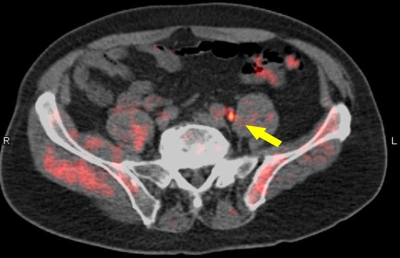
Fused PET/CT 18-F-fluciclovine image indicates recurrent prostate cancer in a pelvic lymph node. (Courtesy: Barry A Siegel, Mallinckrodt Institute of Radiology and the Alvin J Siteman Cancer Center, Washington University School of Medicine)
Up to 30% of patients with prostate cancer will develop local or distant recurrence within 10 years of radical prostatectomy or radiotherapy. Determining the location and extent of recurrent disease helps optimize the selection of appropriate management. Current anatomical imaging procedures, however, have limitations in identifying the sites of recurrence.
For the LOCATE trial, 213 men with biochemically recurrent prostate cancer were evaluated with 18F-fluciclovine PET/CT, after having negative or equivocal findings on conventional imaging, such as a bone scan, CT or MRI. Results showed that 59% of patients had their clinical management changed by the 18F-fluciclovine findings, with 78% of these changes classified as “major”, meaning a change in treatment modality.
“Selecting appropriate treatment for men with recurrent prostate cancer is critical,” explains Austin Pantel of the University of Pennsylvania. “Many options are available, and additional information, such as that provided by 18F-fluciclovine PET/CT, may help tailor personalized treatment plans.”
Alpha emitter targets wide range of solid tumours
In 2017, researchers from Memorial Sloan Kettering Cancer Center developed a novel approach to pretargeted radioimmunotherapy (DOTA-PRIT) that demonstrated, preclinically, complete responses in several solid tumour types using the beta-emitting 177Lu-DOTA-hapten. Now, they have expanded this approach to 225Ac, an alpha-emitting isotope.
225-Ac-proteus-DOTA hapten can be targeted in vivo to GPA33-expressing human colorectal cancer xenografts using anti-GPA33-DOTA-PRIT, with minimal radioactive uptake in critical radiosensitive tissues. (Courtesy: S M Cheal et al, Memorial Sloan Kettering Cancer Center)
“Targeted alpha radiotherapy has shown considerable promise for patients, especially for those with advanced castration-resistance prostate cancer,” explain Steven Larson and Sarah Cheal. “By combining DOTA-PRIT with 225Ac-proteus-DOTA hapten, we can potentially target a wide array of cancer types for which we have validated DOTA-PRIT bispecific antibodies.”
DOTA-PRIT has a major advantage over other forms of radioimmunotherapy because of its high ability to deliver radiation to tumours while sparing normal tissues, such as kidney and bone marrow. The researchers synthesized proteus-DOTA, radiolabelled it with 225Ac, and conducted in vitro and in vivo studies of a mouse model with colorectal cancer. They also performed a toxicity study in tumour-free mice with varying doses of 225Ac-proteus-DOTA.
The team found that the new approach, 225Ac-proteus-DOTA, mimics the behaviour of 177Lu-DOTA-hapten, with high tumour uptake, minimal accumulation in normal tissue, good whole-body clearance, no acute toxicity and no chronic radiation damage. It also offers greater versatility for treating a wide variety of solid tumours.
Graphene could be the two-dimensional nanomaterial for the future, but exploiting it will require further advances in methods to fabricate it. With a high carrier mobility, stability, and rigidity under atmospheric conditions, its electronic, chemical, and structural properties are unparalleled. In particular, AB stacked bilayer graphene, which consists of two graphene sheets stacked in an offset configuration, has an impressive tuneable bandgap ideal for usage in high performance device fabrication. However, growth of defect-free graphene and large-scale transfer from its native growth substrate, a necessity to bring such applications to fruition, remains challenging. Researchers Hussain Alsalman et al., under the direction of Michael Spencer, hope to overcome these challenges with their novel growth and dry transfer method.
High-quality graphene growth
Large-scale, uniform, defect-free graphene samples are difficult to obtain. Traditional exfoliation methods, where flakes of graphene are mechanically scraped off of graphite, are limited to small areas. Chemical vapour deposition (CVD) performs slightly better regarding area but it can take hours to achieve AB stacked sheets.
To overcome these obstacles, researchers at Cornell University utilized SiC epitaxy to grow large areas of graphene up to hundreds of microns,. Essentially, they annealed a prepared layer of SiC and the Si atoms sublimated to leave a layer of carbon that rearranged into graphene. Hussain Alsalman, a graduate student working on the project, notes, “SiC epitaxy sublimates the silicon atoms across the entire SiC wafer surface at once, giving it a clear advantage in the speed of single crystal synthesis.”
They then obtain bilayer graphene through high-temperature hydrogen intercalation. As Alsalman et al. point out in their report of the work, “SiC graphene on the Si-face is not limited by the orientations randomness that challenges CVD graphene.” This control over the AB stacking is another clear advantage over traditional synthesis methods.
A novel transfer method
Since SiC epitaxy produces graphene on an undesirable SiC substrate, the next step is isolating it from its native growth substrate, and this is not trivial. Alsalman et al. resolved this issue by transferring the graphene layers to more functional materials.
First, they deposited a layer of gold onto the graphene/SiC stack and spin-coated the stack to deposit a layer of acrylic polymer on top of the new gold layer. They can then mechanically remove the original SiC substrate thus allowing transfer of the graphene to the new target substrate. The acrylic and gold layers they remove via acetone and etching, respectively. Lastly, they rinse the isolated graphene/substrate stack to obtain the final product.
Structural and electronic characterization
To emphasize the success of their dry transfer method, Alsalman et al. employed a range of characterization techniques. Transmission electron microscopy (TEM) data show the classic atomic honeycomb structure of graphene and reveal high crystallinity within the monolayers even after transfer, which the researchers describe as “a testament to the robustness of epitaxial graphene.”
Although Raman spectroscopy revealed few defects within the graphene structure and uniform AB stacked bilayers, these results were not as impressive as those from monolayer growth via SiC epitaxy, most likely due to the intercalation method used to generate the second graphene layer. In their report of the work Alsalman et al. state, “This limitation for bilayers might be mitigated by carefully selecting for growth of bilayer on SiC and not using intercalation…However, this process needs optimization and would be a topic of further research.”
Field effect transistors (FETs) constructed from graphene grown with this technique also highlighted discrepancies in the quality between monolayer and bilayer graphene. While monolayer FETs showed field effect mobilities of approximately 1700 cm2/Vs, bilayer devices lagged behind this value at 250 cm2/Vs. Annealing the graphene before device construction increased the mobility values, which the researchers attribute to release of trapped hydrogen gases. The increase was limited to around 25%, and these generally low mobility values may be due to defects caused by step edges present on the SiC wafers. Control of bilayer growth, as previously discussed, may also increase the mobilities.
TLM devices gave contact resistance values of approximately 585 Ω μm for monolayer samples and 2310 Ω μm for AB stacked samples. Such a result is unsurprising, as contact resistance directly depends on the quality of the graphene, which was worse for the bilayer samples compared with the monolayer samples. Carrier mobilities also affect resistance.
Limitations aside the researchers conclude, “And while the performance figures might fall short from that of CVD graphene, transferred epitaxial graphene does currently have an advantage in synthesizing large area AB stacked bilayer graphene where in principle it is only limited by the size of the SiC wafer.”
Dong-Woo Cho, director of the Intelligent Manufacturing Systems Lab at the Pohang University of Science and Technology (POSTECH) in Korea, is working together with his colleagues to apply mechanical engineering know-how to biofabrication. Potential applications for the team’s work include the repair of complex bone and cartilage injuries, as well as non-transplant based treatments for end-stage liver disease.
Cho has long been interested in the use of 3D printing technology to fabricate micro-sized structures, and in the early 2000s he chose to make the jump into biofabrication. “We had the chance to discuss our research with medical doctors and there was a hint that microstructures could have a dramatic impact on cell activity,” he recalls. “That motivated us to work with biocompatible materials.”
Bone regeneration in rabbits
The group’s core expertise is 3D printing-related manufacturing technology, which involves system design as well as the development of printing processes and software technology. But that’s only part of the picture. “Collaboration with experts in the field of biology and medicine is essential for the application of biofabrication,” Cho comments. “Research on this topic becomes more meaningful when cellular and clinical expertise is reflected in the work.”
Over the last 15 years, the group has been proactive in building networks domestically and internationally to link its mechanical engineering expertise with biologists and physicians. Cho is excited by prospects for the technology, such as the potential of 3D bioprinting technology to boost patient-specific medical care.
“Traditional medical practice typically relies on products manufactured with a mass production paradigm,” he explains. “However, by using 3D bioprinting technology, we have the opportunity to personalize not only the outer shape of a device, but also its inner structure.”
Taking tissue scaffolds as an example, 3D printing could produce structures that are a much better fit for patients and could also enable cells to be printed at the histologically desired location for organ regeneration.
Recently, Cho and his team highlighted this benefit by examining the regeneration of tissue in rabbit knee joints. In the work — published in the journal Biofabrication – scaffolds that had been tuned to match cartilage and bone regions were able to smoothly regenerate tissue in as little as eight weeks. “As a follow-up study, we will investigate localizing not only the extracellular matrix, but also tissue-specific cells at the desired location,” Cho adds.
Applying this tailored approach towards the repair of organs such as the liver, the team has also shown that bioprinting can be used to establish and maintain a co-cultured 3D environment of multiple types of cells. “In order to regenerate liver tissue, it is very important to realize the hepatic histological structure as it is,” Cho points out. “In other words, you need to promote the growth of hepatocytes and hepatocyte surrounding cells, as well as blood vessels.”
Adding to the challenge is the need to realize a solution in three-dimensions to better match the performance of the native micro-environment, since the regeneration will then be more efficient.
Building-up structures layer-by-layer also allows researchers to introduce porosity where needed to encourage vascularization deep inside the biocompatible construct – which in turn will help to enhance tissue formation. Indeed, Cho’s team has already used 3D patterning-enabled porosity to good effect in studies on bone repair and on liver regeneration.
Another design feature being investigated in the lab is how to optimize the scaffold’s strength so that its form persists during treatment. In earlier work, the team integrated a mechanically stiff polymer (polycaprolactone) as a framework material, but it hopes to simplify the design for clinical applications.
Ideally, the researchers would prefer to 3D-print their constructs using bioink that’s able to maintain its shape without the support of an additional framework. Suitable materials are now being evaluated by Cho and his group as they work towards their goals in 2018.
Read our special collection “Frontiers in biofabrication” to learn more about the latest advances in tissue engineering. This article is one of a series of reports highlighting high-impact research published in the IOP Publishing journal Biofabrication.