Scientists in the USA have developed a new deep brain stimulation (DBS) method to treat the symptoms of Parkinson’s disease. While DBS for Parkinson’s is currently delivered continuously, the new approach uses adaptive DBS, in which the stimulation amplitude is modified in real time in response to neural signatures of motor impairment or of stimulation-induced adverse effects.
To test their approach, the researchers trialled adaptive DBS in two patients with Parkinson’s disease, using a fully implanted neural prosthesis enabled to use brain sensing to control stimulation amplitude (J. Neural Eng. 15 046006).
DBS can be an effective treatment for Parkinson’s disease, but it has limitations that reduce efficacy for individual patients and hinder more widespread use of the technique. For example, trained clinicians must programme the implants. It can also be time consuming and, for some patients, satisfactory settings are never achieved.
“This is the first demonstration of adaptive DBS in Parkinson’s disease using a fully implanted device and neural sensing,” said senior author Philip Starr from the University of California, San Francisco. “Our approach uses an algorithm to measure the patient’s neural feedback from the brain surface and change the stimulation in real time. This way, we avoid the stimulation being too intense when it is not needed, which can cause adverse effects such as involuntary movement, known as dyskinesia.”
The device works by using a cortical narrowband gamma oscillation (60-90 Hz) associated with dyskinesia as a control signal. An adaptive DBS algorithm reduces the stimulation voltage when gamma oscillatory activity is high (indicating dyskinesia is likely) and increases the voltage when it is low.
In addition to testing the adaptive DBS system, the researchers also completed an open-loop DBS control session. They observed that, in both patients, the total energy delivered by adaptive stimulation was substantially less than that of open-loop stimulation, while maintaining therapeutic efficacy. The algorithm performed as expected, appropriately detecting changes in gamma band power and triggering voltage reduction when the gamma threshold was exceeded.
“Reducing the stimulation current without losing the therapeutic benefit could reduce stimulation-induced adverse effects. It could also extend battery life, or allow the relatively large pulse generators we currently use to be made smaller,” explained first author Nicole Swann. “Additionally, some of the Parkinson’s disease patients most in need of DBS are also among the most difficult to successfully program: those who alternate between extreme states of dyskinesia and bradykinesia with little in-between time. Adaptive DBS could be very effective for them.”
“This study is a demonstration of the feasibility of adaptive DBS,” said Starr. “Now, further work is needed with a larger-scale trial.”
Remote sensing: Image of Earth, taken by the Envisat satellite, showing volcanic ash erupting from the Icelandic volcano Eyjafjallajökull in 2010. (Courtesy: ESA)
Telescopes and satellites are complex pieces of equipment, expensive to build and to operate. Consequently, astronomers and space scientists demand – and are prepared to pay for – sensors that maximize what they can achieve scientifically. Astronomers, especially, are looking at faint objects that are usually very far away, such as distant galaxies, stars and exoplanets. To capture light from these objects, they need telescopes with both large collecting areas and highly sensitive detectors.
One aspect of sensitivity is the quantum efficiency of the detectors – a measure of how well you can collect photons and use them. The other aspect is the noise associated with reading that photon signal into your system. A third important parameter is effectively the telescope’s operating efficiency, which depends on how quickly you can read out the sensors. These facilities may cost hundreds of thousands of dollars per night to run, so it is important that they spend most of their time capturing light and as little as possible transferring data into a computer.
These three requirements – high quantum efficiency, low noise and high operational efficiency – have long helped to drive improvements in detectors built for the astronomy market. More recently though, we are seeing the benefits of these improvements in areas outside astronomy as well. Here are a few examples.
Quantum efficiency
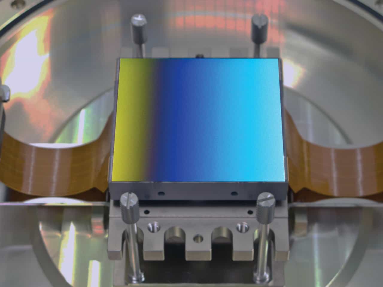
Silicon sensors have electrodes on their front surface. These electrodes are partly transparent, but even so, a “front illuminated” sensor is only around 50% efficient at recording photons. Since the 1980s, however, we have been able to remove most of the silicon material, tip the sensor upside down and illuminate it from the back side instead. This allows the photons to hit the silicon without any intervening layers, giving you very close to 100% efficiency. Back illumination also extends the wavelength range, making it possible to use sensors at soft X-ray and ultraviolet wavelengths to collect photons that would otherwise be absorbed in the surface layers of silicon.
Back-illuminated sensors are more expensive and time-consuming to make, but in the astronomy and space market that trade-off is worthwhile for all of the reasons mentioned above. More recently, though, this technology has become increasingly prevalent and valuable in other areas as well. For example, all modern mobile phones include CMOS sensors in their camera chips, and there is a huge drive to make these chips smaller. We are now at the point where the pixel size of the sensors is down to ~1 µm, and as the pixels shrink, their collecting area gets smaller, lowering their sensitivity. That’s a disadvantage, but within the last 10 years, manufacturers have begun to compensate by using back-illuminated technology. Back illumination is also becoming more common in sensors for other markets, such as drug discovery, DNA sequencing and life sciences more generally.
Red sensitivity
Many recent cosmological studies of dark energy and dark matter have involved measuring distant objects with very red-shifted light. However, chips made with standard materials, designs and processes have only a modest red sensitivity. There is a good reason for this: the response of the human eye begins to roll off at wavelengths in the near-infrared, so if you are making a chip for a digital camera, you probably want it to have a similar sensitivity. But astronomers need to measure all wavelengths, so at Teledyne e2v we have developed techniques (such as making the silicon thicker so that it absorbs near-infrared light better) for making our sensors more red-sensitive.
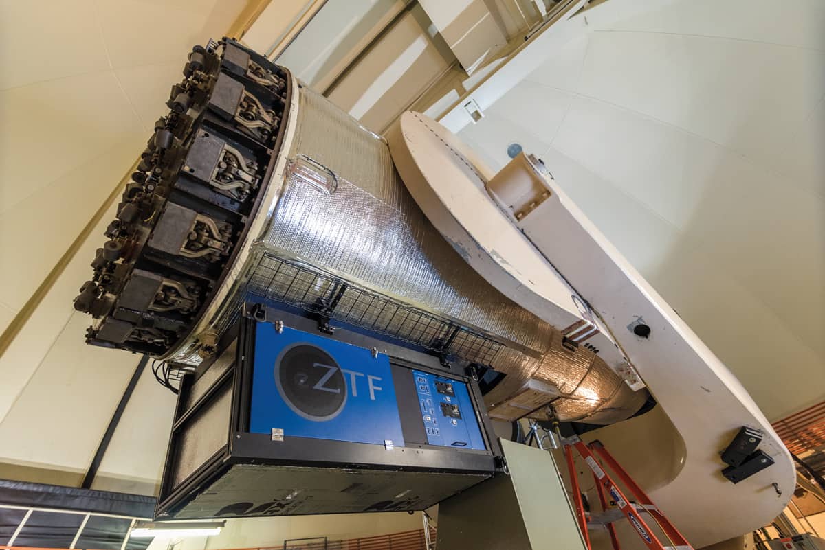
Sensitive instrument: A large-area mosaic camera lies at the heart of the Zwicky Transient Facility (ZTF) instrument installed on the 1.2m diameter Samuel Oschin Telescope at Palomar Observatory in California. (Courtesy: Caltech Optical Observatories)
We have supplied these sensors to the astronomy market for a long time, but red sensitivity is also starting to be advantageous in sensors used in ophthalmology thanks to a technique called optical coherence tomography. This technique uses near-infrared illumination to penetrate a little bit deeper into cells, so red-sensitive sensors have obvious benefits.
Adaptive optics
The basic purpose of adaptive optics is to correct for the turbulence and disturbance caused by the Earth’s atmosphere. This doesn’t apply to space telescopes, of course; once you’re up there, you don’t have to look through air. But all ground-based astronomers do, even if they’re high up a mountain, and that imposes a limit on how sharp their images can be.
To fix this, you need to point the telescope at a “reference star” of some sort, measure the wavefront coming from the reference star, capture it with a wavefront-sensing detector, read out the detector in 1–10 ms, and feed the signal into a closed-loop system with a deformable mirror that essentially corrects for what the atmosphere has done. The result is an image that will be sharper than it would have been without the technique.
At the moment, we are working on an adaptive-optics system for the Extremely Large Telescope (ELT), which is under construction for the European Southern Observatory on their telescope in Chile. The ELT is a 39 m diameter telescope that will cost about €1bn and will be the largest telescope in the world when it’s finished in 2024, but without adaptive optics it would be no better than smaller, older instruments built a generation or more ago.
The usefulness of adaptive optics is not limited to astronomy. There are also some emerging applications in laser communications – for example, in optical communications between ground and satellite – and medical applications are becoming more widespread. For example, if you want to capture images of the human retina, you can build a very-high-resolution microscope, but when you use it to look through the front of the eye, the cornea and the lens will disturb the sharpness of the retinal image. Clinicians want to see the blood vessels, the structure, and even the photoreceptors – the cones and rods – in the retina. To do that they are starting to use adaptive-optics instruments that can correct, 100 times a second, for the modulations or imperfections of the eye. It’s a powerful technique, and it is not limited to big research instruments anymore: it is also used in the medical diagnostics found in hospitals.
Multispectral imaging
Astronomers have always wanted to make measurements with spectrographs that cover as large a wavelength range as possible. That kind of spectral range used to be much less common for instruments that look down from satellites to the Earth, but now there are many hyperspectral satellites up there scanning the atmosphere, the sea or the land. These newer satellites use more complex sensors than the old broadband filters, and that means they can get good spectral resolution and good spatial resolution at the same time. Because of this, they can distinguish, for example, how well trees are growing, what type of foliage is in an image or what types of minerals are on the Earth’s surface. With broadband filters, you can get some of that information, but it’s rather crude.
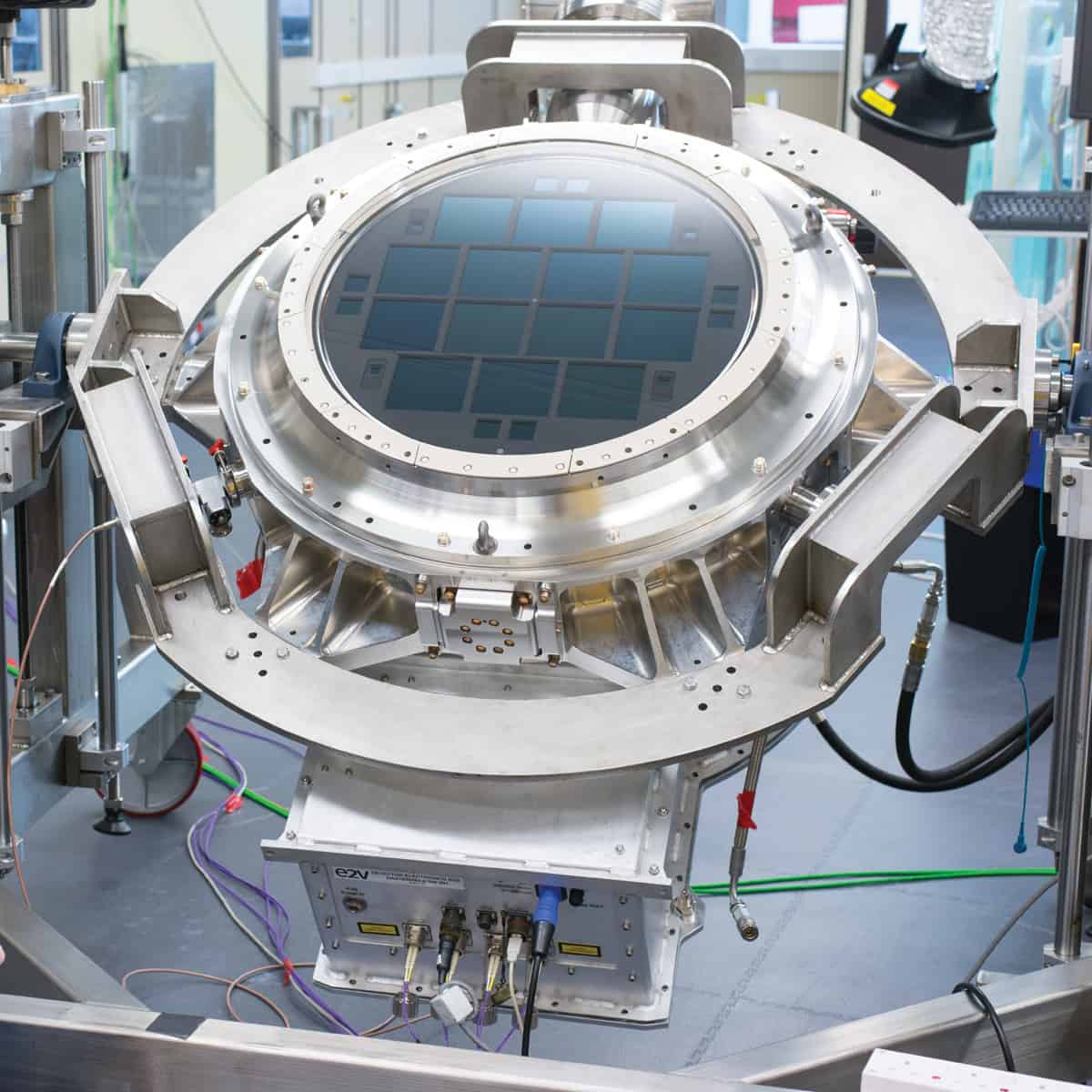
Full spectrum: Example of a multispectral sensor used for astronomy. (Courtesy: Teledyne e2V)
This technology is now being extended to less expensive instruments. It’s being used on drones, for example, that fly over farmland to conduct agricultural surveys, and in food processing, where you might use it to detect whether your potatoes have a fungus. With high-resolution spectral information you can really see what the problem is, as opposed to just detecting a slight change in colour.
Curved sensors
For most optical systems – including our own eyes – the focal surface is either spherical or curved. Despite this, essentially all sensors that have ever been made commercially are flat. This is because when you make sensors, you typically start off with a silicon wafer that has been polished so flat that you can see your face in it, and all the manufacturing equipment is designed to operate with a flat wafer. For example, the big, expensive optical lithography machines that project patterns onto silicon to create the structure of the circuit will only focus properly if the surface of the wafer (150–200 mm across) is flat to a precision of 1 µm, This means that the finished devices are only flat and cannot be curved when first made. However, there is a lot of development work being done on technologies that allow you to curve flat wafers after they have been made. This is difficult to do because silicon is a crystal and it doesn’t like being bent – you impose stresses in it when you bend it – and we are now learning the limits of how much you can do that and still have it function in an imaging system.
Red eye on the sky: A large-area cryogenic camera built by Teledyne e2v using red-sensitive CCDs. (Courtesy: Teledyne e2V)
There is a lot of interest in making curved detectors or sensors because it would reduce the number of optical elements in a system. At the moment, when people design imaging cameras or spectroscopic instruments, they have to put in extra optical elements to focus the light onto a flat detector. Those elements cost money, take up space and introduce both extra complexity and some degradation in design quality. On the ELT, for example, some instruments are the size of an entire room and the optical elements are correspondingly large. If you could cut out a few of them, you could save millions. The same principle applies for space-based instruments, where cutting out optical components would give you a better system that is also smaller and lighter.
Space agencies are paying for the development of these technologies because they can see their benefits, but other, more “down to Earth” companies – Sony, Apple, Microsoft and others – are also starting to do research on curved sensors. As with back-illuminated sensors, the driver here is the miniaturization of mobile-phone technology. If you want to make a small camera for a mobile phone, you generally have to put a tiny lens (or multiple lenses) in front of it. A curved sensor would make the optics of these cameras simpler. For example, to make a compact camera with a wide field of view – like a fish-eye camera – you need a huge and difficult-to-manufacture piece of shaped glass to make a lens that will accomplish that with a flat detector. But if you can make the detector curved, the components in front of it can get smaller and cheaper, and so can the device as a whole.
And one for the future
One thing that we are seeing now is a transition from CCD technology to CMOS technology. These are both silicon technologies, and if you had asked me 20 years ago what an astronomer would need, the answer would probably have been CCDs. But the chips in mobile phones are CMOS-based, and we are now making more and more CMOS devices for use in both ground-based and space-based astronomy. CMOS chips have several advantages for astronomy and space applications. One is that they are “harder” against radiation – they tolerate being irradiated better than CCDs. They are also more integrated in the circuitry that’s inside them, so they are easier to operate and you don’t need as many other electronics around them to get the data out and back to wherever you’re storing it.
Overall, we see two pulls for the types of technology development we do. One pull comes from customers, and curved sensors are a good example – various people have asked for that, so we are working on it. But the other pull is our internal knowledge of what’s possible. We create a road map of technology developments each year, and curved sensors, new CMOS sensors and improved wavelength sensitivity are all on it. We are pushing the boundaries as much as we can all the time, because as astronomers know well, nothing in this field ever stands still.
A round-up of the latest international patent applications in radiation therapy.
MRI/PET-guided system verifies dose-deposition
Researchers from Alberta Health Services have designed an MRI/PET-guided radiotherapy system that can determine the in vivo dose deposition of a treatment beam in real time (WO/2018/023195). The system includes a bi-planar MRI apparatus that comprises a pair of spaced apart magnets, one of which has a hole in the centre. A radiotherapy source is configured to generate a treatment beam and transmit it through the hole in the magnet. A patient support positions the patient within the system such that the treatment target is proximal to the radiotherapy beam. A PET detector, configured to obtain PET data from the beam impacting the patient, is positioned such that a transverse section of the patient including the treatment target lies between opposing portions of the detector.
Treatment planning system exploits machine learning
Elekta has published details of systems and methods for developing radiotherapy treatment plans via machine learning approaches and neural network components (WO/2018/048575). A neural network is trained using one or more 3D medical images, one or more 3D anatomy maps and one or more dose distributions, to predict a fluence or dose map. During training, the neural network receives a predicted dose distribution that is compared to an expected dose distribution. The comparison is performed iteratively until a predetermined threshold is achieved. The trained neural network is then utilized to provide a 3D dose distribution.
HIFU ablates large volumes, protects critical structures
SonaCare Medical has developed a method for delivering high-intensity focused ultrasound (HIFU) to large tissue volumes while protecting critical structures (WO/2018/057580). The approach includes positioning at least one focal zone of a transducer in an ultrasound probe proximate to the targeted tissue. The transducer delivers ultrasound energy to a portion of the tissue for a predetermined time to create an initial focal lesion(s). Next, the transducer delivers ultrasound to the targeted tissue continuously and along a predefined treatment path. The first lesion(s) can act as a barrier for subsequent HIFU ablation of tissue located beyond or behind it, thereby protecting this tissue from unintended ablation.
Phantoms offer QA for biologically-guided radiotherapy
RefleXion Medical has created phantoms for calibration and quality assurance of radiation therapy systems, including biologically-guided systems that deliver dose in response to real-time detected PET lines-of-response (WO/2018/081420). The modular phantoms comprise a cylindrical housing with a number of disks stacked within the housing and a number of radiographic films between the disks. The disks may comprise a positron-emitting material. Some disks have a background region and a target region (with a higher level of PET activity) of any desired cross-sectional shape. The disks may be arranged within the housing such that regions of higher PET activity are aligned to simulate the shape of a tumour in the patient.
Ultrasound takes control of neuromodulation
Neuromodulation, using electrical stimulation of the central nervous system, for example, is used to treat a variety of clinical conditions. However, positioning electrodes at or near the target nerves is challenging, as is specific tissue targeting. A team from GE and the Feinstein Institute for Medical Research has published details of techniques for neuromodulation of tissue via application of ultrasound energy into the tissue (WO/2018/081826). This energy causes altered activity at a synapse between a neuron and a non-neuronal cell, in order to achieve a targeted physiological outcome.
The Big Bell Test Collaboration has put quantum entanglement to the test with help from about 100,000 computer gamers worldwide. Run by an international team of physicists, the experiment used decisions by members of the public to close the “freedom of choice loophole” in several different Bell tests – which show that the quantum entanglement of two systems violates local realism.
The idea of quantum entanglement dates to 1935, when Albert Einstein, Boris Podolsky and Nathan Rosen pointed out that two quantum particles can be in a state in which a measurement on one particle instantaneously affects the other – no matter how far apart they may be. This entanglement of particles cannot happen in the world of classical physics because it would require information to travel faster than the speed of light.
Stronger correlations
Since then physicists have shown that entanglement can be determined by looking at correlations between measurements made on the two particles. Entangled particles have much stronger correlations than are allowed in classical physics – a property that can be exploited in quantum computers and other quantum technologies.
In 1964 the Northern Irish physicist John Bell famously calculated an upper limit on how strong these correlations could be if they were caused by classical physics alone – what has become known as Bell’s inequality. Stronger correlations could only occur only if the particles were entangled – and confirming entanglement in this way has since been dubbed a Bell test.
Experiments using photons, ions and other entangled particles have confirmed that Bell’s inequality is indeed violated. However, these experiments are plagued by one or more loopholes that could allow unforeseen effects of classical physics to cause the violation.
Measurement choices
Bell tests usually involve producing large numbers of entangled pairs and making random measurements on certain properties of particles in each pair. For example, either the horizontal or vertical polarization of a photon can be measured. There cannot be any inherent correlations in how these measurements are chosen – and the inability to completely rule out the existence of such correlations in an experiment is called the freedom of choice loophole.
It turns out that this loophole can be closed if the measurement choices are made by humans, rather than by random number generators. This was done by inviting people to play a video game called The Big Bell Quest, which involved players randomly pressing their “0” and “1” keys. On 30 November, 2016 more than 97 million bits were fed to 13 different Bell test experiments worldwide. These tests used a variety of entangled particles and systems including photons, atoms and superconducting devices.
As well as closing the freedom of choice loophole, the collaboration also showed that random numbers can be collected rapidly from large numbers of people. New networking techniques were also developed to allow worldwide participation in laboratory experiments.
Around a third of environmental and social impacts from consumption in wealthy nations is displaced to developing countries, according to the latest analysis. And that trend in outsourcing responsibility is increasing.
“Many citizens of rich countries require the work of up to five poor people to satisfy their consumption,” said Manfred Lenzen of the University of Sydney, Australia. “Rich consumers like us are implicated in pollution and inequality all over the world, and people in poor countries bear the brunt of our large environmental and social footprints.”
According to Tommy Wiedmann of UNSW Sydney, indirect effects facilitated by often complicated supply-chains are mostly hidden from consumers, who generally do not know where the raw ingredients of their purchases stem from.
“Carbon emissions are still accounted for on a territorial basis,” said Wiedmann. “This means that if a country moves from producing goods domestically to importing them from China, its carbon footprint decreases – leading politicians to think that the country is cleaning up its act.”
Lenzen, Wiedmann and colleagues used global multi-regional input–output models (GMRIO) to perform the analysis and untangle complex international trade routes. They found that traded goods embodied a substantial amount of emissions, water, pollutants and resources.
“Our new research clearly points towards a need for so-called consumption-based accounting – where a country’s environmental score includes its imports – and as such leaves no room for loopholes,” said Lenzen.
Although the importance of displacement of carbon emissions – also referred to as carbon leakage – has been known for some years, the researchers say they have now amassed evidence in terms of environmental issues such as air pollution, water scarcity, biodiversity loss, raw material and energy depletion, and nitrogen emissions.
Proton therapy plans rely on estimates of particle range in the patient, typically derived from X-ray CT scans, with the CT numbers converted into proton stopping-power ratios (SPRs) using a generic Hounsfield look-up table (HLUT). But this approach cannot account for differences in tissue composition, or patient-to-patient variations, thus limiting the treatment accuracy. To manage range uncertainties arising from this CT-to-SPR conversion, it was suggested back in 1985 that a safety margin of 3.5% of the total range should be applied for all proton treatments.
Speaking at the recent ESTRO 37 congress, Christian Richter from Helmholtz-Zentrum Dresden-Rossendorf and OncoRay pointed out that in 2018, the majority of proton centres still employ this 3.5% range uncertainty margin. “We currently do not use the full potential of this advanced technology. Provocatively speaking, it’s like driving an aeroplane rather than flying it,” he told the delegates.
We can do better, Richter explained, by using dual-energy CT (DECT) to determine particle range. DECT uses two scans with different X-ray spectra to provide complementary information on tissue composition. But although DECT has been clinically available in radiology for about a decade, its use in particle therapy is far from being a clinical standard.
“At the German National Center for Radiation Research in Oncology (NCRO), partnered by OncoRay in Dresden and DKFZ in Heidelberg, we have set up a project to focus on the translation of DECT into particle therapy,” said Richter. The goal is to demonstrate and quantify the benefit of DECT-based patient individual range prediction (PIRP).
In a first step, the NCRO team validated PIRP in extensive high-accuracy ground-truth settings, in both biological tissue and realistic anthropomorphic geometries. These studies demonstrated the superior accuracy of PIRP/DECT over the HLUT approach. “For example, Christian Möhler from the NCRO team proved that PIRP accuracy in multiple biological tissues was better than 0.2%,” Richter noted.
And since April 2015, OncoRay has been using DECT, along with state-of-the-art HLUT, to calculate treatment plans for the majority of patients in its proton therapy facility in Dresden. This approach provides improved image quality and flexibility for proton planning. And with over 2500 DECT scans in their database now, it also acts as a great research resource.
Reducing the uncertainty
To investigate uncertainty levels in more detail, Richter and colleagues have studied intra- and inter-patient variabilities in CT-to-SPR conversions. In over 100 brain tumour patient, for example, they observed an average 4.5% intra-patient variation in the relationship between SPR and CT number in soft tissue. Such variation is intrinsically considered by the PIRP approach, he noted.
Another study of over 100 head tumour patients revealed that conversion factors also differ according to the patient’s age. In bony tissue, there was a 5% difference in the relationship between SPR and CT number between adults and children, due to differing calcium content in the bone. “This variation is not covered by the one-fits-it-all HLUT approach,” said Richter.
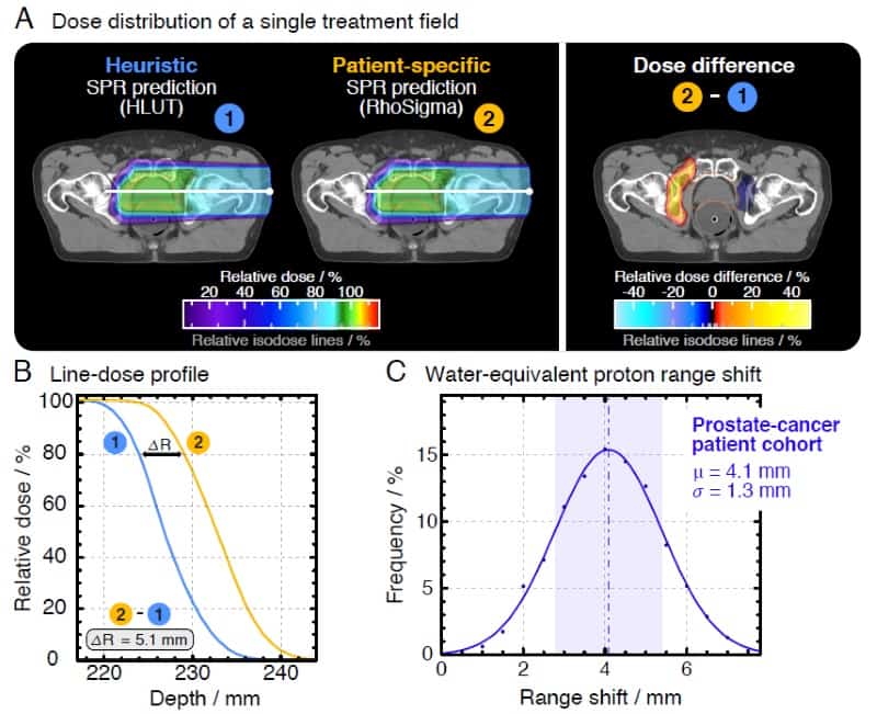
Differences between the HLUT and PIRP/DECT approach. Credit: Patrick Wohlfahrt (OncoRay, HZDR)
The team, led by OncoRay’s Patrick Wohlfahrt, performed a study assessing range differences between CT-to-SPR conversions using HLUT and PIRP, in 25 brain tumour and 25 prostate cancer patients. In the latter group, they observed average range deviations of 4.1 mm (1.7%) for HLUT compared with the more accurate DECT. “This is clinically relevant, this does matter,” Richter emphasized. He added that one approach is to adapt the look-up-table to better match the PIRP result. “This DECT-based HLUT refinement is an important step – for the first time, PIRP has influenced clinical range prediction,” he said.
One potential stumbling block in this technique is that DECT scans are acquired consecutively, and that motion in and between scans may perhaps affect reliability. Richter described a proof-of-principle study analysing the clinical feasibility of using dual-spiral 4D-DECT scans for proton dose calculation in three non-small cell lung cancer patients. No anatomical differences were seen in the 4D-DECT data processed from consecutively acquired scans, validating the feasibility of PIRP.
The higher imaging accuracy afforded by DECT, as well as its intrinsic consideration of patient variability, will ultimately reduce uncertainty in range prediction for particle therapy. DECT also benefits applications such as identifying and quantifying metallic implants, or providing improved inputs for Monte Carlo calculations. “I see only advantages without major drawbacks,” said Richter. “So why don’t we use DECT more routinely in radiation oncology?”
The NCRO team is now working to achieve this goal, collaborating with a manufacturer to develop a prototype DECT system for clinical implementation and creating a dedicated imaging protocol for proton therapy scans. “I think this is a game changer – after 30 years we should finally be able to reduce the margins,” Richter concluded.
If you go to a sandy desert or beach on a blustery day, you might see a range of topographic features created by the wind. Looking down at your feet, you may notice centimetre-scale periodic ripples made by hopping grains of sand. You could also explore much larger sand dunes that form on length scales of tens of metres.
Ripples and dunes are thought to form in very different ways and in most sandy environments you will not see periodic structures at intermediate length scales – often referred to as the “forbidden region”. There are exceptions to this rule, and unusually-large “megaripples” are found in some places. Now, researchers at the University of Leipzig in Germany and Ben-Gurion University of the Negev in Israel have worked out that these structures form in much the same way as larger sand dunes.
Sand ripples are thought to be caused by a synchronization of the hopping of grains of windblown sand with emerging waves in the sand bed. The ripples are rarely larger than a few centimetres wide, and tend to be regularly spaced, with about 10 cm between peaks.
A mighty wind
Dunes result from the asymmetric way the wind blows over obstacles: “If you stand on top of a hill, the strongest wind is not exactly at the top: it’s slightly upwind of this position,” explains Leipzig’s Klaus Kroy. The maximum mass of a particle that can become airborne increases with the airflow rate, so, as the wind slows down slightly at the top of a dune, it deposits more sand, making the dune bigger.
Normally sand does not fly continuously in the wind but travels in discrete hops several centimetres long. For a dune smaller than about 10 m in length, this hop length is longer than the distance the point of strongest wind is displaced from the dune crest. Therefore, sand picked up from the windward side of a dune is not deposited on the crest and the dune is eroded by the wind.
These rare intermediate structures are known as megaripples and have, until now, lacked any theoretical explanation. As the name implies, they have generally been studied as unusually large ripples, but this has only deepened the mystery. For example, ordinary ripples tend to be periodic, and are neatly characterized by the distance between successive peaks. However, says Kroy, megaripples “don’t care where the nearest neighbour is”. Furthermore, megaripples tend to be covered with coarse sand: “People were saying to me things like ‘It’s really hard to understand why the coarse grains always accumulate on them,'” says Kroy.
Bimodal grains
Treating megaripples as dunes explains why they are not periodic, but raises other questions. They are often not much more than 30 cm in length, so how can they be stable? The researchers present a mathematical model of how, under specific geological conditions, the distribution of sand grain masses can become bimodal: fine, light grains perform large jumps in the wind and heavier, coarser grains creep along the sand bed in small jumps, pushed along by repeated impacts from the lighter grains.
These heavier grains respond to changes in wind speed on much shorter length scales than the lighter grains, so they can build much smaller dunes. Unlike large dunes, however, megaripples are transient features prone to destruction by storms. This, say the researchers, is because the coarser grains of sand will make large jumps in very high winds, making small dunes unstable.
The team confirmed its hypothesis by studying cross sections of megaripples in the southern Negev desert in Israel. They also used previously published data: “We show people how to look at the available data and reinterpret it in a different way,” explains Kroy.
Kroy suggests that the analysis technique could be further developed for use in other fields. “[Researchers have] found small ripples on the surface of the comet that passed near Earth recently, even though there’s no atmosphere,” he says, “Under such conditions, you can immediately see that there must – quite unexpectedly – be some wind.” He also says that studies of petrified megaripples could tell us something about conditions long ago on Earth when the features formed.
Geoscientist Nathalie Vriend of the University of Cambridge is impressed with the research: “There have been observations of megaripples before, but this is one of the first papers that really puts some physical modelling behind them,” she says. She cautions, however, that the researchers’ dynamical ideas about the effects of various wind speeds on sand grain size need field testing.
Over the past 40 years significant strides have been made towards tackling cancer. During this relatively brief period, survival has doubled in the UK and 50% of people will now survive the disease for a decade or more. At the core of this progress is research, bringing advances in screening techniques, diagnostic tests and treatments that together are helping to transform the outlook for this disease.
Despite these developments, now is not the time for complacency. Progress has not been uniform and for several cancers, such as oesophageal and pancreatic, survival has lagged far behind and seen little improvement.And for most patients whose disease has spread, achieving a cure remains a distant prospect. To ensure advances continue rather than stagnate, there is a clear case for innovation in the way we approach and carry out cancer research.
That is why in 2015 Cancer Research UK launched the Grand Challenge – an ambitious £20m programme to support scientists to solve some of the greatest problems facing the field. The grants not only seek to overcome major obstacles that stand in the way of progress, but also challenge more traditional ways of working. Where the increasing competitiveness of research funding may have sparked a trend towards safety as opposed to innovation, Grand Challenge seeks to encourage bold thinking and reward novel ideas and approaches to research.
A worldwide approach
With the complexity and scale of the issues at hand, from unravelling disease biology to overcoming treatment toxicity, the programme is not exclusive to the UK but open to researchers worldwide – encouraging collaboration across an exceptional pool of international talent. But collaboration requires more than the sum of many minds.
To tackle such significant problems we need a breadth of knowledge and expertise. This is why Grand Challenge is designed for the physical and life sciences to come together, forging truly multidisciplinary teams that can bring forward and capitalise on the range of unique skills that each discipline offers. By breaking down both geographical and disciplinary barriers, Grand Challenge ultimately seeks to transform cancer research and to beat cancer sooner.
While cancer research may traditionally be placed among the life sciences, physics has been pivotal to some of the greatest advances in treatment and diagnosis to date. It was physicists who pioneered the field of radiology as far back as the 1800s. The serendipitous discovery of the X-ray, by German physicist Wilhelm Röntgen in 1895, triggered a global change in diagnostics virtually overnight, turning previously invisible maladies into diagrams of ill health.
And it was the subsequent surge of interest worldwide that soon identified another purpose for radiation in medicine: radiotherapy. Now a cornerstone treatment that has helped save millions of lives, radiotherapy has had a century of research and refinement to turn it into the incredibly sophisticated and precise technique that it is today. Yet the work is not over. As new technology allows the limits of what can be achieved to be pushed further and further – not just in these fields but across the board – opportunities arise to explore previously uncharted territories in cancer science, making now a more exciting time than ever to be involved.
The inaugural round of Grand Challenges resulted in four groups being funded. One winner was Josephine Bunch from the National Physical Laboratory, who was awarded a £16m grant to map tumours and get down to their metabolic nuts and bolts at the molecular and cellular level. Bunch and colleagues will use an array of new mass-spectrometry techniques, and other methodologies they have pioneered, to detail everything from whole tumours down to individual molecules within the cells that comprise them. This ambitious project will involve physicists, chemists, biologists and technology innovators who all bring their own unique skills. Hopefully it can spur new developments in treatment and diagnostics that could ultimately lead to more lives saved.
Seed funding
For the programme’s second round, eight new challenges are looking to be solved. These range from developing artificial intelligence to help detect cancer earlier to defining mechanistic rules for treatment combinations. Ten teams have been shortlisted, which have each been given £30,000 of seed funding to get their ideas off the ground. The winners will be announced later this year.
Though applications for the current round are now closed, the 14 challenges that have so far been set are compelling evidence that applicants for round three will be no less audacious. The stakes are high, but the rewards are even greater. We hope that more physicists will take this opportunity to lead groundbreaking science that will revolutionize the field of cancer science.
Physics World published a special issue on the physics of cancer in July 2013
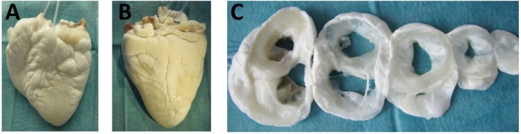
Decellularization of tissues for regenerative medicine applications, such as cartilage repair, is the subject of much research effort. The principle of naturally derived extracellular matrix (ECM)-based tissue engineering is to remove cells from the tissues without affecting their ECM, leaving an intact niche for cell repopulation and, hence, enabling enhanced healing (see: Decellularized and recellularized grafts repair injured cartilage).
Taking this a step further, the decellularization of whole organs offers the possibility of achieving the same goals in a complete structure such as a heart. This would revolutionize the fields of transplantation and regenerative medicine.
Whole organ decellularization is achieved through perfusion, which delivers the decellularizing agents (detergents and enzymes) using the tissue’s own vasculature. Obviously, this is a complex process and there are many variables that can influence the decellularization and affect the microstructure of the organ. In addition, research in this field has mainly focused on small animals like rats (which have much smaller organs), and is now more and more striving towards human-sized organs. Thus, the field of tissue engineering needs further research to understand the processing of whole organs by perfusion, particularly in large animal models.
Standardizing and optimizing
To shed some light upon the decellularization of whole hearts, Jörn Hülsmann, Hug Aubin and colleagues from the working group of Payam Akhyari and Artur Lichtenberg at Heinrich Heine University Düsseldorf have published a study in which they assess the processing of small and human-scale heart models. They performed decellularization of rat and ovine hearts and analysed the process by-products (perfusates or the solution that runs through the organ) and the effects on both organs. As result, they observed how the process presented varying characteristics that may help further its understanding and optimization (Biomed. Mater. 13 035014).
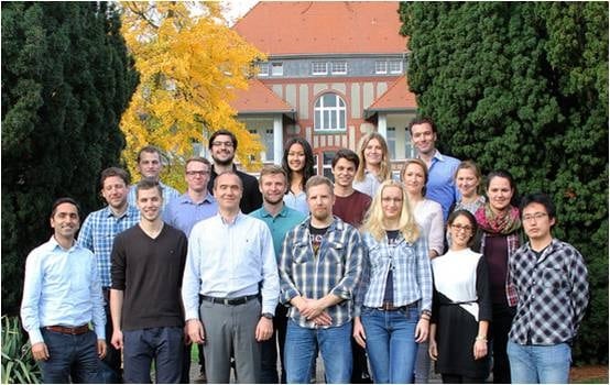
The research group of Payam Akhyari and Artur Lichtenberg at Heinrich Heine University Düsseldorf.
First, they confirmed the efficacy of the decellularization using macroscopic observation, histology and quantification of DNA and cell markers, which demonstrated a high removal of cellular material, while maintaining the tissue ultrastructure and extracellular matrix architecture. Later, the researchers investigated the dynamics of this process via analysis of the by-products or perfusates.
Ovine hearts decellularized by perfusion.
What are we losing?
Ideally, decellularization should only remove the cellular material, which comprises around 19% of the organ’s wet tissue weight. This can be observed after phase separation, drying and weighing of both hearts. Additional exposure to detergents harms the scaffold and, finally, may lead to its de-functionalization.
Surprisingly, after analysing the perfusates, the researchers observed that the detectable protein content was much higher, particularly in rodent hearts, where it represented 71% of depleted mass versus 37% in the larger models.
These differences between small and large organs may be attributed to varying physico-chemical interactions, potentially due to a size effect or to physiological differences (the lipid fraction in ovine hearts is higher, which can affect the action of the decellularizing agents). Furthermore, within the ovine hearts, important differences appeared between individual’s hearts. The researchers related such differences to different discharge dynamics (the speed at which a component is eliminated) produced by effects such protein interactions, debris formation or inherent organ characteristics that complicate the distribution of the solutions. These discordances are evidence for the need to further illuminate and optimize the decellularization of human-like organs.
A more detailed approach
From another point of view, this study presents a more detailed follow-up on the perfusion decellularization of organs. The combination of the analysis of discharged proteins and the viscosity of the decellularizing solutions offers added value that could be implemented into current protocols and improve the control, reproducibility and outcomes.
In future, such a protocol could improve the assessment of the efficacy of the decellularization process and the effects on the organ structure, and help optimize the process during its different phases. Furthermore, it could be a first step towards an automated set-up for production screening or industry production of decellularized whole organs. However, the authors point out that some limitations must still be solved with, for example, the use of more sophisticated and accurate detection systems.
How did you get involved in commercializing science?
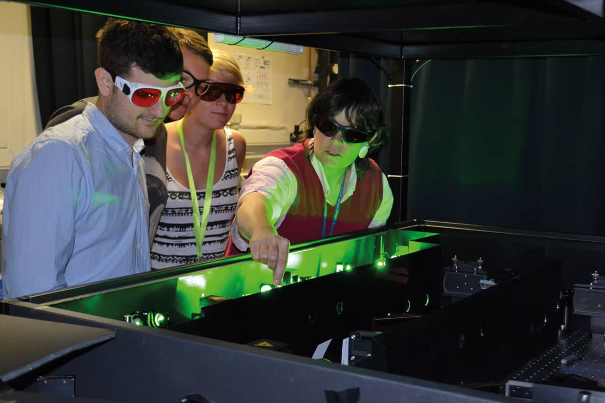
When I moved to New Zealand in 2007, I had only ever done fundamental science – looking at molecules and how they absorb light. But in New Zealand, the funding system is such that if you want to buy a million-dollar piece of equipment, you have to find a way to cover the depreciation, which means your budget starts at $100,000 a year before you even get to do anything. That meant I had to get funding from applied research, so we started a lab called the Photon Factory to expose New Zealand’s scientists to the exotic laser pulses that we use. But of course, you’re not going to commercialize femtosecond spectroscopy directly. Nobody’s going to open a spectrum store. So I learned to go out and listen to what people need, rather than talking about what I can do.
How did Engender get started?
A dairy investor said, “Hey, you want to go grab a coffee? I have some ideas of things that needs to happen.” He told me there were five problems facing the dairy industry, and out of those five I picked sperm sorting because it seemed like it might have a physical solution. Then I went back to my lab and gave the first four students I encountered 24 hours to come up with six ways of sorting bull sperm into male and female. Four of their ideas were really stupid – I know there are no stupid ideas, but these were close – and of the other two, we chose the one that would damage the sperm cells the least. Then my postdoc and I did the background work, and when we’d finished I told the investor and our university tech transfer office. They put in a little bit of money and we started a company from a drawing on a piece of paper – and some due diligence, of course.
Why is sperm sorting important?
It enables dairy farmers to accelerate the genetic gain in the top half of their herd. Let’s suppose that one of your cows, Maisy, makes a tremendous amount of protein and fat in her milk. Naturally, you want to breed Maisy against a top dairy bull, and you would love it if she had a girl calf every time. Without sperm sorting, though, it’s a 50/50 chance. The other thing is that although dairy cows have to have babies to give milk, not all of those babies are equally valuable. In fact, it can be cheaper to kill the bobby calves, the males, for meat than it is to raise them. What Engender does is to allow dairy farmers to breed the bottom half of their herd for males, but against a beef bull, and that adds tremendous value to those bobby calves, boosting animal welfare. Finally, if you look at the developing world, right now it takes three Indian cows to make the same amount of milk as one New Zealand cow because they haven’t been using artificial insemination for as long. With sperm sorting, Indian dairy farmers might be able to produce the same amount of milk, but with a third of the cows, which would reduce the impact on the environment.
How does Engender’s technology work?
The incumbent technology uses flow cytometry, which basically takes the cells, squirts them through a nozzle, puts them in droplets, charges the droplets and uses an electric field to sort them. You tell them apart by staining the DNA content with a fluorescent dye: because the X chromosome is a little bit bigger than the Y chromosome, the females are a little bit brighter than the males (which I love; it’s only about 3%, but it makes a big difference). Anyway, squirting the cells through a nozzle damages them because it puts shear stress on the membranes, and they don’t like the electric field either. So instead, we use a microfluidic chip, where the laminar flow means there’s not as much shear stress. We also use a laser instead of an electric field to do the sorting. One complication is that the sperm cells are shaped like flat discs, 5 × 10 × 1 µm. That means the biggest difference in fluorescence is actually an indicator of cell orientation, not gender. However, because the cells are asymmetric, you can generate a torque by shining light on them, so we have a three-step process where we use one laser to orient the cells, a second to do the fluorescence measurement and a third to nudge the unwanted cells into different flow streams on the chip.
Where are you now in developing the company?
Engender was founded in 2012, and towards the end of 2017 we proved that we could sort and enrich sperm collections by either X chromosome or Y chromosome. We are now in the middle of raising series B funding of about $18m and that will get us to a full commercial product. At the moment, the device is still a prototype in our lab, but the business model is to get all the pumps and lasers into a box about the size of a large desktop printer, and also to provide consumable chips, one per bull – or, rather, one per ejaculate. You start using some unusual words for a physicist when you talk about this project, by the way.
Not following the herd: For Cather Simpson (right) and colleagues, transforming a chip in a lab into a truly portable “lab on a chip” is the next challenge. (Courtesy: University of Auckland)
You sound enthusiastic about it, though. Are you sure it was just New Zealand’s funding system that pushed you into applied research?
I used to be in the “fundamental science is everything” camp, and when I gave lab tours I would say things like: “We do all this applied stuff to fund our basic science habit.” What changed my mind was a project I did with a US company called Intuitive Surgical, which makes a robot called Da Vinci. By chance, their chief research officer heard me say that femtosecond lasers can cut anything and her response was, “Can they cut bone?” So we travelled to Intuitive Surgical and all the people they funded were there talking about what they were doing. I just thought, “Holy crap! This isn’t about figuring something out that might be in a textbook in 15 years; I might actually be able to help real people in three years.” It was a transformative moment for me. Of course, we can’t do without fundamental science: you can’t apply something unless you understand it. But at the same time, I look at the world around us as a challenge-rich environment. Our job, as scientists, is to help meet those challenges.
What do you know now that you wish you’d known when you were getting started?
I wish I’d known more about how the commercial world works. Early on, I couldn’t have told you the difference between series-A and series-B funding, and I had no idea that there were so many ways for the people on the team – mostly students, postdocs and engineers – to reap financial benefits. Engender started with an idea and a tiny amount of “okay, prove it might work!” funding. The university was a major shareholder, a venture capital group put together most of the rest, and as the chief scientist, I have some founding shares as well.
I had no idea that a founding shareholding was different from any other shareholding, but it turns out that the university’s policy is to give a third of what the university owns back to the inventors. That means that our first-phase team will get a third of the revenues from whatever the university eventually realizes on Engender. In contrast, the second phase, where the team showed that each of the individual steps could all happen on the same chip – a really important job – was done as a research contract. Members of that second-phase team got paid for their work, of course, but it wasn’t until afterwards that I realized that their “skin in the game” was different from the first crew’s. Now that Engender’s more established, there are option-related and milestone-linked incentives for the R&D team, and I had to learn how those worked. Luckily, it all worked out so that the R&D team – including me – is properly rewarded and motivated to make sure Engender succeeds, and the intellectual property is linked to the inventors’ properly all the way along. But I still wish I’d had a business class, or that I’d done the reading and picking the brains of experts before starting a company.
Once you step past the money stuff, I think the hardest lesson to learn is about failure. In the real world, you can fail for reasons that have nothing to do with your science. For example, not understanding timelines and deliverables can lead you to fail because you’re not connected with what the real world is anticipating for your business. And failure is not always met with the same equanimity as it is in science. You can’t go to the board of your company and say, “Oh, I tried to sort sperm, but it didn’t work, so I went down this other interesting pathway instead,” like you can with, say, a National Science Foundation grant. That happened to me on a grant earlier in my career and I called up my programme director and said, “Look, you know how I had this whole elaborate five-year plan? There’s no point because my first hypothesis was wrong.” And he said, “Oh, well, that’s all right. Come up with another one.” That would not happen in the business world. I’d be fired. It wouldn’t be my project anymore.
That brings me to a second hard lesson. Right now, Engender is going from a laboratory prototype to a commercial effort. Sometime over the next year, I might not be in charge of the D part of the R&D anymore. As we grow, the company might hire someone who has more experience with that stage. And I’ve been surprised by how much I care about that. You’ve taken this thing, and you’ve put your heart and soul into making it work, and suddenly you realize that actually you don’t get to make the decisions anymore. So, success means that I’m not needed in some ways. That is a little bit hard.
Do you have any advice for anybody thinking of starting up a company in photonics and optics?
Find someone in the business world who you trust and ask lots of questions. The assumptions that people go in with are amazing. I have colleagues who are trying to start companies, or who are trying to do company-facing research, who assume that because it’s their idea, they should own 100% of the intellectual property. But that’s not how business works. As academics we tend to think, well, I wrote the paper on that, therefore I should get the credit. But it’s not about getting credit: it’s about risk and reward, and as soon as you wrap your head around that, you understand the language and the conversations a lot better. You also understand whether it’s for you or not because, for a lot of people, it’s not.