

SuperNova Green deactivates proteins on the nanoscale

SuperNova Green (SNG) is an effective new tool for the deactivation of proteins within cells. Developed by Yemima Dani Riani and the Nagai lab at Osaka University, the tool consists of a genetically encoded protein that reacts to light by generating damaging reactive oxygen species (ROS) in a small area around the molecule. These singlet and superoxide radicals damage neighbouring proteins enough that they become deactivated. Such a tool joins a growing arsenal of so called “optogenetic” actuators: genetically encoded light-activated proteins that can switch on or off spatial signalling pathways within cells, locally, on the nanoscale (BMC Biology 16 50).

Why destroy proteins with light?

Signalling pathways in cells depend on multitudes of molecular machines convening, accumulating and splitting apart again, in a vast molecular communication network. Super resolution microscopy has confirmed that there is a nanoscale component to this regulation that allows cells to elicit highly adaptive control over cell function. Direct links to such function have so far been hard to discern, and one reason is that the tools for altering such pathways on the nanoscale don’t exist. Dani Riani and colleagues have now introduced a new iteration of optogenetic tool, which can be hardwired into cells, fused to a protein and targeted for deactivation using laser light.

Depending on the size of the area illuminated with blue light, the length scale of protein deactivation can be altered. By concentrating the beam, single groups of proteins can be targeted, and by widening the beam, entire cells can be ablated, while their neighbours remain intact.

Yemima Dani Riani

In the study, the Nagai group showcased whole-cell ablation. By selectively targeting the mitochondria in cancer cells, SNG could kill cells outright. The probe is relatively sensitive, so 2 min of illumination was all that was needed to kill the cells. This whole-cell application could be useful for studying the elasticity or mechanics of cells in the developing spinal cord (neural tube), for example, as an alternative to high powered laser ablation, which tends to damage nearby cells and makes measurement difficult.

The group showed that selective nanoscale protein inactivation is possible without significant damage to non-targeted proteins. By testing the effect of SNG on two tagged proteins that were very close to each other in the cell membrane, they demonstrated that only the SNG-tagged protein was affected. In this case, they targeted pleckstrin domains – short lengths of protein that bind to lipids in the cell membrane. When the pleckstrin domain is inactivated, the protein moves away from the membrane and into the cytoplasm. Sure enough, in these experiments, the signal in the cytoplasm only increased for SNG-tagged proteins, due to ROS-based destruction of the pleckstrin zone.

Why is SNG better?

Imaging multiple targets in cells allows for elements of signalling pathways to be visualized down a microscope. Much effort has gone into linking function to signalling, and one powerful method is to view both cause and effect at the same time. The authors point out that using SNG allows you to do this, by deactivating proteins with a short excitation wavelength (blue), while imaging something else, for example calcium dynamics, using a probe at a higher wavelength. This demonstrates a clear advantage of this new probe.

Additionally, SNG can deactivate proteins without flavin mononucleotide (FMN), which is present in many cancer cells, but is not always present in all somatic cells at the concentrations required by other optogenetic protein deactivators. SNG is also much simpler than other protein inactivation strategies, such as “anchoring away” (using a signal sequence to mislocalize a protein away from its functional location in a cell) or photo-induced oligomerization (engineering special proteins that bind to each other upon irradiation, and then fusing them to two separate proteins-of-interest).

Another method of deactivation is the use of the CRY2 system, which causes oligomerization of two protein targets, separately tagged with the two optogenetic components of the CRY2 system. The oligomerization causes non-physiological clusters, ablating protein function. The authors argue that their system is superior in its simplicity: only one laser is required for only one optogenetic protein, overcoming the barriers of cumbersome dual protein tagging, which can affect cell function and health.

Is SNG deactivation irreversible?

In the paper, the authors describe irreversible deactivation of a protein by SNG as an advantage for long-term study. This is of course true in theory – ROS-based damage to proteins affects covalent and ionic bonds, and is therefore irreparable. In practice, however, Dani Riani confirms that the protein has potential to be used for studying transient effects on very labile proteins such as cofilin – an actin regulator protein targeted by previous optogenetic tools developed in the Nagai lab.

Cytoskeletal regulation of nanocluster characteristics is still largely unexplored, as is the study of cell-wide changes resulting from the perturbation of a fast moving spatial signalling network. By using SNG to perturb such a protein (cofilin, for example), in conjunction with super resolution imaging of a related signalling protein (integrin, for example), researchers may have a powerful new tool to link nanoscale machinations to cell function.

 

Exploiting mussel technology to glue tissue

Surgical tissue adhesives, biomaterials capable of “gluing” tissues such as skin wounds, are the subject of much research effort. Such adhesives work by forming crosslinks (stable chemical bonds) between the proteins of the tissue. Many of the new strategies to develop tissue adhesives try to imitate the crosslinks that mussels employ in nature to attach to surfaces. However, the current chemical approaches in surgical glues that mimic mussel crosslinks still present major drawbacks, such as inflammation or toxicity.

One strategy to overcome these limitations is the use of enzymes produced by mussels to create such crosslinks; but their purification and use also present several complications. To address these issues, Nathaniel Hwang and Byung-Gee Kim, together with their group at the Seoul National University, have developed a system to produce hyaluronic acid and gelatin gel crosslinked with tyrosinase, an enzyme implicated in the formation of mussel adhesions. Here, however, the tyrosinase was produced in genetically modified bacteria. With this novel technology, the researchers were able to develop a tissue adhesive system that is sprayable and injectable (Biomaterials doi: 10.1016/j.biomaterials.2018.04.057).

Fabricating tyrosinase-loaded systems

The team’s first task was to produce and characterize the enzyme. To this end, the researchers developed two genetically modified bacteria cell strains (Streptomyces avermitilis and Bacillus megaterium) to produce two different types of tyrosinase. They purified the enzymes and then compared them to a third commercial tyrosinase derived from mushroom. The tyrosinase from the modified bacteria Streptomyces avermitilis presented the highest activity (acceleration of chemical reactions). This only happened in the presence of hyaluronic acid or gelatin, which the researchers related to a better “docking” of the reactive molecule in the enzyme. In addition, the hyaluronic acid-gelatin hydrogels containing tyrosinase did not cause any harm to cells in vitro, overcoming the cytotoxicity limitation.

Improved enzyme activity

Secondly, the researchers fabricated hyaluronic acid and gelatin hydrogels with both tyrosinases. Again, the tyrosinase from Streptomyces avermitilis was the fastest at forming the gels, and had the most favourable mechanical properties, confirming previous observations. This capability to create fast (50 s) and stable crosslinks is an attractive feature in a surgical adhesive. Furthermore, these properties can be tuned as desired by adjusting the quantity of hyaluronic acid or gelatin.

Good enough to “glue” tissues?

Once the researchers had proved the efficiency of the enzyme at creating crosslinks, they tested how it performed as a tissue adhesive. During mechanical tests, the hyaluronic acid and gelatin hydrogel glued mouse skin much better (adhering for a longer time and enabling greater stretching) and showed impressive results when compared with studies employing other tissue adhesives.

To create an injectable system, they added salt to the hydrogel to reduce its viscosity. The hydrogel recovered its properties after injection into a liquid environment, due to diffusion of the salt. Furthermore, when injected under the skin of mice, the engineered hydrogel caused no inflammation. After 28 days, it was fully resorbed and replaced with host tissue. Moreover, it was also able to recruit cells. These results suggest a positive effect of the adhesive hydrogel in the healing of wounds.

Sprayable hydrogel

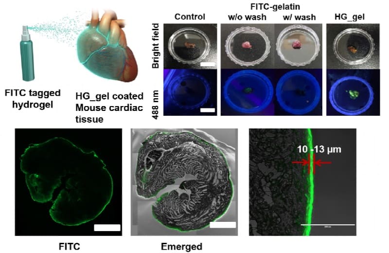
As a proof-of-concept, the researchers also developed a sprayable aerosol system employing the same solution as for the injection. The system effectively coated glass surfaces and recovered its properties in wet conditions, as seen in the injectable system. Taking a step further, they showed that the aerosol could coat a mouse heart, creating a stable film.

A potential new surgical tool

The authors have developed a tissue adhesive based on a novel enzymatic crosslinking system. The technology can be employed as a sprayable or injection-based system, which provides even more potential for further applications. In addition, due to its good compatibility with cells and after injection, the adhesive also shows potential as a system to carry and deliver cells for cell therapy strategies. All of these properties show promise for creating a future surgical tool for tissue engineering and regenerative medicine.


Polymer nanofibre could help improve solar cells

Materials that can transport excitons (photo-generated electron-hole pairs) over distances of more than 100 nm are useful for a range of light-harvesting and optoelectronics devices, including solar cells and photodetectors. 100 nm and beyond is the optical absorption depth – a measure of how deep light can penetrate into a material. Such materials are difficult to make, but researchers in the UK say they have now developed polymer-based semiconducting nanofibres in which excitons can move extremely quickly and travel over distances as long as 200 nm.

“The ability to control the assembly of these relatively large and remarkably well-ordered nanofibre structures is really exciting and opens up a whole new landscape of possibilities in light-harvesting,” says Richard Friend of the Cavendish Laboratory at Cambridge University, who led this research effort together with George Whittell and Ian Manners at Bristol University.

Good contacts

The researchers made their nanofibres from a core of well-ordered short chains of an organic semiconductor polymer (crystalline poly(di-n-hexylfluorene), or PDHF). “The chains stack to create very good contacts between them. These contacts allow the excitons to move extremely quickly and travel over distances as large as 200 nm,” according to our measurements,” explains Friend. “The important point here is that 200 nm is thick enough to absorb all incident light, so we can envisage using these materials as light harvesters for solar cells and photodetectors,” he tells Physics World.

The nanofibres also contain a solvated, segmented corona consisting of polyethylene glycol (PEG) in the centre and quarternized polythiophene (QPT) at the ends. The excitons transfer with a diffusion coefficient as large as 0.5 cm2/s from the core to the lower-energy polythiophene corona in the end blocks.

A “real game-changer”

“We have been using polymer crystallization from solution as a method to make nanoparticles with complex structures and controlled size and shape for the past 12 years now,” add Whittell and Manners. “The fact that this same technique also allows us to produce particles with enhanced optoelectronic properties, compared to the same polymers processed in a different way, is a real game-changer for the field.”

Spurred on by their new result, the researchers say they are now planning to use their technique to prepare structures thicker than the optical absorption depth. “We would like to find out whether the large exciton diffusion lengths we observed in our present study can be preserved,” explains Friend. “The next step would be to construct bilayer junctions that may function as structurally simple but very efficient solar cell devices. We are also looking to prepare more complex structures, which will allow us to harvest the energy from light to promote chemical reactions.”

Full details of the research are reported in Science.

Reducing the contact resistance in 2D semiconducting transistors

2D transition-metal dichalcogenides (TMDs) such as molybdenum disulphide (MoS2) could be ideal for making next-generation optoelectronics devices. Those made from the materials so far suffer from low ON-currents, however, because of the large contact resistance between the metal electrodes in the devices and the TMDs. Researchers in the US, Korea and Singapore have now overcome this problem and have made high-performance field-effect transistors from monolayer MoSfilms containing graphene bottom gates that are one atom thick.

The devices boast a low contact resistance of just 2.3 kΩ·μm between the MoSand electrodes made from nickel, have excellent switching characteristics and a high channel conductance (of more than 100 μS/μm). They also have short channel lengths and a fully transparent channel region which means that they might be useful for various optoelectronics applications such as biosensors, photodetectors, and display backplanes.

Transition-metal dichalcogenides

MoSis a single-layer material and belongs to the family of TMDs. These are quasi-two-dimensional materials with the chemical formula MX2, consisting of an atomic plane of a transition metal M (Ti, Nb, Mo, Re) sandwiched between the atomic planes of a chalcogen X (S, Se or Te). TMDs go from being indirect bandgap semiconductors in the bulk to direct bandgap semiconductors when scaled down to monolayer thickness. These monolayers efficiently absorb and emit light and so might find use in a variety of electronics and optoelectronics device applications.

There is a problem, however, in that the ON-current in transistors fabricated from 2D semiconductors thus far has been considerably lower than that of silicon devices. This is because it is difficult to reduce both the contact resistance and channel length in devices made from atomically thin materials.

Cleaner and flatter graphene surface

The new MoS2-based FET was made by James Hone of Columbia University in New York and colleagues at the US Army Research Laboratory in Maryland, Nanjing University in China, Sungkyunkwan University in Suwon, Korea, and Nanyang Technological University in Singapore. It consists of a monolayer MoS2 channel contacted by nickel/gold metal electrodes atop a monolayer graphene back-gate with a high-k dielectric layer.

The graphene was grown by chemical vapour deposition on copper foil and then electrochemically delaminated onto an oxidized silicon substrate. The delamination step is quicker and cheaper than conventional wet-etching transfer techniques and has the added advantage of producing a substrate that contains fewer impurities when it is adhered to graphene, thus making for a cleaner and flatter graphene surface.

The single-atom thick graphene also shrinks the height of the gate electrode and its smooth surface is ideal for growing ultrathin high-quality dielectric layers. The graphene gate can then be used to highly dope the monolayer MoS2, to a carrier density of 4.6 x 1013cm-2. This allows the researchers to control the electrostatic properties of the contact region between the MoSand the nickel electrode, which is how they can reduce the contact resistance down to just 2.3 kΩ·μm in this region.

Short channel lengths

Hone and colleagues made devices with channel lengths as short as 50 and 14 nm with a 5-nm-sized gate dielectric that have excellent switching characteristics – including a near-ideal subthreshold slope of 64 millivolts per decade, low threshold voltage (of around 0.5 V) and a high channel conductance (of more than 100 μS/μm). The fact that the graphene gate makes the device highly transparent on flexible polymer substrates means that it might be used in a host of flexible optoelectronics applications too.

To summarize, reduced contact resistance improves both the device’s switching characteristics and reduces its ON-state current to levels close to those defined by the International Technology Roadmap for Semiconductors (ITRS), say the researchers. “Thanks to the enhanced electrostatic gating effect, we found no significant degradation in the device’s performance upon scaling the channel length down to 14 nm. Our work thus provides a chemical-free, energy-friendly, and scalable method of achieving high doping levels of 2D materials, which may benefit device fabrication.”

Full details of the research are reported in Nano Letters.

Physicists propose space mission to test quantum gravity

A global team of researchers is planning to use the International Space Station (ISS) to test the fundamental nature of quantum mechanics. The Space QUEST proposal aims to investigate, for the first time, whether gravity can affect a quantum state of light over large distances by firing entangled pairs of photons from a ground station to the ISS. If the European Space Agency (ESA) gives the proposal the green light, the experiment could begin by the early 2020s.

The work is based on a theory, developed by Timothy Ralph from the University of Queensland and colleagues, based on the notion that quantum states should behave differently than classical counterparts under the influence of gravity. “We have tested quantum mechanics and general relativity separately to incredible precision, but they are so fundamentally different that it is hard to reconcile the two in a theory of quantum gravity,” says Siddarth Joshi from the University of Bristol who is part of the team. “This theory is one of a few that are actually testable with current technology.”

This is a measure of how excited and interested the community is in this proposal

Siddarth Joshi

The experiment, which is described in the New Journal of Physics, involves creating entangled photons at a ground station and sending them to the ISS where they are detected. The ground station will first send photons that are not part of an entangled state before quickly sending up photons that are. If there is an effect due to gravity on the entangled pair, they should arrive at a random time. Ralph’s theory predicts that there will be a change in the arrival time of approximately a few percent of the photons, with Siddarth adding that the experiment should be sensitive such changes.

Going through the phases 

The new experiment has its roots in a proposal sent to ESA in 2008 to use the ISS to test quantum com­munication. The idea was to use the ISS to generate entangled pairs that would then be sent to two ground sta­tions across the globe. Studying the effects of gravity on these entangled states was meant to be a side pro­ject to that mission. But when the proposal went through initial study rounds, reviewers felt that this “sec­ondary objective” was much more compelling, which resulted in the collaboration revising the scope of the mission.

To reduce the new mission’s cost, the complexity of the experiment will be in the ground station. It is planned that the ISS would host four single-photon detectors at varying degrees of polarization – vertical, horizontal, +45° and –45°. The ground station will be used to generate the single photons using a faint pulsed source. Siddarth says that while one ground station would be enough, more would be better, and while the location for the first ground station has not been agreed, it will likely be at La Palma or Tenerife.

ESA proposals usually undergo a Phase 0 study to show that a mission is feasible followed by Phase A and then B, which involve a cost evalu­ation and prototype development, respectively. Phase 0 was completed recently, but ESA then decided to join Phase A and B together to accel­erate the process. “I think this is a measure of how excited and inter­ested the community is in this pro­posal,” says Siddarth, who expects the Phase AB study to be complete by the end of the year.

Siddarth says that if the experi­ment is successful and it verifies Ralph’s theory, it would be a “mas­sive” result that would mean quan­tum mechanics is even stranger than we realize. However, he says that even a negative result would also be useful as it would place limits on the effect of such gravitational effects on quantum systems, which could be used to rule out some theories.

More tests to come

Siddarth and colleagues’ experiment is not the only one searching for quantum gravity. Another proposal from Sougato Bose and colleagues from University College London, for example, involves entangling a mass with a second identical mass via the gravitational field. To do this, the two masses would first be prepared using two adjacent, identical inter­ferometers. If gravitational fields are truly quantum in nature, the gravitational attraction between the two masses would become entangled once they have left their respective interferometers.

Siddarth says that such set-ups are much more complex and there­fore harder to accurately determine if there is any effect due to gravity and that they cannot be used to test theories similar to those devised by Ralph and colleagues. Indeed, Bose himself admits that the ISS proposal is “one of the simplest experiments one can do” to test quantum fields interacting with gravity adding that most of the technology is ready.

“Certain degradation of the cor­relations between entangled photons is predicted due to general relativity, which will be a very novel effect to check,” says Bose. He is not clear, however, what conclusions about quantum gravity could be drawn from the results. “While experi­ments exploring such regimes may indirectly shed some light on quan­tum gravity, it is perhaps not a priori that clear whether that will be the case or precisely how it will illumi­nate that path,” he adds.

Scintillating sounds of science, carnival physics, Han Solo’s blaster, neutron coffee

What does physics sound like? To answer that question, Colin Hunter of Canada’s Perimeter Institute for Theoretical Physics has put together a collection of sounds associated with physics experiments and theory. Not surprisingly, the collection includes the now-famous “chirp” of gravitational waves produced when two black holes become one (see above video). Also in Hunter’s collection are the bird-like song of electrons whizzing through Earth’s van Allen radiation belt and audio simulations that highlight the differences between two theories of gravity

A carnival procession is one event where you are guaranteed to hear a cacophony as different bands march past. The physicist Michael Schreckenberg at the University of Duisberg and colleagues are big fans of the Cologne Rose Monday parade and have even written a paper about its dynamics. Using GPS data from marchers, they worked out that people at the end of the parade moved along the route much faster than those in the lead – something that goes against conventional wisdom about how traffic jams form. They explain why in a paper in EPL: “Traffic dynamics of carnival processions”.

Real-life blasters

Before you watch Solo – the latest instalment of the Star Wars saga – you might want to read “Versions of Han Solo’s blaster exist”, the latest attempt to get people interested in physics by appealing to their love of Star Wars. Written by Martin Archer, who is a space plasma physicist at Queen Mary University of London, the article explains how the “blasters” ubiquitous in Star Wars films could be firing blobs of plasma – and how similar systems have been created here on Earth.

I bet Han Solo gets revved up for a day of adventures with a double shot of espresso. Physicists at the Paul Scherrer Institute in Switzerland have used neutrons to make a fantastic movie of coffee being made in a mokka.

OPERA’s final act features five new tau neutrinos

Five more tau neutrinos have been uncovered in a fresh analysis of data taken by the OPERA neutrino detector in Italy, bringing the total number of detections to 10. The analysis establishes the “oscillation”, or changing, of muon neutrinos into tau neutrinos at a statistical significance of 6.1σ. This is well above the level normally required for a discovery in particle physics (5σ). It is also much better than OPERA’s previous result of 5.1σ, which was published in 2015 based on the detection of five tau neutrinos.

Located deep within a mountain at the Gran Sasso National Laboratory in Italy, OPERA ran between 2008 and 2012. Researchers there studied  a beam of muon neutrinos that had been created at CERN in Switzerland before travelling 730 km under the Alps to arrive at Gran Sasso. Along the way, some of these neutrinos changed their flavour and became tau neutrinos. This neutrino oscillation indicates that neutrinos have mass, which is not predicted by the Standard Model of particle physics. Gaining a better understanding of the properties of neutrinos could therefore provide an answer to the mystery of why there is so much more matter than antimatter in the universe.

Writing in Physical Review Letters, OPERA physicists describe how they have used their new result to calculate the absolute value of the square of the mass difference between the two neutrino mass eigenstates (m2 and m3). This is the first such calculation based on measurements of the appearance of tau neutrons and agrees with the mass difference calculated from experiments that measure the disappearance of muon neutrinos.

Completely new strategy

“We have analysed everything with a completely new strategy, taking into account the peculiar features of the events,” explains Giovanni De Lellis spokesperson for the OPERA collaboration. Based at the University of Naples, De Lellis adds: “We also report the first direct observation of the tau neutrino lepton number, the parameter that discriminates neutrinos from their antimatter counterpart, antineutrinos.” He also points out that OPERA’s performance has exceeded the collaboration’s expectations.

The collaboration has also made its data available to the public on the CERN Open Data Portal. This is the first time that data not associated with the Large Hadron Collider has been made public in this way.

OPERA hit the headlines in 2011 when it appeared that neutrinos were travelling from CERN to Gran Sasso faster than the speed of light. If correct, superluminal neutrinos would have been the discovery of the century – however, the anomalous measurement turned out to be the result of previously-unknown problems with the detector. In the audio interview below, the former OPERA spokesperson Antonio Ereditato explains how the experiment works and reflects on the how the collaboration dealt with the superluminal result.

Defending scientific integrity

Self-healing composite could make resilient robot skin

A new metal-elastomer composite automatically self-heals by creating new electrical connections that bypass damaged areas. The material, which repairs itself in a process that is somewhat analogous to plasticity in nervous tissue, might be used to make stretchable circuits, soft machines, bio-inspired robots and wearable electronics that won’t suddenly fail when mechanically damaged.

“I’ve always been fascinated by neuroplasticity and the way that neurons can form new synaptic networks to bypass diseased or damaged tissue,” says Carmel Majidi from Carnegie Mellon University in the US, who led this research effort. “While we haven’t fully mimicked neuroplasticity here, this process was a helpful source of inspiration for developing and understanding our material.”

Soft and deformable circuits

Researchers have made great strides in developing soft and deformable circuits, mainly from soft conductive polymers, conductive fluids in gels and soft microfluidic channels. These materials can be used to make highly flexible, stretchable and conformable electronics that work as artificial skin and nervous tissues in a wide range of innovative applications, from second-skin wearable computers to bio-inspired robots. However, the problem is that these soft elastic structures can easily tear, puncture or mechanically fail, which ultimately means loss in electrical conductivity.

The new material is made of a soft silicon rubber embedded with micron-sized droplets of a gallium-indium based metal alloy that is liquid at room temperature. “To trace circuits in this structure, we use a pen plotter that ruptures the droplets and makes them coalesce into electrically-conductive lines along the pen path,” explains Majidi. “Since the material is mechanically compliant, the resulting circuit is also highly stretchable and as soft as natural skin.

New conductive pathways

“When a circuit trace is damaged, the liquid droplets around the damaged area rupture and form new conductive pathways to bypass the damaged area and re-route electrical signals without interruption. The new pathways form spontaneously on their own and allow the circuit to remain functional. Such electrical ‘self-healing’ is especially important for circuits that are soft and stretchable since, like natural biological tissue, they need to be resilient to cuts, punctures and bruising loads.”

The material could be useful for robust circuit wiring in stretchable textiles and inflatable structures, Majidi tells Physics World. Some examples include sensor-containing garments for wearable computing, or even inflatable houses and airships that are integrated with damage-resistant electrical wiring.

Towards artificial nervous tissue

“Looking longer-term, we believe it might be used as artificial nervous tissue for soft machines and robots that mimic soft natural organisms able to physically interact with humans in a safe way. As with natural nervous tissue, the self-healing circuitry would be resilient to damage and allow the robot to withstand extreme loads and real-world conditions.”

Indeed, the researchers say they have already demonstrated the unprecedented robustness of their material in a self-repairing digital counter and a self-healing soft robotic quadruped that continues to function even after being damaged quite seriously.

The Carnegie Mellon team now plans to engineer and study soft conductive materials capable of both spontaneous electrical and mechanical self-healing. “While electrical self-healing is an important property, our material cannot repair itself mechanically after damage,” says Majidi. “A structure that can do both would bring us even closer to fabricating machines and electronics that are as extraordinarily resilient as natural biological tissue.”

The electrically self-healing liquid metal-elastomer composite is detailed in Nature Materials.


Ingestible device could help diagnose disease

MIT researchers have built an ingestible sensor equipped with genetically engineered bacteria that can diagnose bleeding in the stomach or other gastrointestinal (GI) problems. The prototype device, dubbed IMBED (ingestible micro-bio-electronic device), could eventually be rendered small enough for a human patient to ingest, enabling physicians to better manage or diagnose a range of gut-related diseases (Science 360 915).

The IMBED features bacterial cells designed to sense disease biomarkers, placed in individual wells covered by a semipermeable membrane. When target molecules diffuse across the membrane they activate the bacteria, causing them to generate light. This light is detected by photodetectors underneath each well, and the resulting electrical signals are transmitted wirelessly to an external radio or mobile phone.

"By combining engineered biological sensors together with low-power wireless electronics, we can detect biological signals in the body in near real-time, enabling new diagnostic capabilities for human health applications," said MIT's Timothy Lu.

As an initial proof-of-concept, the team developed a version of IMBED that detects bleeding in the GI tract. They engineered a probiotic strain of E. coli to express a genetic circuit that causes the bacteria to emit light when they encounter haem, and placed the bacteria into four wells on the sensor.

The sensor, which is a cylinder about 3.8 cm long, requires about 13 µW of power, enabling a 2.7 V battery to power the device for about 1.5 months. It could also be powered by a voltaic cell sustained by acidic fluids in the stomach, using technology that the team previously developed.

The researchers tested the IMBED in pigs and showed that it could correctly determine whether any blood was present in the stomach. They anticipate that this type of sensor could be deployed for one-time use or remain the digestive tract for several days or weeks, sending continuous signals. To help move toward patient use, the researchers plan to reduce the sensor size and to study how long the bacteria cells can survive in the digestive tract.

In addition to blood sensing, the researchers adapted the IMBEDs to sense two other molecules: thiosulfate and acyl-homoserine lactone (AHL). Thiosulfate is linked to inflammation and could be used to monitor patients with Crohn's disease, for example, while AHL can serve as a marker for gastrointestinal infections.

"Most of the work we did in the paper was related to blood, but conceivably you could engineer bacteria to sense anything and produce light in response to that," explained co-lead author Mark Mimee. "Anyone who is trying to engineer bacteria to sense a molecule related to disease could slot it into one of those wells, and it would be ready to go."

The sensors could also be designed to carry multiple strains of bacteria, allowing them to diagnose a variety of conditions. "Right now, we have four detection sites, but if you could extend it to 16 or 256, then you could have multiple different types of cells and be able to read them all out in parallel," said co-lead author Phillip Nadeau.

Less is more for conducting graphene composites

Gold nanoparticles grown in situ on graphene flakes

Polymers have many attractive features but traditionally conductivity isn’t one of them. Now a lot of work is going into making polymers that are conductive to exploit their mechanical flexibility and plasticity in electronic devices.

“We decided to work on graphene – it’s a natural material and it can really change many physical properties of plastics – mechanical , electrical and thermal,” says Athanassia Athanassiou, senior researcher responsible for the Smart Materials Group of the Istituto Italiano di Tecnologia (IIT) in Genoa. Athanassiou and her group are not alone in turning to the all-carbon conducting wonder material to bring smart properties to plain polymers, but it turns out that with graphene you can have too much of a good thing.

“The point is if you want to work with graphene you need to overload the matrix,” Athanassiou tells Physics World Materials, as she explains how anything short of overloading the matrix leaves you with a composite that is little better at electrical conduction than the original polymer. “But if you overload the graphene to make the polymer conductive then you might get to loadings that counteract the mechanical properties – making the matrix brittle.” In fact while graphene can enhance plasticity at specific loadings, going past that can have the opposite effect on the mechanical properties.

Going for gold

The solution the IIT team worked on was to supplement the graphene-polymer composite with gold nanoparticles. Again they were not the first to try this either but where their approach had the magic touch was by growing the gold nanoparticles in situ.

“Graphene is not just electrical but thermally conducting,” explains Athannasiou. This means that when they add a precursor of gold to the polymer and graphene flakes and heat it, the gold nanoparticles grow directly on the graphene flakes. While electrons cannot move through pure polymer or a polymer composite at such low loadings of graphene or gold nanoparticles alone, the in situ grown nanoparticles surround the graphene flake, making conduction possible.

The in situ growth also prevents clustering, which can be a real problem when trying to enhance polymer properties with gold nanoparticles. Graphene flakes are also increasingly prone to clustering the higher the concentration used. With the in situ  grown gold nanoparticles the concentration was just 1 wt% - as opposed to the 3 wt% or more usually needed - so the graphene flakes do not cluster either.

As well as preserving the mechanical properties and avoiding clustering, there are cost savings associated with using less graphene as it is still an expensive material. “We really hoped it would work like that and somehow we got the experiment just right - usually that doesn’t happen,” says Athanassiou.

The future of nanoparticle enhancements in situ  

Athanassiou points out that there may be useful enhancements to the thermal properties too that they have not yet checked, since thermal and electrical properties often go hand in hand. Other future work will focus on improving the method and materials that reap the enhanced effects.

The researchers worked with PMMA a common polymer that is widely used and well known. Next they plan to develop an extrusion method for producing graphene PMMA composites with in situ grown gold nanoparticles. Solvent processes require a lot of protective equipment for workers so for industry extrusion processes that produce composites from pellets are preferred. They are also looking at whether different metal nanoparticles and polymer matrices may give improved results, as well as the effect of using graphene or carbon nanotubes for the carbon additive, or a mixture of both. For future work they are very focused on the use of biodegradable bioplastics.

Full details are reported in Nano Futures.

Copyright © 2026 by IOP Publishing Ltd and individual contributors