Take a teacher and a pupil

The dialogue as a method of discourse on philosophical matters has fallen out of favour these days, but that’s a recent development. As Frank Wilczek points out in his introduction to physicist Clifford Johnson’s The Dialogues: Conversations About the Nature of the Universe, the format was preferred by Plato (who often included his teacher Socrates as a protagonist), and was also chosen by Galileo for his epoch-making Dialogue Concerning the Two Chief World Systems. Although Galileo’s tract was a self-conscious post-Renaissance echo of the classical form, framing a rational explanation of the world as a discussion between an experienced teacher and an eager but naïve pupil has been popular throughout the history of Western thought. English philosopher Adelard of Bath used it for his Questiones Naturales in the 12th century, one of the books that belies the myth of an irrational Middle Ages. Jane Marcet’s 1805 book, Conversations in Chemistry, in which a teacher-governess instructs her two female pupils, not only challenged the notion that science was a male pursuit but also proved a pivotal influence on the young Michael Faraday, who never forgot the debt.

The appeal of the dialogue form is easy to appreciate. It allows multiple points of view to be explored and interrogated. This is the approach Galileo championed to debate the merits of the Ptolemaic/Aristotelian and Copernican world views, although his bias is painfully clear – Simplicio, who defends the Ptolemaic model with the blinkered mulishness that his name implies, was suspected by Galileo’s opponents of being a lampoon on Pope Urban. And the pupil, whether in Adelard or Marcet, represents the reader, expressing confusion, incredulity or enthusiasm in the face of the teacher’s calm authority.

The ancient and the modern formats turn out to be made for each other

In The Dialogues, Johnson demonstrates how useful and versatile this novice/expert structure can be in conveying complex scientific knowledge, in the form of 11 conversations, on topics ranging from inflation and relativity to simple discussions of experimentation and geometry. But he reinvents the dialogue for our times as a graphic novel. It’s an inspired choice, for the ancient and the modern formats turn out to be made for each other. Not only does the question and answer structure lend itself to dramatization, but the interaction comes alive when the characters are situated in time and space. In the short narratives of Johnson’s book, they meet at a costume party in a natural history museum, or they are siblings at home working out how to conduct a simple experiment to find the answer to a question, or they are two scientific colleagues chatting over a coffee, and so on.

Using the comic-book approach to talk about science isn’t new in itself. Mathematician and illustrator Larry Gonick was one of the first to bring the cartoon to science – most famously in his multi-volume The Cartoon History of the Universe, begun in the late 1970s, and also in his regular “Science Classics” strip for Discover magazine. Sydney Padua’s The Thrilling Adventures of Lovelace and Babbage, first conceived in 2009, offered a hugely popular, knockabout version of the “invention of the computer” by Ada Lovelace and Charles Babbage in the 19th century. These and other explorations of scientific themes in comic form were, however, more humour-driven than Johnson’s book, which aims for something more sober and adult-oriented in the tradition of the “mature” graphic novel, the genesis of which is often traced to Art Spiegelman’s Maus (1980–1991), a story of the Holocaust.

Johnson says he was also inspired to use this approach to science popularization by the realization that it is perfectly suited to the material. While mostly his stories adhere to a realist pictorial style, from time to time they mutate into the diagrammatic. In The Dialogues, characters assemble giant pieces of a jigsaw puzzle bearing the fundamental equations of physical law: we segue from a sidewalk to a vista of the deep universe; a character unfolds a protoplanetary disc from between her outstretched hands. This is perhaps the most successful feature of the project, and it makes the traditional illustrations of popular-science books look rather dry and creaky.

The visual innovations are all the more impressive given that Johnson, a specialist on string theory and general relativity at the University of Southern California, taught himself illustration from scratch. It’s easy to discern his passion for the classic comic-book tradition in his use of cinematic perspectives – the close-ups, panning shots and unusual viewing angles – which no doubt also benefitted from Johnson’s experiences as a scientific consultant for Marvel movies such as Thor: Ragnarok. In this regard, the book is just the latest instalment of Johnson’s extensive outreach activities in physics.

Occasionally the ambition of The Dialogues somewhat exceeds the delivery. Even the “naïve” characters tend to have a rather firmer grasp of physics than the average general reader is likely to, and while a readiness to delve into actual equations is a refreshing departure from the orthodoxy of science popularization, it may well prove a step too far once characters start saying things like “So we write their field as Ψ, and working with A and V allows us to write an equation describing how electrons interact with E and B fields”. As that quote suggests, the speech is also rather stilted at times, the visual friendliness not always quite managing to compensate for a lecture-like tone.

But this is, after all, an experiment, with room still for refinement. The Dialogues presents a new and exciting way to communicate sophisticated ideas, full of potential. I can’t help fantasizing now about seeing papers in Physical Review written this way – a thought that will no doubt horrify some, just as the first serious graphic novels horrified folks convinced that the only “true” literature can be words on paper.

  • 2017 MIT Press 246pp £24hb

4D smart scaffolds for tissue engineering

While 3D bioprinting is widely used to construct complex biocompatible structures, researchers are now attempting to extend the technique into the fourth dimension. Here, 3D printed objects can be made to “self-transform” over time, which means that they can take on different forms or functions when exposed to physical stimuli such as osmotic pressure, heat, current UV light or other energy sources.

Researchers in the US have now shown that they can fabricated 4D hierarchical micropatterns by exploiting natural soybean oil as the bioink material. The work, which is published in Biofabrication, describes how the micropatterns can be used as biocompatible, shape-changing scaffolds for use in tissue engineering and regeneration applications (Biofabrication 10 035007).

“We used smart natural lipids, that is, soybean epoxidized acrylate (SOEA), as an ink material to fabricate biocompatible, topographical and 4D dynamic shape-changing tissue scaffolds,” says team leader Lijie Grace Zhang of The George Washington University in the US.

Such 4D materials have already been demonstrated in other fields. One of the best-known examples are shape-memory alloys, in which a change in temperature triggers a change in shape of the alloy. Although still in its infancy for tissue engineering applications, 4D printing may ultimately allow for tissue transplants that better mimic biological tissues and organs, which are highly organized and contain structurally anisotropic components.

This structural organization affects the way the cells arrange themselves in a biological structure, since they respond to “cues” that depend on the biostructure’s surface topography. Although researchers have developed many techniques to manufacture nano- and microstructures that can regulate the behaviour of cells, it is still difficult to fabricate biomimetic tissue scaffolds that respond to topographical cues in the same way as their natural counterparts.

Complex surface micropatterns

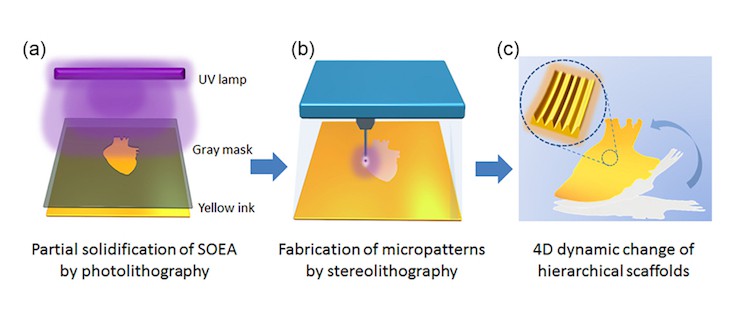
What’s needed, says Zhang, is a technique that generates surface structures on a macro-architecture. “The technique we employed, photolithographic-stereolithographic-tandem strategy (PSTS), relies on sequentially treating the same ink feedstock with photolithography and stereolithography,” she says.

The photolithographic-stereolithographic-tandem process

The team used SOEA as the ink material, which is derived from soybean oil – a natural, renewable source that has been attracting much attention as a biomaterial in recent years. “We were able to make a 10 micron-thick liquid SOEA film in a matter of seconds using the photolithography step,” says Zhang. ”This film then further solidifies after stereolithography to generate complex surface micropatterns, thanks to a layer-by-layer process.”

Shape-shifting constructs

The fabricated scaffolds can change shape when stimulated by an external trigger, which means that they can better mimic natural biostructures that adapt to their morphology. The subtle surface patterns could also be used to regulate the behaviour of human stem cells, such as human bone-marrow mesenchymal stem cells (hMSC). “The images that we obtained and analysed (using National Institute of Health software) show that hMSCs actively grow and highly align along the micropatterns, forming an uninterrupted cellular sheet,” Zhang told Physics World.

As a proof of concept, the researchers made a 4D patch for regenerating heart cells that showed significant growth of heart tissue – as confirmed by immunofluorescence staining and a technique called qRT-PCR analysis. “This study shows the great potential of these smart patching scaffolds as implantable materials for future tissue and organ regeneration,” says Zhang.

The team, which includes scientists from the University of Maryland in Baltimore, says that it is now planning to 4D bioprint light-sensitive smart biomaterials and reprogrammable architectures for use in heart regeneration and bionanorobot applications.

  • Read our special collection “Frontiers in biofabrication” to learn more about the latest advances in tissue engineering. This article is one of a series of reports highlighting high-impact research published in the IOP Publishing journal Biofabrication.

High-resolution direct laser writing exploits scruffy edges

While not the first to be plagued by fine webbing effects spoiling their laser-written structures, Joel K W Yang’s group have been the first to exploit it to achieve feature sizes below 10 nm. “A lot of interesting physical phenomena such as quantum confinement and plasmonic field enhancements occur at these length scales,” Yang points out.

Yang, an associate professor at the Singapore University of Technology and Design (SUTD), had been focusing his research on high-resolution nanofabrication methods and their applications. Having worked with electron-beam lithography, where he was routinely able to achieve sub-10 nm feature sizes, he turned his attention to direct laser writing (DLW), keen to see how far he could push the resolution he could achieve with this technology.

DLW has the advantages of speed and simplicity over ion- and electron-beam lithography, which require vacuum technology and are slow and fiddly to implement. However, DLW is traditionally limited to feature sizes of hundreds of nanometres, around 10 times bigger than ion- and electron-beam lithography. Smaller feature sizes are possible by modifying the equipment and incorporating stimulated emission depletion lithography, but Yang was interested in pushing the basic method to its limits. So when his student complained about webbing structures that he could not get rid of, Yang suggested, “What if instead of getting rid of them we investigate how to control them.”

Intensity matters

DLW exploits two-photon absorption to create cross-linking molecules from a resin, which then link up where the laser beam “writes” the structure. The remaining resin is then washed away. Yang and his co-workers used a 780 nm wavelength laser. As the resin absorbs light with a wavelength below 400 nm, it will only absorb light at the focus of the beam where the light intensity allows absorption of two lower-energy 780 nm photons.

The problem is that fine filaments still form outside where the laser focus has written. “Above-threshold laser powers can cross-link polymers in the wake of the beam but lower powers lead to lower concentrations of crossilinking molecules in the wake,” says Yang. “We think at low power you need a percolative pathway for cross-linking to happen, so you have regions where the concentration is just high enough.”

To exploit these effects Yang and colleagues used a laser at half power to control the spread of these filaments from the wall of one structure to another. This way they could write 20 nm features with a yield of 80% and with some filaments as narrow as 7 nm only 33 nm apart. The filaments grow with residual tensile strain that pulls the walls down if they are not well anchored to the substrate. However, Yang points out that this strain is actually helpful for making the filaments thinner.

“There’s a game where you pick up drinking straws – you stretch it to make it really thin, and use it to saw one another’s straws to see whose straw would break first. It’s a similar effect here,” says Yang.

Pushing DLW further

With improvements to the chemistry of the resin so that the cross-linked material has greater structural integrity to resist fragmenting, Yang thinks even smaller feature sizes should be possible. The team has already experimented with writing a series of filaments that are vertically displaced so that closer lateral spacing is possible without the features merging. It may also be possible to pattern filaments at off-normal angles to the anchoring structures, although Yang expects the angles may be limited to within 10° of the normal as that is the shortest percolation path.

Yang is still keen to better understand the mechanism of the sub-threshold intensity ultrafine DLW and improve the uniformity of the features. At present the filaments grow thicker towards the adjoining wall, most likely due to thermal effects.

Possible applications include use as a template resist for plasmonic structures and 4D printing, where the strain can act as an actuator for printing dynamic structures defined in space and time.

Full details are reported in Nano Futures, and for more research on sub 10 nm nanofabrication visit the Nanotechnology focus collection.

Inorganic semiconductors can be more flexible in the dark

Despite the useful electronic properties that sparked the semiconductor industry, the brittle mechanical properties of semiconductor materials have become an increasingly limiting factor in developing new applications. Now experiments under cover of complete darkness suggest that semiconductors that appear brittle may be capable of extraordinary plastic deformations when the lights are out.

Yu Oshima, Atsutomo Nakamura, and Katsuyuki Matsunaga at Nagoya University in Japan tested the response of single crystal ZnS samples under applied stress when illuminated with white light, UV and in complete darkness. The electrical and optical properties of ZnS have already found use in luminescent and infrared optical devices as well photocatalysts, and it is readily available in large crystals that are convenient for deformation tests.

Behaviour changes with the lights out

The researchers found that although under white light and UV illumination the structures fractured at strains of just a few per cent, in total darkness they withstood strains of up to 45%. The bandgap of the crystal was also affected by the presence or absence of illumination giving crystals deformed in the dark a more orangey hue.

Oshima, Nakamura and Matsunaga attribute the difference in mechanical properties to the difference in the dislocations induced with and without the presence of light. Illumination can excite electrons into bandgap states at the dislocation edge so that the dislocation is charged and less mobile, inhibiting plastic deformation.

“It is interesting to find out that the inorganic semiconductor can exhibit extraordinary plasticity when it deforms in complete darkness,” they conclude in their report. “This suggests that the mechanical strength and fracture properties in inorganic semiconductors maybe controlled by exposure to light.”

They also highlight the ramifications for materials processing. The behaviour of dislocations plays a critical role in the synthesis and processing of most crystalline materials, including film synthesis and epitaxial crystal growth. The results suggest light exposure may also affect these processes.

Full details are available in Science.

Iron in Earth’s core might be cubic, not hexagonal

A long-standing debate about the structure of solid iron at the centre of the Earth looks set to be reignited following new laboratory tests carried out by scientists in the US. The researchers say their results imply that iron crystals in the Earth’s inner core have a body-centred cubic (BCC) arrangement – in contrast to the hexagonal-close-packed (HCP) structure pointed to by many previous results.

Scientists have good evidence that the exceptionally high pressures that exist in Earth’s inner core dictate that the iron there is solid – in contrast to the molten iron present in the outer core. That evidence comes in the form of data from earthquakes. By plotting the paths of seismic waves through the Earth, seismologists have concluded that shear waves – which cannot propagate in liquids – do not travel through the outer core but do travel through the inner one.

However, for several decades debate has raged about how the atoms in that solid iron are arranged. At room temperature and pressure, iron has a bcc lattice, which means that every atom is surrounded by eight others – four in each of the layers immediately above and below it. But at higher pressures, iron atoms form the slightly tighter hcp structure, such that each atom has 12 neighbours – six in its own layer and three in those above and below it.

Diamond anvils

The situation at very high temperatures and pressures, however, is not well understood. Several experiments using iron heated inside high-pressure diamond anvil cells have shown that here too the structure is hcp. These experiments involve squeezing tiny samples of iron between the tips of diamonds, heating them with a laser beam and at the same time illuminating them with X-rays from a synchrotron source to determine the crystal structure from the X-ray diffraction pattern.

In fact, according to Guoyin Shen of the Carnegie Institution of Washington, most Earth scientists are persuaded that the iron in the inner core does in fact have a hexagonal-shaped lattice. “Within the community,” he says, “many think that it is a done deal – that the structure of iron seems to be hcp.”

Seismic anisotropy

There is a problem, however. Seismologists have established that shock waves from earthquakes travel more quickly through the Earth when they go from pole to pole than when they go along the equator. This “anisotropy” ought to be reflected in the structure of the iron, such that it is significantly more elastic at right angles to its atomic layers than it is parallel to them. In other words, that the spacing, and hence the give, between adjacent layers should be significantly greater than that between neighbouring atoms within the same layer. But theorists have calculated that very high temperatures should flatten hcp iron – making the difference in spacing too small to account for the seismic anisotropy.

In the latest research, Shen and his Carnegie colleagues Ross Hrubiak and Yue Meng show experimentally that the inner core iron might in fact have a bcc structure. They did so after realizing that the diamond cell experiments could be giving misleading results. These “in-situ” measurements provide X-ray diffraction patterns of the iron during the brief period that it is heated by the laser. But because the X-ray beam is not much narrower than the hot spot created by the laser there is a chance, says Shen, that the high-temperature data become “contaminated” by data from regions at lower temperatures.

To try and get round this problem, Shen and co-workers have developed an alternative technique in which they compress samples of iron in a diamond anvil cell but this time take X-ray diffraction images before, during and after each heating pulse. Using the HPCAT beamline at the Argonne National Laboratory’s Advanced Photon Source, they find that at relatively low pressures and temperatures the diffraction patterns are as would be expected from hcp-iron grains.

Two distinct axes

However, once the group raised the pressure above about 10atm and the temperature well above 4000 K it discovered that the crystal grains became oriented along two distinct axes. Because the grains’ orientation might be preserved across phase transitions, the researchers say that this “bi-axially aligned microstructure” is evidence that the iron adopts a bcc structure at high temperatures and then transforms back into hcp when it cools down.

Buoyed by these results, Shen and colleagues then carried out fresh measurements at inner-core like conditions. They realized that, as they and other groups had found previously, many of the spots in the diffraction pattern would be due to hcp-iron. But they predicted that they should also see one spot due to bcc-iron in a particular orientation – and say that they have observed it. “In earlier experiments we considered that diffraction spot as noise,” says Shen, “but now it becomes evidence that supports our view.”

Shen acknowledges that others in the field are likely to be sceptical, given potential doubts about his group’s interpretation of the microstructure data and the fact that it has so far only seen one family of diffraction planes that it can attribute to bcc-iron. He says that he and his colleagues are now focused on trying to improve the in-situ measurements, either by reducing the X-ray spot size or increasing the X-ray energy to boost chances of finding more diffraction information.

The research is reported on the arXiv server.

Learning from the ozone solution

In the May episode of our Physics World Stories podcast, Andrew Glester is in conversation with the Nobel laureate Mario Molina. The Mexican researcher shared the 1995 Nobel Prize in Chemistry for his work on understanding formation and decomposition of ozone in the Earth’s atmosphere. He talks about how winning the award transformed his status as a scientist, giving him a unique platform to influence politicians.

The banning of substances (mainly CFC chemicals) that deplete the ozone under the 1987 Montreal Protocol is hailed as a shining example of coordinated global action in tackling an environmental issue. Molina talks about how industries in the 1980s paid attention to the scientists at a relatively early stage, and sought alternative products and processes to CFCs. He engages with the reasons why it is harder today when dealing with the more multifaceted issue of climate change.

Later in the podcast, Glester picks up the story with Lorraine Whitmarsh, a social scientist at the Tyndall Centre – a network of universities seeking sustainable responses to climate change. Whitmarsh is interested in why the general public responds to the science of climate change in particular ways. She is also interested in practical solutions for shifting to a lower carbon lifestyles and offers her top tips for reducing your carbon impact.

If you enjoyed this podcast then you can subscribe via iTunes or your podcast provider. Also check out Physics World Weekly – our news-focused podcast presented by the Physics World editorial team.

Modified Sierpinski triangle makes a good mid-infrared sensor

Fractals are becoming popular for when it comes to designing microwave and radio-frequency antennas thanks to their “self-similarity” that allows the antenna to better collect and focus broadband, multi-frequency light. Researchers at the National University of Singapore have now made a molecule sensor from a modified Sierpinski triangle that works in the mid-infrared range. The device could be used to profile the fingerprints of various biological elements, such as cells and protein monolayers.

Researchers have recently begun to make use of fractal patterns to manipulate surface plasmons, which are quantized collective oscillations of conduction electrons on the surface of metallic nanostructures that interact strongly with light. Such strong interactions allow the plasmons to concentrate light into subwavelength volumes, well below the diffraction limit of light. The applications are many: sub-diffraction focusing, transparent metallic electrodes, improving photovoltaic efficiency, and enhancing molecular fluorescence, to name but a few.

 Modified Sierpinski fractal model

In their work, researchers Chengkuo Lee and Dihan Hasan in the Department of Electrical and Computer Engineering and the Center for Intelligent Sensors and MEMS at the NUS studied a Sierpinski fractal model made of gold and chromium.

“Our original goal was to investigate the properties of this fractal structure once we had tailored its dimensions for mid-infrared sensing,” explains Hasan. “We then came up with the idea of modifying the model slightly to drastically improve its sensing properties in this spectral range.”

The Sierpinski fractal is an equilateral-triangle-shaped fractal with intrinsic “self-similarity” – that is, its constituent triangles are repeated at smaller scales (or “orders”). Self-similarity in electromagnetic structures is important for miniaturizing the size of a device so that it can focus light at a particular frequency. In our work, we slightly modify the existing fractal model without destroying its self-similarity,” says Hasan.

Bow-tie nanostructures

The Sierpinski fractal is particularly suitable for merging with bow-tie nanostructures, he adds. These structures are among the best at manipulating localized surface plasmon resonances and enhancing light fields the most thanks to a “lightning rod” effect at their sharp tips that allow them to act as nanoantennas. These antennas possess “plasmonic modes” that can be tuned to resonate with the optical transitions in molecules nearby. It is these plasmonic modes that increase the coupling between light emitted by neighbouring molecules and the antenna, and means that they can be used as sensors.

Sierpinski fractals have already been used a key element in many high-performance plasmonics applications, such as nanolithography, ultralow power optical trapping, and plasmonic photography film for high density data storage. The problem is that it is difficult to make a pure Sierpinski fractal because of geometric differences at the junctions of the fractal components. What is more, previously-fabricated fractals did not fully exploit the lightning rod effect of nano-bow-ties.

No need for molecule functionalization

Lee and Hasan have now used advanced electron beam lithography to precisely tune the geometric structure of these fractals. By improving their architecture, the researchers have found that the structures can enhance light fields in the mid-infrared range (3000 to 8000 nm). The way the components are arranged also maximizes the lighting rod effect of the nano-bowtie/fractal patterns.

“Conventional resonant sensing focuses mostly on the visible part of the spectrum,” explains Hasan. “Here, we investigate sensing in the mid-infrared spectral range, which is the range in which many biological molecules absorb light.”

Probing these molecular absorptions does away with the need for functionalizing the biological molecules so that they can be picked up the sensor.

Towards on-chip molecule sensors

“Sensing in the infrared will also allow us to more reliably discriminate sequential events occurring in a biological system,” he adds. “And the multispectral fractal platform we describe will ultimately allow for high-throughput multiplexed detection of various molecules on a single platform. This should improve the signal-to-noise ratio of such imaging at multiple wavelengths of interest.”

The broadband and enhanced light absorption made possible by the modified fractalization is promising for sensing biological molecules at mid-infrared wavelengths, he tells Physics World. “But that is not all: the enhanced light-matter interactions that come about thanks to the increased number of light-absorbing ‘hotspots’ also makes the device more sensitive in the optical range. Here, light could be efficiently converted into heat for on-chip electronic readout, something that will help overcome one of the major limitations of mid-infrared sensors – their bulky size.”

The team, which reports its work in the IOP journal Nano Futures 2 025005, says that is now busy working on integrating the platform with 2D materials to make on-chip molecule sensors. “We are actively studying the thermoelectric properties of various 2D materials to this end,” adds Hasan.

Summit examines challenges of developing AI for cancer care

Over 100 radiologists, clinical oncologists, computer scientists, industry experts and funders came together last week for a pioneering stakeholder summit to discuss the challenges of developing artificial intelligence (AI) in imaging and cancer treatment.

The event, “Grand challenges in artificial intelligence in clinical radiology and clinical oncology”, took place at The Wellcome Collection in London on 16 May. The brainchild of Nicola Strickland, President of The Royal College of Radiologists, the summit was organised in partnership with The Alan Turing Institute, Health Data Research UK (HDR UK) and the Engineering and Physical Sciences Research Council.

The packed programme featured a keynote speech from renowned surgeon Lord Ara Darzi, who called on delegates to urgently collaborate to progress AI in clinical practice, both for the benefit of patients and to maintain the UK as a world leader in innovation after leaving the EU.

HDR UK’s Andrew Morris outlined the potential for national data sharing to realise benefits for patients and improve AI training, stressing the timeliness of the meeting given the UK government’s preparations to fund AI initiatives into general diagnostics and cancer detection.

Recurring themes revolved around how academics, funders and technology companies could and should collaborate to accelerate AI, as well as the ongoing challenge of sourcing large, robust sets of data to train algorithms and machine learning programmes.

Delegates were also introduced to an array of developing projects from universities and industry, including AI algorithms and machine learning that detects lung cancer nodules, models pulmonary hypertension and helps oncologists outline tumours ahead of radiotherapy.

“Our key stakeholder meeting demonstrated that the UK has a real opportunity to take the lead on artificial intelligence programmes in healthcare, if clinicians and researchers are brave enough to embrace its potential and work with industry to shape the application of machine learning in practice,” said Strickland.

“For clinical radiologists and oncologists to survive and thrive in the swelling digital revolution, it is vital these medical specialities work in collaboration with funders, AI experts and industry to develop AI that is robustly tested and regulated so it can be confidently put into practice to augment the work of clinicians and bring greater benefit to patients.”

World Metrology Day: Celebrating an underappreciated science

In an ever-changing world the few constants we can rely on become ever more important. The distance to the nearest high street bank may increase, hem lines may rise and fall, but the metre will always measure the same length. Or will it? With several SI units facing redefinition in 2019, the National Physical Laboratory (NPL), the UK’s national measurement standards laboratory, opened its doors to around 5000 visitors for its biennial open house to shed light on what we mean when we refer to everyday units and the work that goes into defining them.

“Measurement is what makes science science,” said NPL researcher Michael de Podesta, when I asked why metrology matters. No surprise then that scientists have discussed ways of defining internationally recognised units of measurements for centuries. However it was not until 1960 that the International Bureau of Weights and Measures first established the International System of Quantities (SI) formerly the International System of Units: the candela, kilogram, metre, second, Ampere, Kelvin, and the mole. This concise list in fact caters for the wide range of measurands across the physical sciences, as other units in current use can be defined in terms of these seven. For example, an Ohm can be defined as kg m2/s3 A2 from the definition of resistance as power / current2.

There are logical explanations for the original definitions of these units: the metre was 1 ten millionth of the distance between the North Pole and the Equator to the best estimates available at its invention in 1799; the second was 1/60th of 1/60th of 1/24th of a day on account of clock conventions. However these definitions have drifted from their original explanations as technological progress has demanded ever greater precision. “A lot of the SI units have been superceded technologically,” said de Podesta. “As technology advances measurements of the fundamental constants keep changing, which is bonkers, so we want to define the units in terms of these constants.”

Constant units

In 1983 the General Conference on Weights and Measures (CGPM) redefined the fundamental reference for the length of a metre – once set by a metre rule in Sevre just outside of Paris. It is now defined as the distance light will travel in a vacuum in 1/299792458 of a second, and that as far as we know is a fundamental constant. As well as the metre, the second has also been redefined in terms of the universal constant of the speed of light in a vacuum. However, there is a noticeable lag between the fundamental reference for other SI units and the technological capabilities of the present day. As Ian Robinson told attendees at NPL’s Open House, the fundamental reference for the kilogram remains a lump of platinum in Sevre, which is taken out every 50 years, weighed, cleaned and then locked away. As Robinson put it, “Not the best way to define mass in these modern times.”

On 16th November 2018 the CGPM will vote to adopt a series of new definitions for the kilogram, Kelvin,  mole and Ampere. Why has it taken a further 35 years to bring the remaining definitions in line with the metre and the second? In fact the technology to define units in terms of fundamental constants is far from trivial.

Robinson, who joined NPL in 1976, worked with Bryan Kibble shortly after the 1975 invention of the Kibble balance, which will redefine the kilogram. The Kibble balance works by putting a current through a coil and measuring the induced force balanced against the gravitational pull on a kilogram. Kibble’s invention removed some of the inaccuracies of its predecessor the Ampere balance, but as Robinson said it is still “like measuring a bag of sugar in a hurricane”.

De Podesta’s work at NPL focuses on different ways of measuring temperature, and has been integral for redefining the Kelvin. “We use electromagnetics, acoustics, precision weighing – it’s a tour de force that brought together teams across NPL,” he says. Their approach uses an acoustic thermometer. As de Podesta explains, the speed of sound in a gas is exactly related to the temperature of the molecules, so he and his colleagues are actually measuring the speed of sound carried by the molecules of the gas with ultraprecision. However having finally achieved this super accurate thermometer, using it in lab proved a struggle. “It was totally humiliating – you say you’ve made the most accurate temperature and then you can’t take the temperature of something in the lab,” says Podesta. “It turned out that it was all the other thermometers that were wrong.” The corrections are small – 0.004 °C at 30 °C – however there are experiments for length measurements for example, where that level of temperature precision matters.

Quantum metrology

Shrinking device sizes also place new demands on measurement science. At NPL’s single electron device lab they are working on an alternative definition for the Ampere. Their GaAs two-dimensional electron gas experiments pump electrons into a sink that holds just one electron at a time. From there electrons hop across into a transistor device in single file, defining the smallest current possible. For larger currents the Ampere can be defined macroscopically in terms of the voltage in a superconducting Josephson junction and the resistance of the quantum Hall effect. However, as devices shrink ever smaller a definition in terms of a single electron device becomes more and more important.

At this scale terms like temperature take on new ambiguities, as their statistical definitions no longer have meaning. “You can describe the temperature in terms of the probability of an electron occupying a particular energy level,” says Masaya Kataoka. “But if this is set then it has no temperature.” In addition, although these devices are devised to define current, at this scale where discrete charges move one by one continuous classical analogue current does not really exist.

Sunday 20th May was World Metrology Day 2018, a celebration of a science de Podesta feels is underappreciated. “When you see physics on the telly there’s a lot of emphasis on boffins and geniuses and maths – some people think physics is maths and that’s completely wrong.” He adds, “Maths is telling a story but what makes physics a science is when you compare with what actually happens.” The measurements for new standard definitions at NPL are checked across the world, with the National Institute of Science and Technology (NIST) in the US also playing a key role. It is reassuring that whatever may be going on in the rest of life, the international Standard of Quantities is something we can all agree on.

Biodegradable pressure sensor eliminates the need for second surgery

Every year in the USA alone, around 14 million people suffer from tendon, ligament and joint injuries. Many of these injuries require surgery, followed by a rehabilitation period to restore the repair site to its pre-injury function. A diagnostic sensor capable of measuring biomechanical characteristics of the affected tissue in real time would represent a significant step towards improved assessment of a patient’s recovery.

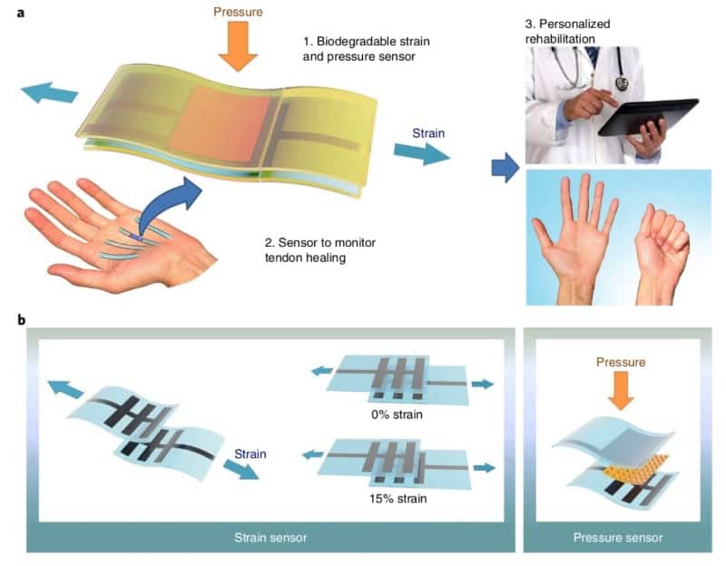
The sensor should, for example, be able to measure typical tendon strains (of less than 10%) after the surgery. However, existing implantable sensors either suffer from inadequate sensing capability or use materials with unreliable biocompatibility. Use of biodegradable materials would avoid the need for second surgery to remove the sensor. With that in mind, researchers from Stanford University devised a biodegradable sensor compatible with living organisms that can satisfy tissue recovery requirements by discriminating strain and pressure stimuli (Nature Electronics 1 314).

The sensor

The device consists of two stacked sensors that can accurately measure strain and pressure independently. For material design, the researchers used two biodegradable elastomers: poly-glycerol sebacate (PGS) and poly-octamethylene maleate citrate (POMaC). Two thin-film comb electrodes are sandwiched between two stretchable elastomer layers and when strain is applied, a change in capacitance is observed as the two electrodes slide relative to each other.

Biodegradable sensor

The pressure signal is monitored with a flexible and highly sensitivity capacitor supported by a thin elastic dielectric layer on both sides. This design allows the pressure sensor to work independently, without inducing any strain signal. The sensor responds in the millisecond range and can discriminate strains as small as 0.4% and pressures as small as 12 Pa (the amount of pressure exerted by a grain of salt).

Biocompatibility study

The team performed an in vivo study, which suggested that the device is compatible with a rat’s body. They tested the sensors by subcutaneously implanting them on the backs of rats. The rats were subjected to strain and pressure stimuli, and afterwards strain signal on tendon was measured. The sensor was observed to be stable for two to three weeks, when compared with a reference non-biodegradable sensor. Testing the sensor in vivo revealed that it was operational inside the body without any side effects and was functional for more than two weeks.

The researchers believe that this sensor – with high-sensitivity, faster response time and biodegradability – can play a valuable role in biomedical applications such as monitoring cardiovascular patches and reconstructive surgery. Sensors transmitting real-time information in vivo will open avenues for refined and personalized medicine where mechanical deformations and pressures can be easily monitored. The research team now plans to develop a biodegradable circuit capable of wirelessly transmitting measured signals through the skin.

Copyright © 2026 by IOP Publishing Ltd and individual contributors