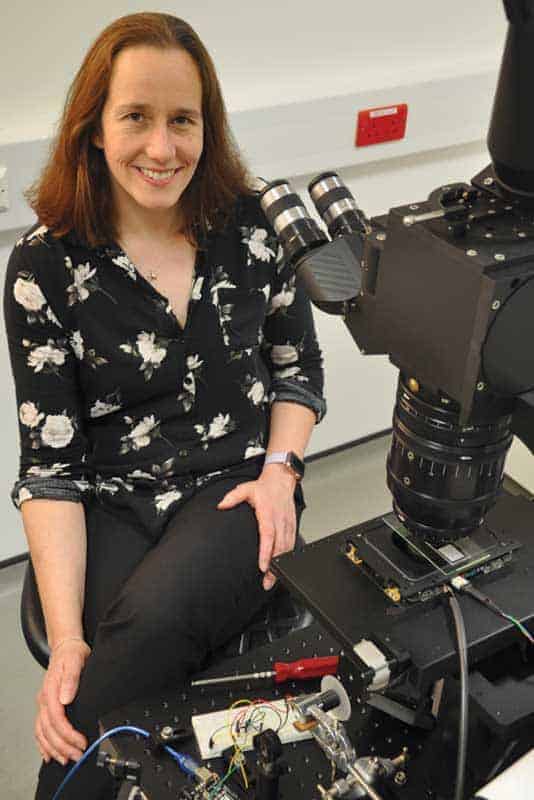
Gail McConnell is an optical physicist at the University of Strathclyde who develops novel optical systems for biomedical imaging. She spoke to Margaret Harris about her recent research and some of the challenges in the field
How did you get into biomedical optics?
After I did my first degree and my PhD in the physics department here at the University of Strathclyde, UK, I had an industry job lined up that had nothing to do with biology whatsoever. But this was around the time of the September 11th terrorist attacks, and the company panicked and rescinded its job offer, leaving me high and dry. At that point my PhD supervisor said, “Well, I’ve got three months’ worth of post-doc salary for work in this newly created ‘Centre for Biophotonics’ – would that interest you?” During my PhD I had spent a lot of time developing lasers using nonlinear optics, and when we wrote papers we’d always include a paragraph mentioning possible biomedical optics applications. But we never actually did any of that future application work, so I thought it might be nice to try.
That three-month post opened my eyes to the possibilities, and then I took a two-year appointment where, as well as engaging in technology development, I was also working with some of the biologists. They would bring their specimen preparations – biological cells and tissue – and I would help them to do some confocal imaging or wide-field epifluorescence imaging. This was a baptism of fire for me because I had never done any biology – I’d never really even done any chemistry, and I had never looked down a microscope until very late in my doctoral studies. But it was also a kind of gateway to another world, and I started to appreciate both what was driving biological and biomedical research, and some of the limitations of commercial imaging technologies. That gave me a sense of how I could use my knowledge in physics to develop better technology for biomedical imaging.
Although I come from a laser development background, as time has progressed I’ve started to appreciate that the laser is potentially just one component within an optical imaging system, and there are other technologies that also need to be developed. This could be optical technologies, addressing questions such as “Can we redesign the microscope objective lens to give more information about the specimen?” But there are also questions like “How can we prepare the biological tissue or specimen in a way that gives more meaningful or revealing information about biological function?” As a consequence, in the last five years we have really developed into more of a biophysics group rather than a physics group who happen to be working in the life sciences.
What are some of the major challenges imaging biological samples?
That very much depends on the biological question that you are trying to answer. This is an extreme example, but behavioural scientists working at the border between biology and neuroscience are interested in animal behaviour, perhaps in response to a reward or some pharmacological intervention. What they need is a video or camera-based system that can track the movement of the whole animal, and that would obviously require quite a different type of optical imaging solution from the type of systems we develop, which are more about understanding sub-cellular activity or structural morphology within individual cells. Biology and biomedical challenges are so diverse that it’s not a case where one solution fits all. That’s why physicists are constantly developing new types of imaging systems. As biologists are using more advanced instruments they are learning more, and as soon as they reach a technological threshold they want to image deeper, they want to collect images more rapidly, they want to see more spatial detail and they want to use the latest fluorophores and photoproteins. At the same time, increases in computational power are making storage and analysis of big data sets possible – it’s a cyclical process that drives the technology development.
Could you give us an example of how this has worked in one of your projects?
The development of the so-called “Mesolens” is a good example. This project was initiated in the mid-1980s by Brad Amos, one of the developers of the point-scanning confocal microscope. Although this instrument can produce 3D images of cells with sub-cellular resolution, and without needing to section the tissue mechanically, he noticed that whenever biologists gave him a large volume of tissue, they were inevitably disappointed with the resolution. So he set about trying to find an optical solution to this problem – how could we accommodate large volumes of tissue without compromising the spatial resolution? I started working on the project around 2009–2010. A prototype of what we now call the Mesolens had been funded by the Medical Research Council (MRC), and Brad had done some preliminary tests, but he was approaching retirement and the MRC rules are that when you retire, you no longer have lab space. After some negotiations, we were able to move the whole project to Strathclyde, and since then we have developed a confocal version of the Mesolens that makes it possible to image up to 120 mm3 of tissue with sub-cellular detail throughout.
This has taken us in directions that we never expected because it’s such a departure from a standard confocal microscope. Initially we thought that the Mouse Atlas community (which is developing a 3D computer model of successive stages of mouse embryo development) would be a key user, and that drove some of the lens design parameters. For example, a mouse embryo at day 12.5 is around 6 mm long and that is why the Mesolens has a 6 mm image field. But while the Mouse Atlas community is engaging with us, we are also seeing applications emerging that we did not expect.
I understand there’s now a Mesolens spin-out company.
Yes, but I have made a conscious decision to not become a part of the company at this time. This is partly because I don’t want to compromise the prospect of academic funding routes, but mostly because I want to focus on the scientific development rather than get diverted into financial management and other technical aspects of the business. I’m a bit of a card-carrying academic, and because my basic training is in physics, I also have a remaining curiosity around what new physics can emerge while we are studying these biomedical processes.
What’s the most frustrating problem in developing the Mesolens?
One of the difficulties we had to overcome was mirror jitter, which wasn’t something we expected to be a show-stopper.
If you take an image with your mobile phone you have maybe 10 megapixels, but for full Nyquist sampling – using the Mesolens to obtain all the available information – we need a 400 megapixel camera. At the moment we have the next best thing, which is a 260 megapixel effective camera where we obtain images by moving the sensor within the body of the camera. But what we really want is to scan a laser beam across 20,000 by 20,000 pixels, and that means we need to accurately sweep the laser spot across many points within our 6 mm image field. At Photonics West we met a company called Nutfield Technology that sells scanning mirrors to the digital video industry (particularly Pixar) so they can project digital images onto a large cinema screen while ensuring there are no gaps.
We were able to work with them and now we have mirrors that can scan our laser spot as accurately as we need to get reproducible data.
Where do you see the field going in the future?
Biomedical research is generating vast data sets. We have excellent microscopes and the biologists are desperately keen to use them to see ever more detail. This might mean using super-resolution techniques to resolve very fine spatial detail, or making very fast recordings of physiological behaviour or intact organs. Either way, it inevitably leads to giant data volumes. So we can generate the data, but are we adequately placed to analyse it? Can we retrieve meaningful measurements and perform appropriate statistical analysis of these data sets that we can now generate? I see a great drive towards every biomedical imaging centre needing a computational biology expert around to help unpack these data volumes so they can provide the biomedical research community with the information that they actually want, rather than what the physicists think they want.
In some ways this is a “luxury” problem because we have these fabulous new tools, and we just need to make sure we are using them to the best of our ability. For example, we were using the Mesolens to image some mouse lung tissue recently, and that generated a 200 GB data file. We can reconstruct that dataset in 3D, but if, for example, we want to count the number of cells within the tissue, that takes quite a lot of computational power. We are starting to become smarter as a community about using computational methods to retrieve meaningful information from these very large datasets, but the datasets are only going to get bigger. I also think that biologists are becoming more keen on quantitative information. Rather than just generating a pretty image, they now want numerical values, and they want to make measurements from within their datasets. This is great for physicists. We can help, through collaboration and interdisciplinary research, to push it along.