
Positron emission tomography (PET) with the radiotracer 18F-FDG provides an important tool for assessing the health of the heart muscle in patients with ischemic heart disease, in which narrowed coronary arteries reduce the heart’s blood supply. Such PET scans help identify the level of damage to the heart muscle and play an important role in clinical decision making.
Current guidelines recommend injecting a 200–350 MBq dose of 18F-FDG. But lowering this tracer dose will decrease the patient’s radiation exposure – an essential goal of any diagnostic procedure – as well as reducing imaging costs and potentially opening up new applications. The downside, however, is that a lower tracer dose may lead to poorer quality images, thereby reducing diagnostic accuracy.
One approach proposed to address this problem is to employ artificial intelligence algorithms to restore image quality. Researchers from Rigshospitalet in Denmark have now investigated the use of deep learning to reduce noise in low-dose PET images. They validated the diagnostic accuracy of this approach using 18F-FDG images of patients with ischemic heart disease, detailing their findings in Physics in Medicine & Biology.
First author Claes Nøhr Ladefoged and colleagues retrospectively examined 168 patients referred for cardiac viability testing using 18F-FDG-PET/CT. Patients received approximately 300 MBq of 18F-FDG and one hour later underwent a low-dose CT scan followed by a thoracic PET scan.
The researchers reconstructed both static and ECG-gated (with eight gates) PET images. They also simulated dose-reduced images with 1% and 10% of the total counts, corresponding to tracer doses of 3 and 30 MBq, respectively. They then trained U-Net, a 3D convolutional neural network developed for biomedical image segmentation, to de-noise the four sets of dose-reduced PET images (static and gated data with the two dose reduction thresholds).
Clinical metrics
Diagnosis of patients with ischemic heart disease is based on several factors, including estimates of end-diastolic volume (EDV), end-systolic volume (ESV), left ventricular ejection fraction (LVEF) and FDG defect extent (deviation from inter-subject normal perfusion). Patients with normal myocardial perfusion usually have low extent scores and high LVEF, although there’s no specific threshold.
The researchers compared full-dose, dose-reduced and de-noised dose-reduced PET images from 105 patients. Using Corridor4DM software, which automatically segments the left ventricle, they extracted values of EDV, ESV and LVEF from the gated images, and FDG defect extent from the static images.
For EDV and ESV measurements, the full-dose and 1% dose-reduced PET images matched well, with a correlation coefficient of above 0.93, which increased to above 0.98 with de-noising. Significantly, for LVEF, de-noising increased this correlation from 0.73 to 0.89. In the 10% dose-reduced images, the team saw excellent correlation across all metrics with only minor improvements after de-noising. They note that none of the de-noised images were significantly different from the full-dose images.
The accuracy of diagnosis, based on European Society of Cardiology guidelines that define normal LVEF as 50% or above, improved after de-noising the dose-reduced images. When using the 1% dose-reduced images, 13 patients had a different diagnosis to that suggested by the full-dose measurement. De-noising improved this to just two patients. For the 10% dose-reduced images, five patients had discordant diagnosis before de-noising and all diagnoses agreed after de-noising.
The researchers note that the FDG defect extent score was, on average, only moderately affected by the dose reduction, with even the 1% dose-reduced images providing similar scores to the full-dose images. This is likely because this metric is measured from static PET images, in which all true coincidence events are used. In contrast, ESV and EDV measurements are taken from gated PET images, which only include one eighth of the counts in each gate, resulting in greater noise.
Deep neural networks synthesize full-dose PET images
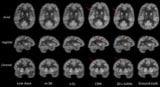
The reduced-dose images also exhibited a marked improvement in image quality after de-noising. Comparing standardized uptake value (SUV) measurements for the 1% dose-reduced and the full-dose static images showed considerable bias in the dose-reduced images. After de-noising, however, they exhibited near-identical SUV. SUV in the 10% dose-reduced images largely resembled those of the full-dose images, but were further improved using the de-noising model.
The researchers conclude that their deep-learning noise-reduction model enables significant 18F-FDG dose reduction in cardiac PET imaging without losing diagnostic accuracy. “A reduction to one hundredth of the dose is possible with quantitative clinical metrics comparable to that obtained with a full dose,” they write. “This dose reduction is important for patients, staff, general radiation protection and healthcare economy.”


