
Preclinical studies play a valuable role in developing novel treatment strategies for transfer into the clinic. The introduction of image-guided preclinical radiotherapy platforms has enabled radiobiological studies using complex dose distributions, with photon energies and beam sizes suitable for irradiating small animals. But there remains a drive to create platforms with ever more complex irradiation capabilities.
Beam shape modulation could improve the conformality of preclinical irradiation, but systems based on fixed aperture collimators cannot easily deliver beam sizes down to 1 mm. Jaw techniques such as the rotatable variable aperture collimator (RVAC) offer another approach, but these are challenging to implement robustly for millimetre-sized fields and, as yet, unvalidated.
A team at MAASTRO Clinic has now proposed another option: synchronized 3D stage translation with gantry rotation for irradiations from multiple beam directions. They demonstrated that this dose-painting technique can improve the precision of radiation delivery to complex-shaped target volumes (Br. J. Radiol. 10.1259/bjr.20180744).
The increased dose conformality that dose painting offers could benefit a range of preclinical studies. “The vast majority of radiobiology studies administered non-conformal dose distributions, which is not realistic compared to clinical practice,” explains senior author Frank Verhaegen. “Also, for studies on normal tissue response, being able to exactly target certain structures can lead to more relevant research results.” He adds that the technique can also be employed to deliver non-uniform doses, to study boost methods, for example, where hypoxic tumour regions receive higher dose.
Paint the target
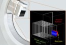
Verhaegen and colleagues used the SmART-ATP planning system to design plans for delivery on the X-RAD 225Cx image-guided preclinical radiotherapy research platform, and simulated the plans using a Monte Carlo (MC) model of the platform.
Dose painting uses heterogeneous irradiations from multiple directions. For each beam direction, a 2D area is defined — based on the projection of the target volume — and divided into many single-beam MC simulations. The team validated their MC model using radiochromic film measurements of the field shape and dose output of several complex fields, including a 225 kVp, 2.4 mm diameter beam.
The researchers considered two scenarios based on a CT image of a mouse with an orthotopic lung tumour. In case 1, the target was the tumour, while case 2 targeted a length of spinal cord. They created dose-painting plans to deliver 10 Gy to the target using the 2.4 mm beam, resulting in 256 beams for case 1 and 280 beams for case 2. Beam-on times were optimized to achieve a D95% and V95% of 100%.
For comparison, the researchers also created plans using a fixed aperture collimator and an RVAC. They selected four gantry angles for case 1, (anterior, posterior, lateral and medial directions) and two opposed lateral angles for case 2. Fixed aperture plans used the smallest available collimator that achieved complete target coverage: a 5 mm diameter beam for case 1 and a 20 × 20 mm beam for case 2. RVAC plans were created using the optimal angle and beam aperture size.
Balancing benefits
All irradiation methods achieved good target coverage and sharp cumulative dose–volume histograms for both cases. For the lung tumour irradiation, the increased conformality of the RVAC and dose-painting methods resulted in considerably lower doses to the left lung, trachea and heart compared with fixed aperture irradiation. Dose painting led to slightly lower dose homogeneity in the target.
For the spinal cord case, dose painting resulted in slightly better dose homogeneity in the target than achieved by the other two methods. Dose painting gave the best conformality, with a slightly larger volume of low doses, but considerably lower volume of higher dose, to both lungs. Fixed aperture irradiation resulted in far higher doses to all avoidance volumes.
These results suggest that for targets that match available fixed aperture field shapes and sizes, dose painting adds limited value. But where no fixed aperture collimators match the required field size, dose painting may surpass the plan quality achievable with RVAC. A major benefit of dose painting is that it can achieve conformal irradiation of concave target volumes. It also provides increased versatility to avoid organs-at-risk and deliver heterogeneous dose distributions to targets.
One disadvantage of dose painting is the greater radiation delivery duration, which can increase the risk of motion errors. Case 1, for example, required a total beam-on time of 1587 s with dose painting, compared with roughly 225 s for the other approaches. This results from the unavoidable trade-off between larger beam sizes with higher dose rates and smaller beams with higher spatial resolution but increased beam-on times. The team note that beam size should ideally be determined by the planning system based on time and plan quality constraints.
Clinical proton system could enable small-animal studies
Dose painting also requires considerably more time for radiation planning and calculation. The authors suggest that with tighter process integrations and further software optimizations, data processing issues should not limit practical implementation of this approach.
The team note that since this irradiation strategy only requires more advanced software and no hardware modifications, it can be used to increase the versatility of current-generation image-guided preclinical irradiation platforms. “The plan now is to implement this new method in our treatment planning system and combine it with beam-optimization methods,” says Verhaegen.


