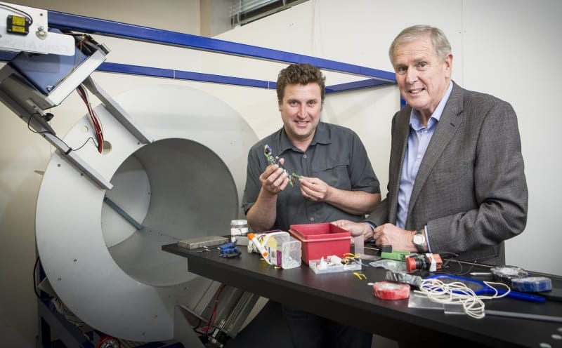
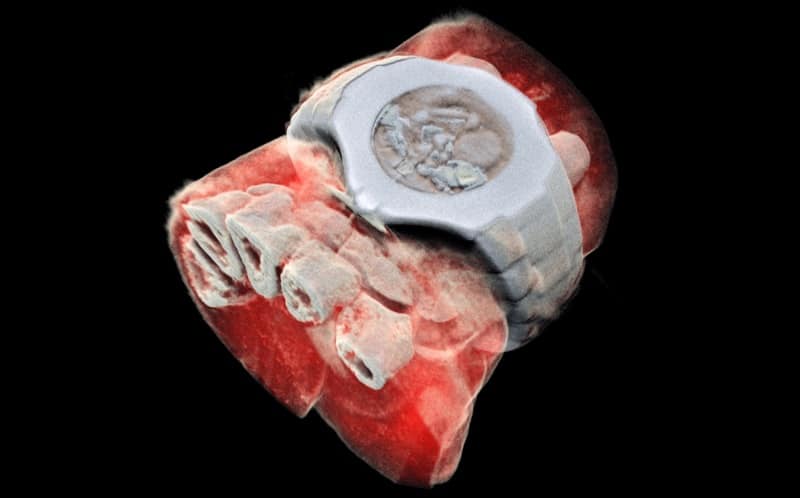
The first human has been scanned with a revolutionary 3D colour medical scanner developed by father and son scientists in New Zealand. Phil Butler, a physicist working at the University of Canterbury, and Anthony Butler, a radiologist at the Universities of Otago and Canterbury, invented the MARS spectral X-ray scanner, which has been commercialized by MARS Bioimaging.
The MARS scanner uses Medipix3 technology developed at CERN to produce multi-energy images with high spatial resolution and low noise. Medipix is a family of read-out chips originally developed for the Large Hadron Collider and modified for medical applications.
The Medipix3 detector measures the energy of each X-ray photon as it is detected. This spectral information is used to produce 3D images that show the individual constituents of the imaged tissue, providing significantly improved diagnostic information.
Phil Butler says that CERN’s Medipix3 technology sets the machine apart diagnostically because its small pixels and accurate energy resolution allow it to record images that no other imaging tool can. “As a new imaging device, a new microscope if you like, biomedical researchers can non-invasively see different kinds of detail inside patients,” he explains.
Small versions of the MARS spectral scanner that can house tissue samples are already in use in research institutions around the world. So far, researchers have used these scanners to study cancer, bone and joint health, and vascular diseases that cause heart attacks and strokes. “In all of these studies, promising early results suggest that when spectral imaging is routinely used in clinics it will enable more accurate diagnosis and personalization of treatment,” says Anthony Butler.
Phil Butler was the first person to be scanned with the MARS spectral scanner, using a larger version to image his ankle and wrist. The next step is an imminent clinical trial, where orthopaedic and rheumatology patients from Christchurch will be scanned. This will allow the MARS team to compare the images produced by their scanner with those generated by current technology used in New Zealand hospitals.
Anthony Butler says that after a decade in development, it is really exciting to have reached a point where it’s clear the technology could be used for routine patient care. “X-ray spectral information allows health professionals to measure the different components of body parts such as fat, water, calcium and disease markers. Traditional black-and-white X-rays only allow measurement of the density and shape of an object,” he says.


