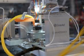
A new optical biopsy technique could improve surgeons’ ability to identify early-stage cancer cells in the liver. Developed by a team at Orel State University in Russia, the approach incorporates a combination of spectroscopy and fluorescence measurements, which can be readily integrated with standard biopsy needles.
Liver cancer is among the leading causes of cancer deaths worldwide. Surgery is one of the most effective methods for treating the disease – but to ensure patient survival, it is crucial that cancer cells are detected as early as possible. To identify tumours in the liver, surgeons must obtain tissue samples using a needle biopsy. If this is successful, they will be able to differentiate between healthy and cancerous cells. Yet in order to do this, they must insert the needle in just the right place. If they miss the tumour even slightly, it could lead to an incorrect diagnosis.
Co-first authors Evgenii Zherebtsov, Elena Potapova and colleagues propose that this approach could become far more effective through the use of optical techniques, which are highly sensitive to the molecular and morphological structures of biological tissues. The system they have developed combines two separate measurements. Firstly, diffuse reflectance spectroscopy measures optical absorption and scattering, by analysing the light reflected by complex molecular structures.
Secondly, lifetime fluorescence analysis measures the average time that a group of molecules will stay in their excited states after being illuminated, before emitting a photon. This lifetime can vary significantly in the presence of different enzymes – which when bound to cell proteins, play widely varying roles in their metabolism. Since the cells surrounding liver tumours will alter their metabolism as a defence mechanism, notable changes in fluorescence lifetime can reliably indicate when a tumour is nearby.
“Although our team as well as others have previously used fluorescence intensity for tissue assessment, studies performed in other parts of the body have shown that fluorescence lifetime is less dependent on experimental conditions,” explains Potapova in a press statement. “Fluorescence lifetime measurements remain more consistent in the presence of blood, when there is non-uniform illumination, or if the contact between the probe and tissue changes due to movement.”
In selecting compact, modern components to build their probe, the team ensured that the 1 mm-diameter device was compatible with a standard biopsy needle. In addition, the probe contains separate channels for the diffuse reflectance spectroscopy and lifetime fluorescence measurements.
Raman spectroscopy guides brain biopsies
Initially, the researchers tested their technique in mice injected with human liver cancer cells. The optical biopsy system produced stable, reproducible results, reliably distinguishing between healthy and cancerous cells, as well as metabolically altered cells surrounding tumours. The group then performed biopsies on human patients with suspected liver cancer, producing similarly promising results.
In enabling surgeons to identify cancer cells in real time, the researchers ultimately hope that their biopsy technique will significantly improve the ability to diagnose liver cancer at the earliest possible stage. Through future research, they will aim to expand their approach to optical imaging of tumours of different types and at different stages of development.
The researchers report their findings in Biomedical Optics Express.