
Researchers in the US have combined artificial intelligence (AI) with an advanced laser-based imaging technique to create a system that can identify different types of brain cancer from surgical samples with a similar accuracy to pathologists, but much, much faster. The test could enable surgeons to bypass the pathology lab and receive real-time diagnostic information during operations, to inform their decision-making (Nature Med. 10.1038/s41591-019-0715-9).
Every year around 15.2 million people around the world are diagnosed with cancer. More than 80% of those will undergo surgery, and in many cases this involves removing and analysing a proportion of the tumour during the operation. This provides a preliminary diagnosis, ensures that the specimen is adequate to achieve a final clinical diagnosis later, and helps guide surgical management.
In the US alone, more than 1.1 million tumours are biopsied annually. These are typically analysed using a histology workflow that is more than a century old. The tissue sample is taken to a laboratory, processed and prepared by skilled technicians, and then interpreted by a pathologist – all while the patient and surgical team wait in the operating theatre. But, both globally and within the US, there is a shortage of pathologists to provide such intraoperative diagnosis.
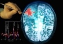
In recent years, Daniel Orringer, a neurosurgeon at NYU Langone, has been involved in the development of a novel laser-based technique that provides rapid, high-resolution images of unprocessed biologic tissue, known as stimulated Raman histology. By using laser light to excite the tissue, this technology generate contrast between the different components, such as lipids and proteins, revealing diagnostic features that are poorly visualized or can’t be seen with other histology methods.
Orringer and his colleagues have demonstrated that stimulated Raman histology can be combined with computer-aided diagnosis to classify brain tumours. However, they believe that matching the accuracy of the pathology laboratory in a clinical setting using this technique could be challenging – particularly with the current shortage of trained pathologists. But, as clinical-level accuracy has been achieved for image classification in other medical fields, such as ophthalmology, radiology and dermatology, using deep learning, they wondered if AI could help.
The researchers trained the deep convolutional neural network behind their AI-tool on stimulated Raman histology images of more than 2.5 million samples from 415 patients. It was taught to classify tissues into 13 histologic categories, focused on the 10 most common brain tumours, and offer a diagnostic prediction.
In a clinical trial at three medical centres in the US, the team tested the system on 278 patients undergoing brain tumour resection or epilepsy surgery. Brain tumour biopsies were collected from the patients and then split so that they could be sent for diagnosis via both the pathology laboratory process and the AI-based test.
The results for the two trial arms were very similar. The AI system classified 94.6% (264 of 278) of the tumours correctly, while the pathologists achieved a diagnostic accuracy of 93.9% (261 of 278). But imaging with stimulated Raman histology and AI diagnosis was significantly faster, providing a classification of brain tumour type in the operating room in less than 150 s. Typically, the pathology laboratory takes around 20–30 min.
There were also interesting differences between the errors made by the pathologists and the AI-based test. The AI correctly classified all 17 of the tumours that the pathologists diagnosed incorrectly, while the pathologists correctly diagnosed all 14 of the cancers that the AI misdiagnosed. The study authors say that this suggests that pathologists could use the new system to help them classify challenging specimens.
Raman spectroscopy guides brain biopsies
“As surgeons, we’re limited to acting on what we can see; this technology allows us to see what would otherwise be invisible, to improve speed and accuracy in the operating room, and reduce the risk of misdiagnosis,” says Orringer. “With this imaging technology, cancer operations are safer and more effective than ever before.”
“Stimulated Raman histology will revolutionize the field of neuropathology by improving decision-making during surgery and providing expert-level assessment in the hospitals where trained neuropathologists are not available,” adds Matija Snuderl, neuropathologist at NYU Langone, who was involved in the study.


