

In less than 100 seconds, Chris Phillips describes how physicists are overcoming the diffraction limit
Watch more from our 100 Second Science video series.
In less than 100 seconds, Chris Phillips describes how physicists are overcoming the diffraction limit
Watch more from our 100 Second Science video series.
By Hamish Johnston
The Institute of Physics has just launched a new blog called physicsfocus that is described as “an online space for the physics community to read about and comment on issues that concern them”.

Researchers in the US have developed a new type of thermal sensor by combining micron-sized “paddles” with nanometre-sized support rods. The device is made using standard silicon-fabrication techniques and is extremely sensitive to infrared radiation – without the need for cooling. As a result, the team claims that it could be used as a thermal and infrared imager that is integrated into electronic devices.
A torsional resonator is a rectangular paddle-shaped device with two thin support rods connecting it to the rest of a chip structure. When a force is applied to the paddle, it vibrates at a certain characteristic resonant frequency. There are two main ways in which the resonator vibrates. These are the torsional mode where the paddle twists back and forth along the rod axis and the flexural mode where the paddle vibrates up and down like a trampoline perpendicular to the rod axis.
Michael Roukes and colleagues at the California Institute of Technology have now made thermal sensors from such resonators. The researchers began by coating the surface of a micron-sized silicon paddle with titanium nitride – a material that absorbs infrared radiation. Next, they applied a time-varying voltage between the paddle and the substrate it was on. This generates a force that sets the paddle vibrating at its resonant frequency.
When the paddle is exposed to infrared radiation, it heats up and this shifts its resonant frequency. “By tracking this change in frequency, we are able to determine the intensity of radiation that hits the device,” explains team member Edward Myers. This change is measured using an optical interference set-up that involves reflecting laser light from the surface of the paddle (see figure).
The paddle supports are made of silicon nanowires and measure just 1 µm long and 50–100 nm in diameter. Thanks to these tiny supports, the paddle is extremely well isolated from its environment. This means that only a small amount of infrared radiation is needed to heat the device by a measurable amount.
The sensor was made from standard semiconductor and metal materials using standard industrial techniques – so in principle, Roukes and colleagues could make large arrays of these paddles for use as thermal imagers.
“Ultimately, we believe that these sensors can perform as well as certain standard infrared sensors that require cooling to cryogenic temperatures,” Myers says. “Our devices can operate without cooling, which makes them potentially useful for portable applications, such as night-vision goggles, home surveillance and perhaps even the next generation of smartphone cameras.”
The researchers now plan to work on making the device smaller and improving the materials used to make it – something that should further improve its thermal characteristics. “We are also looking at scaling up from one device to an array of devices,” reveals Myers. “As part of this plan we hope to integrate these sensors with on-chip CMOS electronics, which will make for easier control and readout of many thousands of elements at once.”
The sensor is described in Nano Letters.
In less than 100 seconds, Martin McCall explains how physicists are pursuing the goal of invisibility.
Watch more from our 100 Second Science video series.
A new type of supernova has been defined by researchers from the US. Designated type Iax, this new class is seen to be less energetic and fainter than previously defined and similar type Ia events – and may even leave behind part of its originating star.
Previously, only two classes of supernovae were recognized – core-collapse events, the explosion of stars 10–100 times as massive as our Sun, and type Ia supernovae, which involve the complete destruction of a white dwarf. Now, a team led by Ryan Foley at the Harvard-Smithsonian Center for Astrophysics has identified 25 members of the new type Iax class based on optical spectroscopic and photometric studies. Its work has shown these stars to be less energetic and with a lower absolute magnitude than would be expected with their type Ia cousin. The team believes that the supernovae of this new class originate from a binary star system comprised of a white dwarf that gathers helium from a companion star, which has lost its outer hydrogen.
“A type Iax supernova is essentially a mini supernova…it’s the runt of the supernova litter,” says Foley. “It turns out that type Iax supernovae are relatively common, but we only recently discovered them because they’re very faint, some only 1% as bright as type Ia supernovae.”
As a result of their absence from elliptical galaxies – which are populated by older stars – it is believed that type Iax supernovae come from young star systems. This observed distribution may be related to the relatively short lifetimes of helium-burning stars, which do not last long enough to be found in elliptical galaxies. The team is uncertain what causes the white dwarf to become a supernova. One theory suggests that the overlying helium shell ignites first, transmitting a shockwave into the white dwarf within, with the opposing hypothesis proposing that the dwarf core might go first, in response to the influence of the surrounding helium.
In some cases, the researchers believe that the supernovae events might be so weak that the white dwarf may even survive the explosion. “It’s a little uncertain exactly what will happen to the white dwarf,” Foley told physicsworld.com, explaining that such a partial deflagration might be expected to leave the star with a lower mass, but with added energy received from the supernova. “Both of these changes should make the star bigger and hotter,” Foley concludes, “so I expect some sort of puffed up white dwarf.”
Unlike other supernovae, members of the type Iax are too varied and faint to be used as standard candles – objects with a known luminosity and observed brightness that can be used to reveal their distance from Earth. However, the researchers believe that the new class could offer insights into supernovae in general. Not only do these events showcase a previously unknown way for a star to explode – with a slower moving ejecta that is easier to analyse – but their physical similarities to, and differences from, type Ia supernovae enable us to focus our models of this existing class of event.
“From an observational point of view, the substantial sample of events that resemble SN 2002cx [the prototypical member of the new class] and the estimate that these sort of events happen [a third] as often as classical type Ia supernovae is compelling,” comments Craig Wheeler of the University of Texas at Austin, US, who was not involved in the research. He adds that while the proposed model – of a fully or partial deflagrating white dwarf with a companion helium star – fits well with the observed properties, such models warrant careful consideration before any firm conclusions are reached.
“If this is the appropriate interpretation, then these events come with a promise to teach us more about the nature of thermonuclear explosions,” Wheeler adds. “Elucidating the nature of these events also holds great promise to aid us in better understanding stellar evolution in binary star systems in all its great variety.”
Next, Foley and colleagues are hoping to address the possibility that the white dwarves might, in some cases, survive going supernova. In order to work on this theory, the team simply needs to observe more supernovae, Foley explains, adding the caveat that this kind of observation will only be possible for the closest supernovae. “The current sample is relatively small,” he says, adding that “with a larger sample, we can hopefully unlock additional mysteries about the frequency, energy, asymmetry and other properties of these supernovae.”
The work is to be published in The Astrophysical Journal. A pre-print of the research is available on arXiv.

By Hamish Johnston
Deep in the North Woods in Minnesota the snow is starting to melt, and the giant NOvA Far Detector is coming to life. Designed to register the arrival of neutrinos that will be created 810 km away at Fermilab near Chicago, the detector has recorded its first 3D images. These are not of neutrinos, but of the trajectories of fast-moving particles that are created in a process that begins with a cosmic ray colliding with Earth’s atmosphere.
Knots are very much a part of everyday life. In some cases, they are extremely useful: sailors and climbers, for example, need them for their own safety or to secure boats and equipment. But in other cases, knots are a nuisance. You may have found this yourself the last time you tried to reel in a long extension cable after, say, vacuuming your attic or mowing the lawn. Invariably, there is a knot in the cable when you want to store it away, and in order to un-knot it, you need to repeatedly pull one of its ends through the entangled region – a rather dull and lengthy procedure, as anyone who has ever tried to untangle headphone wires or Christmas tree lights can confirm.
The theory of how knots form, and how different knots can be distinguished, began as a rather abstract mathematical discipline. Developed by a trio of Scottish physicists – Peter Guthrie Tait, James Clerk Maxwell and William Thomson (later Lord Kelvin) – in the late 19th century (see September 2010 pp35–40), it was originally intended to support the now-abandoned idea that atoms might consist of knotted tubes of aether. Since then, however, physicists have also found practical applications for knot theory. One important example concerns the possible configurations of DNA molecules. Like extension cables, the filaments that make up DNA can become knotted in all sorts of ways, and research into the kinds of knots formed by DNA is telling us some interesting things about the molecule’s properties.
For example, one might imagine that DNA knots will make it difficult for proteins to read, transcribe and replicate the knotted portion of the molecule. Excessive DNA knotting can even prevent cell division and lead to cell death. Moreover, the DNA inside bacteria and human cells is so long (millimetres and metres, respectively) that one would expect to observe knots very frequently. However, certain enzymes, known as topoisomerases, have evolved to solve this problem by cutting DNA and gluing it back together. On the other hand, within some very small viruses, where there is space only for the DNA itself, knots are inevitable because of the tight confinement – yet the knots that form do not stop the virus from infecting its host. A better understanding of DNA knots may therefore give us some insights into these important cellular processes.
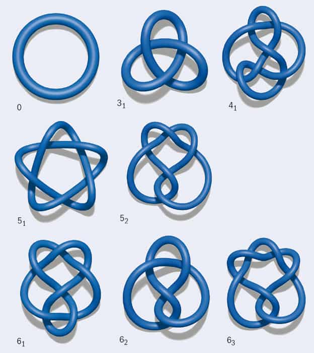
Before going into more detail about DNA knots, we first need some basic information about knot theory. Knots are grouped according to the minimum number of crossings they contain, and each knot in a group is given a number. For example, the simplest non-trivial knot, the trefoil knot, is known as 31: it has three crossings, and it bears the subscript 1 because it is the first and only possible knot with three crossings. Figure 1 shows representations of the trefoil knot along with several other simple knots. Note that in diagrams like these, it is crucial to retain information about which segment is on top at a crossing. For instance, if you flip the top and bottom segments in just one crossing of the trefoil knot, the resulting curve no longer contains any knots at all: it has become an unknot (0).
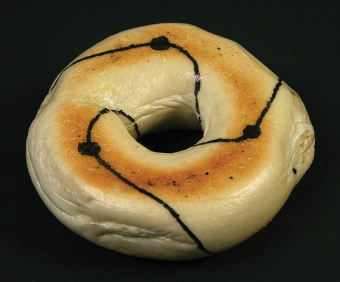
Now imagine trying to draw a knot as a closed curve on a surface. If the surface is a sphere, such as a balloon, the only knot that can be drawn without any self-intersections is the unknot. However, if the surface has the topology of a torus, such as a doughnut or a bagel, an infinite number of knots can be drawn on it (figure 2). These are called torus knots, and examples include the trefoil and 51 (cinquefoil) knots.
A second important family of knots is twist knots. To create one such knot, take a rope and hold it fixed at its two extremities. Then take the middle of the rope and twist the whole loop an integer number of times. Soon, the rope should start to look like a heavily used office telephone cord, a phenomenon known as “supercoiling”. Finally, take one of the ends, pass it through the top loop of the rope, and then link the ends so that the rope is now knotted. By increasing the number of twists, you can create a trefoil (which, notably, is a twist knot as well as a torus knot), a figure of eight (41), a three-twist (52) or a Stevedore’s knot (61).
Of these two families, twist knots are more commonly found in nature. This is because when unravelling a twisted cord, it is easy to inadvertently pass one end of the cord through a loop – and the same is true if some natural process rather than a human is doing the unravelling. A more rigorous way of saying that twist knots are easy to produce is to note that their “unknotting number” is 1. The unknotting number is the minimum number of times that a knot must be passed through itself in order to untie it. Equivalently, it is equal to the minimum number of crossings that must be flipped to obtain the unknot. With just a bit more drawing and thinking (and perhaps the aid of your headphone wires), it is fairly easy to show that the unknotting number of a 51 torus knot is 2. This knot is more difficult to form by mistake, as the cord has to pass through itself twice, rather than once.
Based on these arguments, it seems logical to suspect that DNA molecules will frequently form twist knots, and that 52 twist knots will appear much more frequently than 51 torus knots. But how can this hypothesis be tested? After all, a DNA molecule is only about 2 nm (2 × 10⁻⁹ m) wide. When it is inside a cell or in solution, it occupies a space measured in microns; within viruses, it is even more compact. Given the tiny size of such a molecule, how can we determine which knots have formed in it?
Of course, it is possible to obtain images of a particular knotted DNA molecule directly, using electron microscopes. But if we want to know the probabilities of forming different types of knots – the “knot spectrum” – we need information about an entire population of molecules. One way to obtain this knot spectrum is through simulations. DNA molecules in solution can be made to form loops (by making their loose ends attractive to one another), so we can model them as fluctuating looped elastic rods, the bending rigidity of which matches that of DNA.
However, it is also possible to determine the knot spectrum experimentally thanks to a rather clever trick. DNA molecules are negatively charged, so one can use a weak electric field to drive them through a dilute substance called agarose gel, which is a jelly-like material filled with microscopic holes (a bit like a squishy bath sponge). As one might expect, small DNA molecules will pass through these holes more quickly than large and cumbersome ones, since bigger molecules become tangled up with the polymers that make up the agarose gel. In a simple model, one can treat the velocity of the DNA molecule in the gel as being inversely proportional to the size of the sphere that best approximates the molecule’s average shape. If the DNA under consideration is highly knotted, it will be crumpled up and smaller in size, so it will move more quickly through the gel than it would if it were unknotted. Remarkably, it is possible to separate knots with up to roughly 10 crossing numbers by looking at how fast they move in agarose gel – the first group of molecules to pass through the agarose will have 10 crossings, the next nine and so on.
This measurement technique enabled researchers to confirm in the 1990s that in solution, looped DNA molecules a few microns in length do indeed form twist knots, and that “simpler” knots with lower unknotting numbers are more probable (see, for instance, V V Rybenkov et al. 1993 PNAS 90 5307). Indeed, the statistics of the knots formed in the simulations and in the experiments are pleasingly very similar, which suggests that DNA loops could potentially be used as a test bed for probing ideas on knot theory.
However, a new twist in the field of DNA knots arrived in 2005 when Javier Arsuaga and collaborators studied DNA knots in a type of virus called a bacteriophage. Also known as phages, bacteriophages (literally “bacteria eaters”) are viruses that infect bacteria. They work in a remarkably simple fashion. Using a molecular motor, phages tightly pack DNA in their interior so that it reaches almost crystalline density. The phage then exploits the high pressure that results from this tight packing to fire the DNA into the cytoplasm of its bacterial host as soon as the protein shell containing the phage DNA (the capsid) is “untapped”.
One might naively expect that knots would be a bad thing
The only difference between phage DNA and the previously studied DNA in solution is the degree of confinement, which is much more dramatic in phages since they are so small: about 40–70 nm across. But does this confinement affect the knots that can form within the capsid?
The research of Arsuaga et al. suggests that it very much does: they found that knots occurred far more frequently in the phage DNA than in “unconstrained” DNA in a solution. More specifically, in natural or “wild-type” infective phages, about half the DNA molecules were found to be knotted. The researchers also discovered that the viral DNA often contained torus knots, while twist knots were virtually absent – quite unlike the pattern observed for DNA in solution.
Do these results tell us something more about DNA structure, at least within phages? To find out, it was necessary to extend existing models of fluctuating elastic loops to the case in which the loops are confined within a sphere of radius R – which, to be relevant for the case of phage DNA, must be much smaller than the contour length of the DNA molecule itself. These simulations did reproduce the intuitive result that confinement increases the likelihood of forming knots: the DNA molecule in the narrow space of the phage has more opportunities to cross itself several times, so knots can readily form. However, the simulations were unable to reproduce any bias in favour of torus over twist knots and, unlike in experiments, the number of 52 twist knots observed was always larger than the number of 51 torus knots. Moreover, when R was set to a realistic value of 25 nm, the DNA configurations found by simulations were very disordered and the knots formed very complicated, with far more crossings than found experimentally.
Was the disagreement between simulation and experiment the result of an incomplete understanding of DNA–DNA interactions within the bacteriophage? Or was the bias in favour of torus knots a result of some other effect – such as the “molecular motor” inside the phage, which may rotate the DNA while it is packaged, thereby potentially affecting its conformation?
Our group, as part of an interdisciplinary collaboration, found a possible answer to this question in 2009, when we simulated the packaging and ejection of DNA molecules inside a modified type of phage called a P4 cosmid (PNAS 106 22269). Though still infective, cosmids are mutants that contain only half the genome of the natural, wild-type phage. This makes them more treatable computationally, and experiments have shown that the knot spectra of P4 and P4 cosmids are qualitatively similar. To create our simulated DNA segments, we began with the fluctuating elastic rod models described earlier, but added an extra ingredient: the new simulations contained an “orienting” interaction between model DNA segments that kicked in whenever two segments got close to each other. This orienting interaction was designed to favour a small twist angle between the contacting segments. You can think of it as like the interaction that occurs when two chiral, corkscrew objects, such as pieces of fusilli, lock at an angle when forced into close contact (figure 3a).
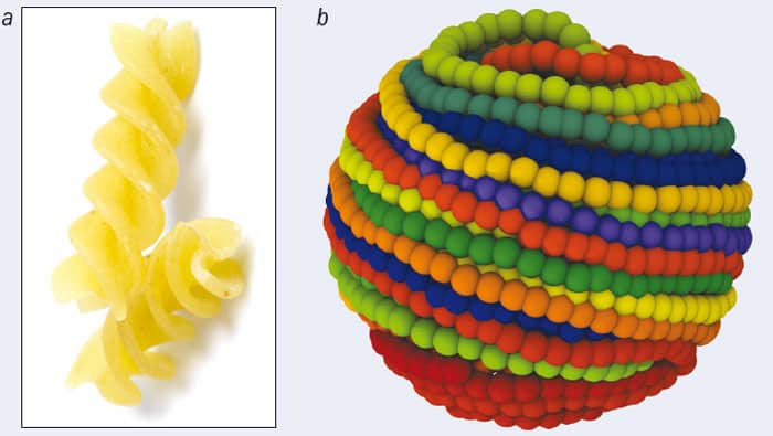
For the fusilli, the origin of the orienting interaction is geometric. For DNA, it may be mainly electrostatic, but whatever its ultimate origins, the effects of adding this interaction to our models were dramatic. When we used realistic values of parameters such as DNA rigidity (inferred from studies of DNA liquid-crystalline phases in solution), the simulations showed that, to begin with, the DNA inside the phage capsid is now ordered into a spool-like structure (figure 3b). This agrees with previous models for DNA packing and with electron microscopy pictures of phage DNA. Importantly, the twist knots are now strongly suppressed, and the knot spectrum is much more compatible with the experimental one.
While the puzzle of the twist knot absence was perhaps solved, another big question remained. One might naively expect that knots would be a bad thing for phages, because in order to be infective, they need to eject DNA from their interior into a bacterium, and the presence of knots might cause the DNA to get stuck. So how could the P4 phage have highly knotted DNA and still be infective?
A final answer to this question is still pending, but simulations suggest that the explanation may have to do with the concept of “knot localization”. If you tie a knot in a string and pull it tight, the knot will eventually localize such that it occupies a certain small region. But for DNA confined within a phage, our simulations suggest that the opposite happens: the knot delocalizes, spreading out over the whole DNA filament. In other words, the knottedness becomes a global property of the curve, one that cannot be captured by looking at a small fraction of the molecule.
This is crucial, because during the DNA ejection process, it is exactly the dynamics of such a small fraction (specifically, the bit that is close to the opening in the phage capsid) that dictates the physics of the process. If the knot is delocalized, this fragment will look no different for a torus knot than it will for an unknot, so simulations show that DNA ejection should proceed at approximately the same speed as it would if the molecule contained no knots at all. One can imagine further that torus knots may provide another advantage during ejection, as their toroidal (spool-like) geometry means that they may be readily unrolled, avoiding the formation or accumulation of self-entanglement – much as a spool helps prevent fishing lines from becoming tangled.
In bacteria, by contrast, knot localization seems to have beneficial effects. DNA molecules in bacteria are usually negatively supercoiled: in other words, the DNA helix is twisted less than it would be in a “normal” or undeformed strand, where the helical pattern repeats every 10 base pairs. Negative supercoiling is advantageous for the bacterial cell because it weakens the DNA helix, making it easier to break the hydrogen bonds keeping the helix together – something the cell must do whenever it “unzips” the DNA helix in order to transcribe and replicate it. But negative supercoiling may also help the bacterial cell remove problematic DNA knots. This is because if a knot happens to develop in a supercoiled strand, it normally becomes tight and localized, as shown in recent research by Guillaume Witz and collaborators from various institutions in Lausanne, Switzerland. This presumably makes it easier for the knot-removing topoisomerase enzyme to find, since the enzyme is attracted to regions of high DNA curvature.
The general picture we are developing is that in bacterial and human cells, DNA knots create many more problems than they do in viruses. Hence, control of these knots is very important, and we have discussed how topoisomerase enzymes have evolved to remove them. Usually, the removal of knots is a good thing, but in some circumstances we might actually prefer the DNA to remain knotted. For example, drugs that inhibit the functioning of topoisomerase enzymes have been used to treat cancer, because they prevent cells from dividing. So while topology and knot theory have already provided us with important insights into the physics of phage DNA, if we can uncover even more of the mysteries of DNA knots, it might open up new perspectives in biotechnology or even medicine. But before we get there, a lot more work has to be done.

By Matin Durrani
If you’re a member of the Institute of Physics, the April 2013 issue of Physics World is now ready to view online or through our apps.
This month marks the 60th anniversary of the discovery of the structure of DNA by James Watson and Francis Crick and – to celebrate that milestone – we have a great feature on an unusual aspect of the famous double helix: namely, how knot theory can help us to understand how and why DNA tangles up.
Elsewhere in the issue, we continue the biophysics theme by looking at the damage caused to the human brain by blows from sports injuries or by the shock waves from exploding bombs. This is not traditional physics territory by any means, but surely there is no harm in physicists bringing a fresh perspective to such matters?
Our final feature this month looks at the history of Maxwell’s demon – the tiny being originally devised by James Clerk Maxwell as a thought experiment to evade the second law of thermodynamics. But, as Philip Ball explains, some of the physicist’s contemporaries actually believed it was an intelligent being that could bridge hidden worlds and provide a scientific route to immortality of the human soul.

The first two books in Stuart Clark‘s trilogy of scientific novels (see May 2012) offered up a heady mix of history, physics and intrigue, concentrating on periods when political upheaval coincided with scientific revolution. In the first instalment, The Sky’s Dark Labyrinth, the main characters are Galileo and Kepler; in The Sensorium of God Halley, Hooke and Newton take centre stage. This pattern continues in the trilogy’s concluding novel, The Day Without Yesterday, which begins in 1914 with Albert Einstein fretting over relativity in Berlin while the cosmologist Georges Lemaître dodges bullets in the trenches of his native Belgium. Einstein is the obvious choice for a central character and, initially, Lemaître makes a highly effective foil. A devout Catholic who was decorated for his wartime service and later became a priest, he could hardly be more different from Einstein, a non-observant Jew whose Swiss citizenship kept him out of the fighting. The wartime setting also gives Clark a chance to explore the rise of antisemitism in Germany and the roles that Einstein’s (mostly Jewish) colleagues played in the war effort. The postwar period presents Clark with a problem, however. By the 1920s Einstein’s resolute opposition to quantum theory was starting to nudge him out of the scientific mainstream. Lemaître, for his part, did not make his seminal contribution to cosmology until 1927, when he published a (largely overlooked) paper suggesting that the universe was expanding. That leaves a gap of several years in which the pairing of Einstein and Lemaître seems forced, and the book loses its way somewhat. Another problem is that the minor characters in The Day Without Yesterday – Arthur Stanley Eddington, Edwin Hubble and Niels Bohr among them – are largely one-dimensional, and prone to disappearing from the narrative as quickly as they are introduced. The exceptions are Einstein’s first and second wives, Mileva and Elsa; as in the trilogy’s first two books, the central character’s family life is described, warts and all, in a way that is both detailed and believable. One might have wished for a stronger conclusion to this generally excellent trilogy, but overall, the strengths of The Day Without Yesterday outweigh its weaknesses.
“When you have eliminated the impossible, whatever remains, however improbable, must be the truth.” As a scientific motto, Sherlock Holmes’ famous aphorism takes some beating. Indeed, there are plenty of scientists out there (your reviewer among them) who trace their interest in the subject back to a childhood spent reading and re-reading the adventures of Arthur Conan Doyle’s great detective. Couple this reservoir of scientific interest with the recent resurgence of Holmesian characters in popular culture, and a book that explains Sherlock’s scientific side with wit and flair ought to find a ready audience. Unfortunately, The Scientific Sherlock Holmes is not that book. For starters, author James O’Brien takes quite a long time to get on to his actual subject. Instead of beginning with the science, he devotes the first 47 pages (nearly a third of the book’s main text) to a largely irrelevant analysis of how the 60 stories in the Holmes “canon” differ from previous works of detective fiction, followed by a flabbily repetitive summary of the main characters. The latter might be useful for readers less familiar with the canon, but O’Brien’s copious references to past works of Sherlockian scholarship will put many newcomers off long before they get there. Likewise, anyone unversed in Holmesian abbreviations such as BOSC (short for The BOSCombe Valley Mystery) or CROO (The Adventure of the CROOked Man) will quickly tire of flipping back to the table in which they are defined. Hard-core enthusiasts won’t mind either of these things, of course – but if they are O’Brien’s real audience, then why frustrate them with such a laboured introduction? Once the book gets going, it is possible to see glimpses of how good it could have been. For example, the chapter on chemistry includes a superb explanation of how Holmes deduced that a character in The Musgrave Ritual had been dead “some days” before being discovered in a tiny sealed chamber. By estimating the volume of the chamber and using the ideal gas law to estimate the quantity of oxygen present, O’Brien, an emeritus professor of chemistry at Missouri State University, is able to show that the man must have expired at least three days before Holmes finds his body. There are several such passages, but not enough to sustain a whole book.
Researchers in the US have made a graphene loudspeaker that has an excellent frequency response across the entire audio frequency range (20 Hz–20 kHz). While the speaker has no specific design, it is already as good as, or even better than, certain commercial speakers and earphones in terms of both frequency response and power consumption.
Loudspeakers work by vibrating a thin diaphragm. These vibrations then create pressure waves in surrounding air that produce different sounds depending on their frequency. The human ear can detect frequencies of between 20 Hz (very low pitch) and 20 KHz (very high pitch), and the quality of a loudspeaker depends on how flat its frequency response is – that is, the consistency of the sound it produces over the entire 20 Hz–20 KHz range.
“Thanks to its ultralow mass, our new graphene loudspeaker fulfils this important requirement because it has a fairly flat frequency response in the human audible region,” says team leader Alex Zettl of the University of California, Berkeley. He told physicsworld.com that the fact that “graphene is also an exceptionally strong material means that it can be used to make very large, extremely thin film membranes that efficiently generate sound”.
Because the graphene diaphragm is so thin, the speaker does not need to be artificially damped (unlike commercial devices) to prevent unwanted frequency responses, but is simply damped by surrounding air. This means that the device can operate at just a few nano-amps and so uses much less power than conventional speakers – a substantial advantage if it were to be employed in portable devices, such as smartphones, notebooks and tablets.
The Berkeley researchers made their loudspeaker from a 30 nm thick, 7 mm wide sheet of graphene that they had grown by chemical vapour deposition (CVD). They then sandwiched this diaphragm between two actuating perforated silicon electrodes coated with silicon dioxide to prevent the graphene from accidentally shorting to the electrodes at very large drive amplitudes. When power is applied to the electrodes, an electrostatic force is created that makes the graphene sheet vibrate, so creating sound. By changing the level of power applied, different sounds can be produced. “These sounds can easily be heard by the human ear and also have high fidelity, making them excellent for listening to music, for example,” says Zettl.
The researchers have already tested their device against high-quality commercial earphones of a similar size (Sennheiser® MX-400) and found that its frequency response over the 20 Hz to 20 kHz range is comparable, if not better.
The Berkeley team says that its CVD technique for fabricating the speaker is very straightforward and could easily be scaled up to produce even larger-area diaphragms and thus bigger speakers. “The configuration we describe could also serve as a microphone,” adds Zettl.
A preprint of the research is available on the arXiv server.