Meta-design: language models generate novel quantum experiments

A workflow for designing quantum experiments

Earlier this year a group of researchers led by Sören Arlt of the University of Tübingen set out to stretch the limits of how far artificial intelligence (AI) can contribute to scientific discovery. In work published in Nature Machine Intelligence, they developed a language model capable of generating classes of blueprints for quantum optics setups that produce specific families of quantum states. Their model was able to design several experimental configurations that successfully generated desired, and in some cases previously unknown, constructions within the limits of its training.

Beyond this immediate technical achievement, the implications of this approach are striking. In principle, a researcher could ask a system like this to propose experimental setups for a desired quantum state without spending months or years exploring possible configurations. Such capabilities could accelerate research in areas like quantum computing and quantum communication, where specially engineered quantum states serve as key resources. Although the system still has clear limitations – it cannot always guarantee that the produced state perfectly matches the target and it sometimes fails to find a solution – this study demonstrates that machine learning can already contribute meaningfully to scientific discovery, even in the design of physical experiments.

Earlier attempts had already hinted that AI could assist in designing quantum experiments. In 2016, researchers from Mario Krenn‘s group (in which Arlt is a doctoral student) demonstrated that automated search methods could propose previously unknown quantum optics experiments capable of generating complex entangled states. Since then the field has grown rapidly, with tools such as PyTheus producing candidate experimental designs and revealing physical mechanisms that researchers had not previously recognised.

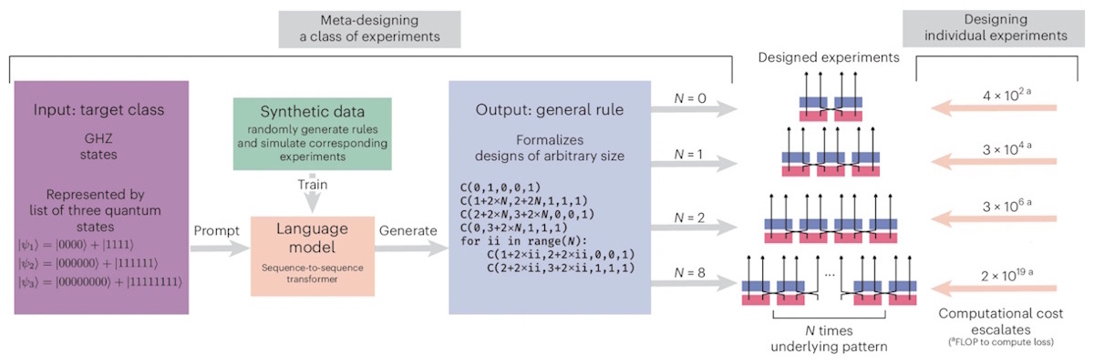
This time, instead of searching directly for a single experimental setup, the researchers trained a transformer-based language model on a dataset linking target quantum states to experimental blueprints. Given a desired state, the model generates Python code describing how to build a corresponding experiment. Based on the same transformer architecture used in modern language models, the system translates a quantum state into a program that constructs it experimentally. This output can be interpreted directly by researchers, allowing them to run the proposed construction and understand the design rules that the model discovered.
Evaluating the codes

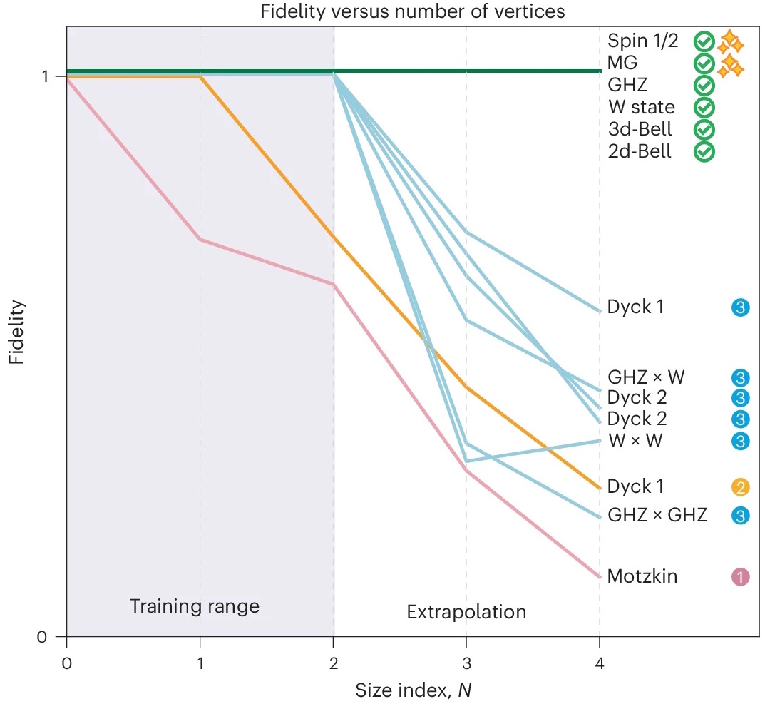
Using this approach, the researchers constructed 20 classes of quantum states of interest, among them well-known entangled states, such as GHZ, W and Bell states, some of which had no known experimental construction rules. Out of these, the system generated valid construction rules for six classes: four corresponded to already known solutions, while two corresponded to genuinely new construction rules for generating particular classes of entangled quantum states.

Rather than discovering entirely new states, the system identified previously unknown ways of assembling optical components that produce states with the required entanglement structure. The team verified these constructions computationally by simulating the resulting quantum states and comparing their fidelity with the target states. Although the experiments have not yet been carried out in the laboratory, the proposed setups provide experimentally testable blueprints.

The practical implications of tools like this are already prompting debate. Some see them as accelerating scientific discovery by exploring vast experimental possibilities, while others raise concerns that increasing automation could sideline experimental intuition. The key advance over previous approaches lies in generalization: rather than producing a single design, the model generates a program capable of constructing experiments for an entire class of states. “Instead of designing a single experiment for one target, this approach generates a general program that produces valid experiments for a whole class of targets,” Arlt explains.

The researchers chose to explore states that are physically relevant across different areas of quantum physics, allowing them to probe entanglement patterns relevant to quantum simulation, communication and computation. In this sense, the system expands the experimental toolbox available to physicists.

In some cases, the system uncovered patterns that the researchers had not previously identified. “We discovered two construction rules that we did not know of before,” Arlt notes. In another case, it generated a different construction rule for a class of states that had already been solved, following a completely different experimental strategy.

Rather than replacing physicists, the authors see AI changing how experiments are conceived. Instead of manually assembling setups, researchers may define the space of possible configurations and allow algorithms to explore it. As Arlt describes it: “instead of thinking about how do I put these components together so my experiment works, we think about what should the space of possible configurations look like so my computer can explore it efficiently”.

Despite the use of machine learning, the system is relatively modest in scale, with roughly 100 million parameters. While this keeps the computational cost manageable, it also constrains the range of experimental sizes and resources that the model can handle. The model also does not verify the correctness of its own outputs, requiring explicit fidelity checks of the generated states.

Looking ahead, the team hopes to extend this approach to other domains of physics and combine it with additional discovery methods.

All in all, tools like this suggest a future in which computers assist not only with simulations, but also with proposing new experiments and uncovering patterns in physical systems. Rather than replacing physicists, such systems may increasingly act as collaborators, helping researchers explore experimental designs that would otherwise remain inaccessible.

Scientists find a new critical point in supercooled water

Researchers at Stockholm University in Sweden have found experimental evidence of a long-predicted critical point in water at -63 °C. The result, which they obtained by supercooling liquid water and probing it with ultrafast laser pulses before it could freeze, provides further evidence that liquid water exists in two distinct phases.

Water is a strange substance. Unlike most other materials, its liquid form is denser at ambient pressures than the ice it forms when it freezes. It also expands, rather than contracting, as it cools, and it becomes less viscous when compressed. All told, water exhibits around 60 different anomalous behaviours, and it is especially atypical when cooled below its usual freezing point. This so-called “supercooled” state of water occurs naturally in high-altitude clouds, and it can be produced in a laboratory by applying high pressures as the water is cooled to low temperatures.

In 1992, a computational study led by the physicist Francesco Sciortino (then at Boston University in the US) indicated a further unusual trait. According to simulations by Sciortino, Peter Poole, Ulrich Essmann and H Eugene Stanley, supercooled water can undergo a transition between two different liquid phases, with a liquid-liquid critical point (LLCP) occurring at pressures 2000 times higher than atmospheric pressure at sea level. “These two liquids would coexist on a line in the supercooled water’s phase diagram,” explains Stockholm’s Anders Nilsson, who led the new study. “As pressure is lowered and temperature is increased, the two phases would vanish to leave only one phase.”

At this critical point, where two phases meld into one, theory predicts that fluctuations will arise between the two liquid states. These fluctuations are not confined to the critical point, however. They also occur in a large region of the phase diagram at temperatures above it; indeed, the predicted phase diagram contains further anomalies that persist up to around 50°C. This means that the existence of an LLCP could play a role in the behaviour of water under ordinary conditions. In fact, its presence could provide a straightforward explanation for many of water’s oddities, especially at low temperatures.

Before the new study, though, this LLCP was only predicted, never proven. “It has been difficult to identify because it has not been possible to conduct experiments at the low temperatures at which ice forms very quickly,” Nilsson says.

A role for ultrafast lasers

The key to the latest work, which is detailed in Science, was a new technology. “Ultrafast X-ray lasers allow us to perform such experiments and probe water before it freezes,” Nilsson tells Physics World.

Working at POSTECH University and the PAL-XFEL facility in South Korea, Nilsson and his colleagues studied supercooled water using ultrafast infrared laser pulses followed by x-ray scattering. This allowed them to detect the phases formed before the supercooled water began to turn into ice. “By varying the laser’s fluence, we were able to access liquid states straddling the predicted critical point,” explains Nilsson.

The Stockholm researchers report that they observed a crossover from a discontinuous to a continuous transition, where the system undergoes broad and slow structural changes. Such a pattern agrees well with the existence of critical fluctuations at this point, Nilsson says. They also observed a rapid increase in the material’s heat capacity indicating a critical divergence at 210 ± 8 K, which is coincident with enhanced density fluctuations. “These results suggest that our experiments have directly probed the vicinity of a critical point in supercooled water,” Nilsson says.

Investigating the impossible

Nilsson adds that finding this critical point had long been a “holy grail” for scientists who study water, with many believing it would be impossible for experimentalists to access. As an X-ray scientist, however, Nilsson realized that the new generation of X-ray lasers could make a difference.

“I took on this challenge 15 years ago and the most difficult aspect was to move water through the phase diagram – by changing the pressure and temperature – very quickly and study it on ultrafast time scales (in less than a microsecond), before ice formation occurred,” he says. “It took us many years of planning and testing: we identified the two liquid phases, a result that we also published in Science in 2020, and have now finally succeeded in reaching the critical point.”

The researchers now plan to continue investigating the critical point in detail, with the goal of understanding the timescales of the fluctuations that occur as the pressure and temperature are nudged away from it. “We also need to research the implications of ordinary water becoming supercritical at interfaces that are important for energy applications, such as fuel cells and water splitting,” Nilsson says. “Other important areas to consider [include] how supercriticality is important for water in living cells; water as a solute for chemical reactions; water in geological pores; and water in clouds, which are important for understanding climate change.”

Team member Fivos Perakis adds that the results are “very exciting”, given that water is the only supercritical liquid known to be present under conditions where life exists. “We also know there is no life without water,” Perakis observes. “Is this a pure coincidence or is there some essential knowledge for us to gain in the future?”

  • This article was amended on 27/04/2026 to clarify the roles of the scientists involved in the 1992 study that predicted a liquid-liquid critical point in water.

Ask me anything: Ian Griffiths – ‘While changing jobs is a daunting task, it has always been worthwhile’

Ian Griffiths studied physics at the University of Bristol in the UK, followed by a PhD in transmission electron microscopy (TEM). He remained at Bristol to do an EU-funded postdoc focusing on 3D gallium nitride LEDs, collaborating with academic and industrial partners in Germany, Spain and Poland. He also worked with the University of Oxford and the University of Southampton on an aberration-corrected scanning transmission electron microscope (STEM).

Following a brief period at the South West Nuclear Hub, Griffiths moved back to Oxford as a support scientist in the David Cockayne Centre for Electron Microscopy, where he managed and trained users on the high-end TEM, and supported electron microscopy research in the Department of Materials. In 2023 Griffiths joined microscope and spectrometer provider JEOL UK as a sales executive, supporting the electron microscope business across the south of England.

What skills do you use every day in your job?

Working in a sales role for a multinational company specializing in high-end microscopy equipment often involves collaborating with a wide range of users and customers. Communication and listening are key to ensuring the correct instrument is configured and offered to a customer.

Having been in academia specializing in physics and materials analysis, it’s easy to see electron microscopy as a technique for studying traditional metallic or semiconductor samples. In my current role, however, I interact with a whole spectrum of samples, from geological to future battery anodes to cryogenically cooled biological materials. It is important to be able to adapt my perception of the technology and also see the similarities between the techniques.

Above all, the main skill I use every day is to be approachable and understanding. The nature of the instruments I offer to customers means they are large value items that will form the basis of their work or research for years to come, and they have often put in a personal commitment to the project and are invested in finding the best solution to their problem.

What do you like best and least about your job?

The best aspect of my job is visiting a user to see their new instrument installed at their facility. It’s the culmination of a long process – from initial discussions, to visits and demonstrations, to ordering – and the excitement from the customer as they talk about future work they’ll be doing is great to see. Being part of their journey and helping them achieve it is a huge positive for me.

Another great part of my job is going to conferences and exhibitions to meet users and hear about the latest research. I’m lucky enough to sit on the organizing committee for the Royal Microscopical Society’s annual UK and Ireland electron microscopy meeting. The event aims to not only present the latest community updates, but also highlight the work of research technical professionals and facility staff in academia to give them greater recognition for the work they do in supporting students and researchers.

One of the parts I like least is discussing projects with users who are constrained with budgets and funding, and hearing about university departments that are sadly struggling for funds and being forced to reduce staff levels. Central facilities – both electron microscopy and other analytical techniques – are often key to the research output of a department but are also hard to maintain without effective central support.

What do you know today that you wish you knew when you were starting out in your career?

I wish I’d known earlier in my career that the most important aspect of a role is to enjoy it. If you find yourself no longer being challenged, look for something new to motivate you. I’ve enjoyed the different challenges and roles I’ve done since starting my physics degree, and while changing jobs is a daunting task, it has always been worthwhile.

On another note, I think I underestimated the role and progress that technology and AI would have in everyday aspects of our jobs. These will continue to change and progress, and it’s a good idea to be up to date on the latest innovations in your area.

Multiplexed PET paves the way towards biologically individualized radiotherapy

While modern radiotherapy techniques provide high-precision cancer treatment, cure rates for some advanced cancers have plateaued, with five-year local control rates often remaining as low as 50–60%. Recently, researchers have hypothesized that this clinical resistance may be primarily driven by tumour heterogeneity.

Positron emission tomography (PET) is the gold standard imaging technique for non-invasively mapping biological processes in the body – and could help define tumour regions that may be more resistant to the effects of radiation. Yet conventional scanners remain “monochromatic”, limited to imaging a single radiotracer per session. This physical limitation means that radiotherapy plans are often based on a “one-size-fits-all” dose model that assumes uniform radioresistance across the entire tumour volume.

Multiplexed PET (mPET) is an emerging innovation that offers a significant enhancement by utilizing radiotracers that emit both positrons and gamma photons to detect multiple biological signals simultaneously. The technique holds promise for enabling biologically individualized radiotherapy, allowing for more personalized treatment plans tailored to the unique needs of each patient’s tumour.

Principles of PET

Positron emission tomography (PET) is a widely used functional imaging technique that enables the visualization of metabolic processes within the body. PET imaging relies on electron–positron annihilation, in which gamma-ray photons are emitted when a radiotracer (a pharmaceutical tagged with a positron-emitting isotope, most commonly 18F-fluorodeoxyglucose (18F-FDG)) administered to the patient undergoes beta decay and emits a positron from its nucleus.

This energetic positron travels a short distance (typically less than 1 mm) through tissue until it encounters an electron in the body. Upon collision, the positron and electron annihilate, converting their mass into energy and releasing two 511 keV gamma photons, emitted approximately 180° apart to conserve momentum. These gamma photons are detected by scintillation crystals in the PET scanner, which convert the photon energy into light. This light is then captured by photomultiplier tubes (PMTs) or silicon avalanche photodiodes (Si-APDs) for precise photon event detection.

The fundamental detection mechanism in PET is coincidence detection, which relies on the arrival of the two photons at opposite sides of the detector ring within a very short time window (typically 6-12 ns). Each coincidence event defines a line-of-response (LOR), which connects the two specific points where the photons strike the detectors. By recording these coincidence events from multiple angles, the system reconstructs a detailed image of the radiotracer’s distribution within the body, allowing for the visualization of physiological processes.

Although PET provides excellent sensitivity for visualizing metabolic activity, conventional single-tracer PET is limited to only one biological process per scan. Since all positron-emitting isotopes produce identical 511 keV photons, standard scanners cannot differentiate between multiple radiotracers based on energy alone. This presents a significant challenge for modern clinical oncology, where tumours exhibit inherent heterogeneity. Different regions within a single tumour often have markedly distinct characteristics, such as variations in oxygenation and vascularization (the network of blood vessels developed by a tumour), which directly influence their radiosensitivity.

For example, hypoxic regions (which lack oxygen) within tumours can increase radiation resistance by up to threefold. While a single radiotracer like FDG can identify metabolically active regions, it does not capture hypoxic, radioresistant areas or variations in clonogenic cell density. This limitation forces radiotherapy to rely on a uniform approach, which often fails to address the complexities of tumour biology.

Sequential imaging with different radiotracers provides more insight into tumour biology but is clinically suboptimal, due to increased radiation burden from multiple accompanying CT scans (used for anatomical registration with the PET images) and higher costs. A method to simultaneously track multiple biological processes in a single scan is needed to fully capture the dynamic nature of tumour biology.

The physical principles of multiplexed PET

In standard PET scans, photons produced by positron–electron annihilation are detected when they arrive simultaneously at opposite sides of a detector ring, defining the LOR. Multiplexed PET builds upon these principles. With dual-tracer PET, however, the detection process becomes more complex due to the need to separate the photon signals from different radiotracers.

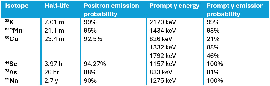
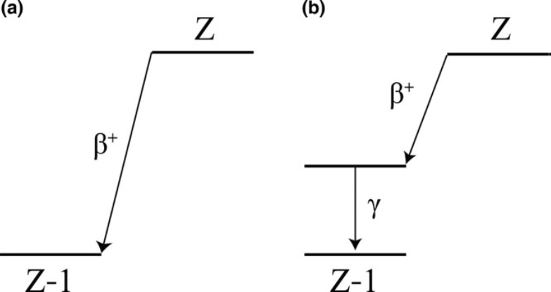
To achieve this separation, mPET exploits positron-gamma emitters such as 124I, for instance, which in addition to emitting positrons, emit an additional prompt gamma photon following the positron decay. Such isotopes decay to an excited state of the daughter nucleus, followed by near-instantaneous emission of a de-excitation gamma photon. This additional photon enables the detection of triple coincidence events, providing more biological information in a single scan.

Decay schemes figure

Using a triple-emitting radiotracer in combination with a pure positron emitter enables mPET scanners to achieve effective signal separation by utilizing an expanded energy window (350–700 keV, for example), which enables capture of both the 511 keV annihilation pairs and the higher-energy prompt gamma photons.

These data are then sorted into two streams: the primary dataset, which includes all detected LORs from both isotopes, and a smaller, tagged dataset containing only the triple coincidences. These triple events are identified via a specific timing selection rule, ensuring that the time difference between the prompt gamma detection and the average detection time of the annihilation photons falls within a narrow coincidence window, typically around 4.5 ns.

Examples of positron-emitting isotopes

To reconstruct the separate radiotracer activity distributions, specialized image reconstruction strategies can be used to address the noise and artefacts inherent in basic subtraction methods. One approach is LOR sorting, which compares line integrals from the initial reconstruction to determine the likelihood that a specific LOR corresponds to one of the two isotopes. Furthermore, triple events can be reconstructed using V-shaped LORs, combining two probable LORs from a triple event into a single geometric unit to more accurately approximate the radioactive origin.

This process requires a spatially variant normalization factor that corrects for the camera’s varying efficiency in detecting prompt gammas across the field-of-view, as certain areas may be shadowed by the scanner geometry. Accurate reconstruction must also account for single-photon attenuation correction for the prompt gamma as it travels through the body.

By generating distinct datasets within a single scan, this method provides perfectly co-registered functional maps, allowing clinicians to simultaneously characterize multiple biological processes within a tumour in a single imaging session.

Towards personalized radiotherapy

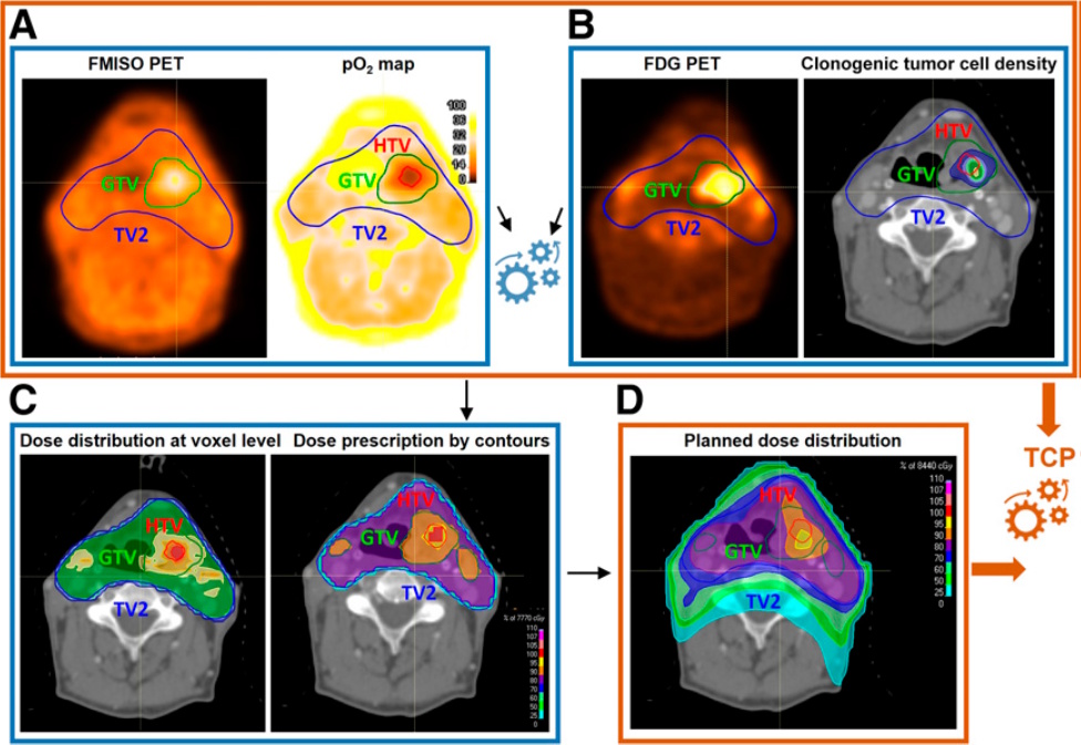
The introduction of mPET facilitates the transition towards biologically individualized radiotherapy, by delivering perfectly co-registered functional maps in a single imaging session. One promising application is the treatment of head-and-neck squamous cell carcinoma, where the radiotracers 18F-FDG and 18F-FMISO have been used to map clonogenic cell density and hypoxia-related radioresistance, respectively.

Biologically individualized radiotherapy

Using radiobiological modelling, radiotracer uptake is converted into voxel-level cellularity maps via linear functions and oxygen partial pressure (pO2) maps via nonlinear sigmoid functions. These biomarkers inform “dose-painting” strategies that strategically escalate radiation to radioresistant areas, such as the hypoxic target volume, while maintaining safe limits for adjacent organs-at-risk. Modelling indicates this synergistic approach could increase tumour control probability from the clinical standard of 60% to a projected 90% or higher.

Researchers have also validated the feasibility of mPET in melanoma mouse models. Here, mPET successfully separated the signals of the triple-emitter 124I-trametinib (targeting proliferation) and 18F-FDG (targeting metabolism). This preclinical trial confirmed that mPET’s ability to separate dual isotopes offers a more detailed and timely assessment of tumour biology.

Future outlook

The clinical translation of mPET represents a significant potential advancement over traditional sequential PET scanning, providing an inherently quicker, cheaper and safer approach. By acquiring dual functional maps simultaneously, the second CT scan required in sequential procedures is no longer needed, roughly halving the patient’s cumulative radiation exposure.

Furthermore, mPET offers the advantage of shorter study duration, as both radiotracers are imaged simultaneously, eliminating the need to wait for the first to decay or wash out before injecting the second. This operational efficiency not only enhances patient compliance but also reduces total costs by minimizing scanner time and overheads. Crucially, mPET is highly viable for near-term implementation as it requires no modifications to existing hardware or acquisition software, when using standard clinical systems, such as the Siemens Biograph mCT, for example.

Despite these advantages, the primary technical pitfall remains the low statistics of the tagged “triples” dataset, which typically represents only a small fraction of total events. This statistical scarcity can introduce significant noise and “shadow” crosstalk artefacts into reconstructed images, potentially affecting quantitative accuracy. To mitigate this, ongoing research into bilateral guided filters and specialized V-shaped LOR algorithms is essential.

In addition, while the physics is compatible with current hardware, many clinical software packages still lack built-in capability for simultaneous multi-energy window acquisition or automated triple-coincidence tagging. This requires the development of manual workarounds that must be standardized for hospital use.

In the next five to 10 years, as the field moves from discovery into prospective interventional trials, the integration of machine learning for multi-parametric analysis will likely refine signal separation and tumour characterization. Looking further ahead, simultaneous imaging is not necessarily limited to two radiotracers: by utilizing multiple positron–gamma emitters and detecting their unique prompt gamma energies, mPET could evolve into “several-colour” imaging, capable or tracking three or more biological processes at once.

Ultimately, if upcoming trials confirm that predicted gains in tumour control probability translate into actual long-term patient survival, mPET may revolutionize oncology by enabling the first truly biologically individualized radiotherapy.

Quiz of the week: how many galaxies and quasars are in the biggest high-res 3D map of our universe?

Fancy some more? Check out our puzzles page.

Dark energy survey unveils the largest 3D map of the universe

The Dark Energy Spectroscopic Instrument (DESI) has created the largest high-resolution 3D map of the universe. The work involved observing more than 47 million galaxies and quasars as well as 20 million stars over a five-year period. Researchers will now use the vast dataset to probe the nature of dark energy.

DESI, which began collecting data in 2021, is mounted on the Nicholas U Mayall 4-m Telescope at the Kitt Peak National Observatory in Arizona. It comprises 5000 robot-controlled optical fibres that send light to an array of spectrographs.

This allows DESI to make an extensive map of galaxies and quasars with the spectroscopic data providing a measure of how fast a galaxy is moving away from us, which is determined by a galaxy’s redshift.

By comparing how galaxies clustered in the past with their distribution today, researchers can trace dark energy’s influence. Work published in 2024 found hints that the acceleration of the expansion of the universe has not been constant.

DESI will now use the expanded dataset to further test whether the “cosmological constant” could be evolving over time with the results expected to be published next year.

DESI director Michael Levi, who is based at the Lawrence Berkeley National Laboratory, says the survey has been “spectacularly successful and is “incredibly exciting”.

“The instrument performed better than anticipated,” he says, “We’re going to celebrate completion of the original survey and then get started on the work of churning through the data, because we’re all curious about what new surprises are waiting for us.”

DESI will now continue observations into 2028 and further expand the map by about 20% to include parts of the sky that are more challenging to observe.

STEM stock rising in quantitative finance

Quantitative trading plays an ever-increasing role in the global financial markets. Automated algorithms analyse millions of financial instruments simultaneously, while mathematical models anticipate price movements on nanosecond timescales.

Susquehanna is a proprietary trading firm, meaning it invests its own capital in the markets. Susquehanna’s quantitative researchers – or “quants” – collaborate with traders and technologists to drive the company’s success. Quants design and implement the complex models and algorithms the firm needs to make rapid, well-informed pricing and trading decisions.

The quant advantage

Lyubo Panchev

Lyubo Panchev, a quant at Susquehanna with seven years at the firm, describes how quants collaborate across a wide range of instruments and problem types. “Our quants are all trying to mathematically understand the world and the financial markets,” he says, “and then we use that information to determine whether we want to make a trade or not.” While the challenges vary considerably across the firm’s different trading desks, that shared mathematical mission is what unites them.

The details of this work can differ from quant to quant, from devising new pricing approaches for financial instruments, to finding patterns in data to turn into trading signals, to developing specialized software to implement new trading strategies.

However, specialist knowledge in specific fields is not what Susquehanna is primarily interested in when hiring a new quant. Instead, the firm is looking for the types of transferrable skills that PhD students in STEM fields often possess. “We want to hire people who can reason through first principles and feel comfortable working in an uncertain environment with open-ended questions to which answers sometimes might not even exist,” says Panchev. “So that’s why we like to hire PhDs.”

A physicist, for instance, brings the skills and intuition for modelling systems with incomplete information – whether that’s modelling interactions in a complex system or inferring signal from noise in a vast dataset. The mental frameworks used by a theorist studying quantum field theory or an experimentalist analysing data translate surprisingly well to pricing derivatives or spotting anomalies in market behaviour.

Life outside academia

Panchev – a three-time International Mathematical Olympiad medallist with a PhD in pure mathematics from MIT – says that the most satisfying part of working at Susquehanna for him is that it preserves what he loved about academia, while at the same time addressing some of the shortcomings.

“The freedom to work on what you want is a unique advantage in academia, over pretty much any industry,” says Panchev. “But what quant researchers do at Susquehanna is close to that spirit.”

Though he enjoyed focusing on challenging questions surrounded by like-minded people, he found working on hyper-specialized academic problems during his PhD a slow, lonely slog. At Susquehanna, quants work on challenging problems, but never in isolation. Quantitative trading problems are invariably interconnected, requiring close collaboration between researchers, traders, technologists and many other experts, to connect all the pieces together.

What’s more, the environment is highly dynamic. “The impact is much more immediate, sometimes instantaneous,” he adds. “You can be looking at the data and then decide to make a change to your algorithm, tweak a few things, and five minutes later, you’re already getting data that’s from the change you just made – it’s a very fast feedback loop.”

When you add a highly desirable salary, benefits package, career development opportunities, and a company culture that values strategy games like poker to hone decision-making skills and apply them to complex financial markets, it is clear to see why a STEM PhD student might choose Susquehanna over a career in academia.

From toy problems to market mastery

To earn a seat at this table, applicants are put through their paces. The first and perhaps greatest challenge they face is getting through the interview process. Quant skills – like original thinking, intuition, and problem-solving – are not easily described in a CV or interview, they need to be demonstrated. But how can an applicant demonstrate those skills in an interview?

“We build interesting toy problems that are representative of what we do,” explains Panchev. “And then we give them time to think and work on it on their own, before reconvening to see how they approached the problem, and what they found out.”

The internship builds solid foundations in finance domain knowledge, machine learning, programming and data analysis

Successful applicants who are hired on immediately participate in a comprehensive 10-week internship – the first step in an intensive front-loaded education program at the company. This internship builds solid foundations in finance domain knowledge, machine learning, programming, data analysis, as well as what Susquehanna’s different quant groups do and how their work all fits together.

Panchev says that a typical direct full-time hire requires five months or more of very structured education, over time, however, the quant will be faced with more open-ended problems and need to chart their own way, free to explore their own ideas and methods.

“There’s a long, steep learning curve but at the end you become an expert,” he adds. “In a way, it’s very similar to how a PhD is structured.” This means that, while the barrier to entry is fairly high, the support system is robust, with a well-organized education program that ensures that everyone is equipped with the tools that they need to succeed.

For the successful STEM PhD student assessing their career options, Susquehanna offers a compelling proposition – the chance to remain a scientist, but on a stage where the stakes are higher, the collaborations deeper and more dynamic, and the results play out in real-time and have real-world impact.

Memristive synapses could reduce AI energy consumption

A new highly stable and energy-efficient memristor based on a hafnium oxide material can emulate the behaviour of synapses in the brain. The neuromorphic device could help dramatically cut the energy consumed by artificial intelligence (AI) hardware, say its developers at the University of Cambridge in the UK.

Today’s AI systems rely on conventional digital computers. These have separate processing and storage units and consume huge amounts of energy when performing data-intensive tasks. As global AI use is exploding, this energy consumption has already become unsustainable, says materials scientist Babak Bakhit, who led this new study.

An alternative way to process information

Neuromorphic computers could provide an alternative way to process information. As their name suggests, they are inspired by the architecture of the human brain. The circuits in these computers are made up of highly connected artificial neurons and artificial synapses that simulate the brain’s structure and functions. These machines have combined processing and memory units that allow them to process information at the same time as they store it, in the same way as a multi-tasking human brain. This means they could reduce energy consumption by as much as 70% compared with their digital counterparts.

Memory-resistors, or memristors, have become a fundamental building block of such neuromorphic architectures. This is because they can be engineered to behave very much like neurons in the human brain, which learn by reconfiguring the strengths of the connections (synapses) between neurons. Memristors excel in this respect as they can bring this learning functionality to the connections in electronic circuits.

First described theoretically in 1971, it was not until 2008 that researchers made the first practical version of a memristor. These devices are special in that their resistance can be programmed and subsequently stored. This is because, unlike standard resistors, the resistance of a memristor changes depending on the current previously applied to it – hence the “memory” in its name. What is more, the device “remembers” this resistive state even when the power is switched off.

Randomness in switching behaviour is a problem

All well and good, but most of today’s memristors unfortunately suffer from randomness in their switching behaviour because they rely on the formation of tiny conductive filaments in the materials making them up. These filamentary devices also typically require high forming and operating voltages and extra devices to avoid uncontrolled current changes that lead to permanent device failure. These challenges make such devices difficult to scale up for real-world applications, says Bakhit.

The researchers, who report their work in Science Advances, claim to have overcome the intrinsic stochasticity of memristive switching by exploiting a completely different switching mechanism – based on carefully engineered heterointerface physics rather than random filament switching. They achieved this by adding strontium and titanium to a hafnium-oxide thin film, which results in the formation of a p-n heterointerface. This junction allows the device to change its resistance smoothly by shifting the height of an energy barrier at the bottom interface through the migration of electro-ionic charges, explains Bakhit.

The new interfacial device has an ultralow switching current of less than or equal to 10-8 A, which is around 106 times lower than those of conventional oxide-based memristors. It also produces hundreds of distinct and stable conductance levels that can be easily modulated, a key prerequisite for analogue “in-memory” computing. And that’s not all: the device can also undergo tens of thousands of switching cycles without losing its programmed states for around a day.

Looking ahead, the researchers say they will now be focusing on translating their material and device breakthrough into a functional computing system. “In particular, we are working on reducing the thin-film growth temperature (which currently stands at around 700 °C) so that it is compatible with standard semiconductor manufacturing (CMOS) tolerances,” says Bakhit. “We will then scale up device arrays to demonstrate large-scale integration.”

Ultimately, the goal is to move from individual devices to fully integrated neuromorphic chips that can compete with, or surpass, conventional AI hardware in both performance and energy efficiency, he tells Physics World.

Word flower puzzle no. 3

How did you get on?

10 words Warming up nicely

16 words Getting hot, hot, hot

22 words Top dog!

Fancy some more? Check out our puzzles page.

Collisional quantum gates created using fermionic atoms

Collisional quantum gates based on fermionic atoms have been realized independently by researchers in Germany and Switzerland. The gates are a long-proposed building block for quantum processors, but had been very challenging to create.

Both teams’ gates achieve entangling operations with a fidelity above the theoretical threshold for quantum error correction – and could potentially be particularly useful for simulations of quantum chemistry.

The potential of collisional quantum gates was proposed in the late 1990s by researchers such as Peter Zoller of the University of Innsbruck in Austria and Ivan Deutsch of the University of New Mexico in the US. The underlying principle is that the states of qubits are encoded into the spin states of atoms in an optical lattice. Then, gate operations between qubits are performed by manipulating interactions between the atoms’ wavefunctions. Experimental attempts followed shortly after, but the technology of the time was insufficient to create practical gates.

Early schemes

“Schemes were developed to move the atoms using state dependent potentials, but the laser light was too near resonant, so it worked in principle, but in practice there was too much heating involved,” explains Konrad Viebahn of ETH Zurich and a member of the Swiss team.

German-team member Petar Bojović of the Max Planck Institute for Quantum Optics in Garching adds that imaging the resulting gates was another problem: “They got some first collisional gates showing proof of principle that this could possibly be done at around the same time as they did [trapped] ions, but they couldn’t move further and scale this up or do many more things with it because there was no way to really see the individual qubits and individual gates”.

Since those early days, much progress has been made in quantum-computing schemes that use neutral atoms held in optical tweezer arrays. During a gate operation, one atom is laser excited to a high-energy, large-size Rydberg state in which its wavefunction easily overlaps with the other atoms – allowing atomic qubits to interact.

There are, however, challenges associated with this architecture. Rydberg states are loosely bound, so the qubits are prone to disruption by classical noise. Furthermore, ensembles of Rydberg atoms tend to be large and this is a barrier to scaling-up the architecture.

Robust collisional quantum gates

Bojović and colleagues at the Max Planck Institute led by Titus Franz and Viebahn’s group at ETH Zurich now unveil independent work on new, more robust collisional quantum gates using fermionic lithium-6 atoms. Lithium has the advantage of being lighter, which allows for faster gates.

Most prior work on collisional quantum gates has used bosonic atoms, explains Viebahn, but using fermions makes the gates more robust because the exclusion principle guards against gate errors: “For our [collisional] implementation, the wavefunctions are allowed to overlap completely, and this amplifies the effects of quantum statistics,” he says.

Both groups produced two-qubit gates, including those able to perform entangling operations, with fidelities of over 99%. The Max Planck researchers controlled the interactions between the qubits by manipulating the potential barriers between them. They utilized an optical lattice among the most stable in the world, together with a quantum gas microscope that allowed single-site resolution.

“There’s been some criticism from other communities,” says Bojović; “Once you get to a regime of ‘ninety-nine point something’ fidelity, you really need to be able to see it precisely in order to characterize it.” The researchers would like to go on to demonstrate all the other gates in a universal quantum gate set, but Bojović says that researchers in quantum chemistry are already intrigued by the potential of the platform to simulate molecular behaviour.

Different protocol

The ETH Zurich researchers used a different protocol involving control of the bias voltage to couple the quantum states of their fermionic atoms rather than manipulation of the barrier height. The researchers have not achieved single site resolution – they are currently working to do so – but Viebahn believes his group’s protocol should prove more robust to noise.

“I would say the key novelty here is that we came up with this more robust way of doing this interaction, which was not part of the original proposals from the 90s,” says Viebahn. “We’re the first to implement this gate where these qubits form this fully overlapped quantum state.”

Both groups’ two-qubit gate fidelities are well above the theoretical minimum required for quantum error correction (QEC) to be possible. However, implementing QEC will be difficult  because creating the required universal gate set involves a complete set of single qubit gates as well as at least one two-qubit gate that can generate entanglement in the system. Nevertheless, Viebahn concludes, “The two-qubit gate is limiting many other quantum computing platforms, and that’s the thing that we’re very good at.”

The collisional quantum gates are described in two papers in Nature: links to the Max Planck paper and the ETH Zurich paper.

Quantum-computing expert Barry Sanders of University of Calgary in Canada says the papers “have two different purposes and both purposes are significant”. The Max Planck paper, he says, is especially impressive because it opens up the potential to simulate the Fermi–Hubbard dynamics of strongly-correlated electronic systems directly in a quantum simulator. The ETH Zurich paper, meanwhile, uses Fermi dynamics to offer gate operation protection against time-dependent sources of error. “There’s a lot of rich physics available with two fermions at a site,” he says.

Copyright © 2026 by IOP Publishing Ltd and individual contributors