Eileen Collins, NASA astronaut: commanding missions and making history

Eileen Collins was the first woman to pilot, and later command, NASA’s Space Shuttle. She logged over 38 days in space and, as the commander of mission STS-93 in 1999, her legacy includes deploying the Chandra X-Ray Space Observatory. The story told in Collins’ autobiography Through the Glass Ceiling to the Stars is one of triumph over adversity, one that can inspire and excite the reader – but there is also a poignancy to reading it at this point in history.

Collins’ debut piloting the Space Shuttle in 1995 was also NASA’s first mission to approach and fly around the Russian space station, Mir (without docking). When Discovery was at the point of closest approach, Shuttle commander Jim Wetherbee said to Mir commander Aleksandr Viktorenko “Together we will lead our world into the next millennium,” to which the Russian cosmonaut responded “We are one. We are human.”

The Shuttle–Mir programme marked the beginning of a period of international collaboration in space, which, for many, reached its zenith with the International Space Station. In Collins’ book, we read stories of the fun that NASA and Roscosmos astronauts had while working together, even as politicians tangled with each other across borders on Earth. It is all a stark contrast to Vladimir Putin’s internationally condemned invasion of Ukraine – a tragedy that is now threatening even this most cherished of international partnerships.

Anyone looking for a spot of escapism to a time of collaboration, or an inspirational read, will find both among the pages of this book. The preface is a compelling description of Collins leaving Earth for the first time on board Discovery as part of that 1995 STS-63 mission. Such is the skill with which we are transported into the shuttle as it escapes the clutches of Earth’s gravity that readers could be forgiven for thinking they’d opened the latest Andy Weir novel. We leave the crew just as the curvature of the Earth comes into view through Discovery’s windows, and re-join them in a later chapter, but first we discover how you get to be the first woman to pilot a NASA Space Shuttle.

It might be tempting to think of astronauts as starting from some position of privilege, but it becomes clear that Collins has succeeded in the face of adversity. Her father was “the worst kind” of alcoholic, which had serious consequences for the whole family. Her mother became suicidal and was institutionalized, leaving Collins to run the home for herself and her two younger siblings.

Collins had a number of jobs to pay her way through an associate degree in maths and science at Corning Community College, including working the counter at a pizza restaurant and selling tickets at a pitch-and-putt golf course. From there her journey to space follows a familiar route to anyone who has read astronaut autobiographies – after training as a pilot she then worked as a pilot trainer before becoming a flight test pilot. While it’s no longer the only way to becoming an astronaut, as it was in the time of the Apollo missions, it is still a tried and tested route followed even by the likes of Tim Peake.

Once the “dream comes true”, the book examines each of Collins’ four journeys to space. As well as her inaugural mission in 1995, Collins piloted STS-84 in 1997, during which the Discovery crew boarded the Mir space station – the sixth such docking. But it was the 1999 mission, STS-93, that saw Collins become the first woman to command the Space Shuttle. The primary objective of this mission was to deploy the Chandra X-Ray Observatory, which still provides data today, having led, for example, to the discovery in March 2021 that Uranus emits X-rays.

Next follows a deeply affecting chapter retelling the tragedy of Columbia, the Space Shuttle that disintegrated upon re-entry in 2003, from the perspective of those in the programme. Collins’ fourth and final trip, STS-114 in 2005, was NASA’s first “return to flight” mission after the accident. To read of the emotional resolve of the astronauts at the centre of that historic time is one of the most compelling segments of the book.

Collins’ determination and strength of character shines throughout this book, with the title pointing to the misogyny that Collins, like so many women, had to confront and, at times, rise above. There is no sense of complaint in the pages, but the reader will become aware of a series of microaggressions that confronted Collins in her rise to the peak of her profession.

I like to believe that my story shows that people who play by the rules and act with character and integrity can indeed finish first

Eileen Collins

There is a common misconception that to get to the top, one must “tread on a few toes” or burn a few bridges. Towards the end of the book, Collins reflects, “I like to believe that my story shows that people who play by the rules and act with character and integrity can indeed finish first.” It is just one lesson to take from a book that I would recommend to anyone with the slightest interest in human space flight, or who is striving to be the best person they can be in any walk of life.

  • 2022 Simon & Schuster £20hb

‘Nanotwinning’ produces stronger metals

When steel, aluminium and other widely used metals or alloys pass through industrial processes such as machining, rolling and forging, their nanoscale structure undergoes dramatic changes. Extremely fast production processes make it difficult to analyse these changes due to the sheer speed and small scale at which they take place, but researchers at the Massachusetts Institute of Technology (MIT) in the US have now succeeded in doing exactly that, pinning down what happens as crystal grains form in the metal under extreme deformation at the nanoscale. Their work could help in the development of metal structures with improved properties, such as hardness and toughness.

In general, the smaller these crystal grains are, the tougher and stronger the metal will be. Metallurgists often seek to shrink grain size by placing the metals under strain. One of the main techniques they use to do this is recrystallization, in which the metal is deformed at high strain and heated to produce finer crystals. In extreme cases, this process can produce grains with nanoscale dimensions.

“Not just a laboratory curiosity”

The MIT team led by Christopher Schuh has now determined how this high-speed, small-scale process takes place. They did this by using a laser to launch copper metal microparticles onto a metal at supersonic speeds and observing what happened when the particles struck it. Schuh points out that such high speeds are “not just a laboratory curiosity”, with industrial processes such as high-speed machining; high-energy milling of metal powder; and a coating method called cold spray all taking place at similar rates.

“We’ve tried to understand that recrystallization process under those very extreme rates,” he explains. “Because the rates are so high, no one has really been able to dig in there and look systematically at that process before.”

In their experiments, the researchers varied the speed and strength of the impacts and then studied the impacted sites using advanced nanoscale microscopy methods such as electron backscatter diffraction and scanning transmission electron microscopy. This approach allowed them to analyse the effects of increasing strain levels.

They found that the impacts dramatically refine the structure of the metal, creating crystal grains just nanometres across. They also observed a recrystallization process that was helped along by “nanotwinning” – a variation of a well-known phenomenon in metals called twinning, in which a specific kind of defect forms when part of the crystal structure flips its orientation.

Schuh and colleagues observed that the higher the impact rates, the more frequently nanotwinning took place. This leads to ever-smaller grains as the nanoscale “twins” break up into new crystal grains, they say. The process could increase the metal’s strength by about a factor of 10, which Schuh describes as non-negligible.

A better mechanistic understanding

Schuh describes the team’s result as an extension of a known effect called hardening that comes from hammer blows in ordinary metal forging. “Our effect is a sort of hyper-forging type of phenomenon,” he says. Although the result makes sense in that context, Schuh tells Physics World that it could lead to a better mechanistic understanding of how metal structures form, making it easier for engineers to design processing conditions to control these structures. “The very small, nanoscale structures we observed in our work are of interest for their extreme strength, for example,” he says.

According to team member Ahmed Tiamiyu, the new findings could be directly applied right away to real-world metal production. “The graphs produced from the experimental work should be generally applicable,” he says. “They’re not just hypothetical lines.”

In the study, which is published in Nature Materials, the researchers focused on understanding the evolution of a metal’s structure during an impact. It would be interesting to study other characteristics, such as how the temperature around an impact site evolves, they say. “We are conducting work in this direction now,” Schuh reveals.

Water harvesting gel works at low humidity levels

Researchers in the US have designed a sustainable polymer gel that can harvest large quantities of water from the surrounding air, even in low-humidity conditions. Created by Youhong Guo and colleagues at the University of Texas at Austin, the low-cost material combines water-absorbing plant fibres with cellulose, which expels water when heated.

Many parts of the world experience some degree of water scarcity and the problem is expected to grow with increasing global warming. Extracting moisture directly from the atmosphere could provide millions of people with vital access to clean water. Researchers have already developed a variety of different porous materials that can capture and release moisture on demand – but these often require humid conditions. In dryland regions, now home to over a third of the world’s population, existing techniques suffer from low water uptake and high energy demand.

To address this challenge, Guo’s team developed a new polymer material, containing a hybrid of konjac gum (KGM) – a plant-based fibre widely used in Asian cuisine — and hydroxypropyl cellulose (HPC). This polymer matrix also contains a uniformly-dispersed solution of lithium chloride – a moisture-retaining salt.

Large surface area

Within the material, hydrophilic KGM has a hierarchically porous structure. This provides a large water-collecting surface area, while also allowing water vapour to rapidly pass through the structure. When heated,  HPC undergoes a phase change, and its previously straight polymer fibres contract into amorphous, twisted shapes. In the process, any moisture in the KGM fibres is rapidly released.

Guo and colleagues have shown that in 14–24 cycles of water uptake and release in arid conditions, 1 kg of the gel can produce more than 6 l of water per day in 15% relative humidity. At 30% relative humidity, up to 13 l per day can be produced.

The researchers also showed that the polymer can be easily produced through a user-friendly casting method, where a gel precursor containing all three ingredients is mixed and poured into a mould. After 2 min, the mixture is freeze-dried and peeled from the mould, ready to be used straight away.

On top of this, the material’s three ingredients are abundant and can be sourced sustainably. Altogether, the ingredients cost just $2 per kilogram. Guo’s team hope that the low cost and simplicity of production will mean that the gel can be produced commercially. They predict that far larger quantities of water could be readily harvested by fabricating thicker films and introducing absorbent beds to the gel.

The research is described in Nature Communications.

Deep learning enables fast and accurate proton dose calculations

Successful radiation therapy relies on the creation of an accurate treatment plan that will deliver radiation dose precisely to the prescribed targets. The accuracy of this plan, however, is only as good as the accuracy of the underlying dose calculations. And for proton therapy, accurate dose calculation is even more critical, as protons deliver a more conformal dose distribution than photons and are more sensitive to anatomical changes.

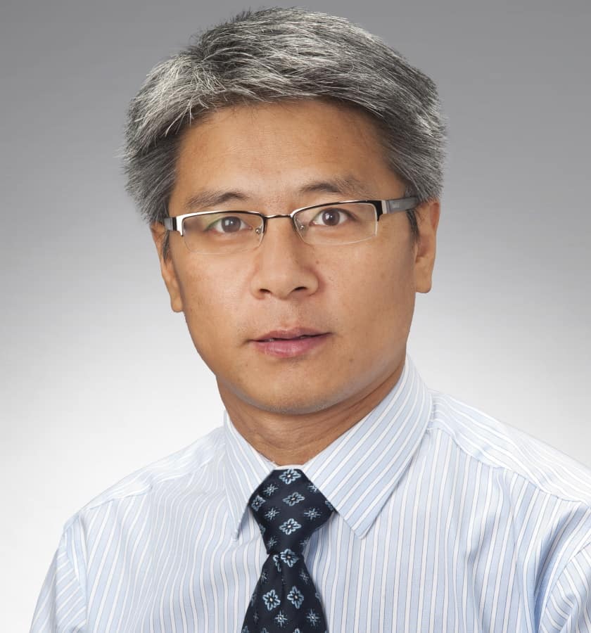
Steve Jiang

Speaking at the Mayo Clinic’s 1st Proton Therapy Research Workshop, Steve Jiang – professor and director of the Medical Artificial Intelligence and Automation (MAIA) Laboratory at UT Southwestern Medical Center – described the key requirements of proton dose calculation – and described ways in which deep learning could help achieve these goals.

As well as high accuracy, Jiang explained, proton dose calculations also need to be fast. For treatment planning this means a few minutes; for replanning prior to fraction delivery in adaptive radiotherapy, a few seconds. Looking further ahead, we may see the introduction of real-time adaptation during treatment delivery. “We don’t do this right now,” he noted. “But at some point we may want to adapt the treatment plan in real time. For that kind of application, we will need dose calculation in milliseconds.”

Currently, there are two main types of technique used for dose calculation, represented by: pencil beam algorithms, which are less accurate but quite fast; and Monte Carlo (MC) simulations, which are more accurate but typically far slower. “But we need accuracy and speed for proton dose calculations,” said Jiang. “So there’s an unmet clinical need: we need to develop an algorithm that is both fast and accurate.”

So how can this be achieved? One approach is to improve the efficiency of MC calculations, using graphics processing units (GPUs) to accelerate MC code, for example, or deep learning-based denoising to reduce the noise inherent in MC-calculated results. Another option is to employ deep learning methods to improve the accuracy of pencil beam algorithms. Finally, it may be possible to develop new, totally different algorithms that meet both requirements; and deep learning could help explore this possibility.

Combining speed and accuracy

GPU-acceleration of MC simulations is already possible. Ten years ago (while at UC San Diego and in collaboration with Mass General Hospital), Jiang and colleagues developed gPMC, a MC package for fast proton dose calculation on a GPU. This enabled calculation of a typical proton treatment plan with 1% uncertainty in 10–20 s. Jiang notes that with today’s faster GPUs, gPMC may offer even higher efficiency.

Working with colleagues at the MAIA Lab, Jiang has also developed a deep learning-based MC denoiser. They created a deep dose plugin that can be added to any GPU-based MC dose engine to enable real-time MC dose calculation. The denoiser runs in just 39 ms, with the entire dose calculation taking just 150 ms. Jiang notes that the plugin was developed for photon beam radiotherapy, but could also be used for MC denoising in proton dose calculations.

Next, Jiang described ways to use deep learning techniques directly for dose calculation. He emphasized that this differs from dose prediction, which assumes a relationship between a patient’s anatomy and their optimal dose distribution, and uses this relationship to build a predictive model. After training on data from historical treatments of the same disease site, the model predicts an optimal dose distribution for the new patient and uses this to guide treatment planning. UT Southwestern has employed this type of patient-specific dose prediction clinically for over two years now.

But dose calculation is more than this. “Here, the relationship we are trying to exploit is between patient anatomy plus machine parameters and the actual dose distribution,” said Jiang. “You know the patient anatomy, you know the treatment plan, now you want to see what is the dose distribution, so it’s a dose calculation.”

Jiang’s team first developed the deep learning-based dose calculation model for photon beam radiotherapy. The model is trained using MC-calculated dose distributions for various patient anatomies and machine parameters. For the model inputs, the team used the patient CT scan and the ray tracing dose distribution for each beam, with machine parameters encoded into the ray tracing. “This makes the whole deep learning process easier and is a good way to incorporate physics into the deep learning,” Jiang noted.

The researchers applied a similar approach for proton dose calculation, using a deep learning model to boost the accuracy of pencil beam dose calculation to that of MC simulations. They trained and tested the model using pencil beam dose distributions and data from the TOPAS MC platform, for 290 head-and-neck, liver, prostate and lung cancer cases. For each plan, they trained the model to predict the MC dose distribution from the pencil beam dose.

The approach achieved high levels of agreement between the converted and the MC dose. “Compared with pencil beam, we see a huge improvement in accuracy, and the efficiency is still very high,” said Jiang. The developed model can be added to the clinical workflow of proton treatment planning to improve dose calculation accuracy.

Jiang also highlighted similar research under way by other groups, including DiscoGAN from Wuhan University, DKFZ’s use of artificial neural networks for proton dose calculation and deep learning-based millisecond speed dose calculation algorithm developed at Delft University of Technology.

Keeping users reassured

While deep learning may appear the obvious way forward for proton dose calculation, Jiang noted that people still feel more comfortable using physics-based models such as pencil beam algorithms and MC simulations. “When the idea of deep learning for dose calculation first came out, people had concerns,” he explained. “Because it’s data driven, not physics-based, you do not know when it’s going to fail; there might be unpredictable catastrophic failures. And because it’s a black box there’s no transparency.”

The answer may lie in hybrid models, such as the examples described above that use pencil beam or ray tracing data as inputs to a deep learning model. Here, the physics (machine parameters) is encoded in the input data, which already has an accuracy of 80–90%. Deep learning can then address effects such as scatter and inhomogeneity to gain the remaining 20% accuracy that’s very difficult to achieve with analytical algorithms. This should provide both the desired accuracy and efficiency.

“I actually think this is a good idea because it can also eliminate unpredictable, catastrophic failures,” Jiang concluded. “I’d feel much more comfortable with the results. Also you’d have some degree of transparency, because you know the first order primary effect that is there is physics-based, and that’s correct.”

Sun NuclearAI in Medical Physics Week is supported by Sun Nuclear, a manufacturer of patient safety solutions for radiation therapy and diagnostic imaging centres. Visit www.sunnuclear.com to find out more.

New cylindrical water scanning system unlocks efficiencies in beam commissioning, annual QA

Faster, easier, hyper-accurate: these are the key benefits of the new SunSCAN 3D Cylindrical Water Scanning System from Sun Nuclear Corporation, a US-based manufacturer of QA solutions for radiotherapy and diagnostic imaging providers. Think faster and easier workflows for medical physicists tasked with beam commissioning and annual and biannual machine QA. Think hyper-accurate dosimetry for the growing demands of stereotactic radiosurgery (SRS) and stereotactic body radiotherapy (SBRT). Think granular beam data and beam model visualizations to ensure the treatment machine is calibrated accurately while also guaranteeing “gold-standard” verification of the delivered dose applied to the patient.

Automation means reproducibility

Building on the capabilities of Sun Nuclear’s 3D SCANNER water tank, with over 1000 systems in use worldwide, the SunSCAN 3D was formally unveiled to the market last month at the ESTRO annual congress in Copenhagen, Denmark.

“Feedback from delegates was phenomenal – whether from existing users of our 3D SCANNER or prospective new customers,” explains Julia Kirchhefer, a medical applications physicist at Sun Nuclear. “SunSCAN 3D’s automated set-up was one of the main conversation topics on the booth,” she adds, “not least with ESTRO attendees accustomed to manual preparation of their water tanks – a time-consuming exercise that can take up to an hour.”

With SunSCAN 3D, form, function and automation come together to make the medical physicist’s job easier to do. Specifically, the AutoSetup routine takes about seven minutes to provide a truly levelled water tank (to within 0.05° and centred within 0.1 mm). AutoSetup is in fact a stepped process comprising Auto Level (three widely set points quantify tank “levelness” with a repeat measurement to confirm set-up); Auto Centre (using profile measurements, fine adjustments in the x and y direction align the centre of the SunSCAN 3D with the beam centre); and Auto Angle Offset (for alignment of the ring centre and angular orientation to the collimator axis).

Crucially, the whole SunSCAN 3D workflow is automated to guarantee a fast and reproducible tank set-up that is independent of the user’s experience. “You could have three different people set up the tank and it will be in the exact same spot each time,” adds Kirchhefer. “Alongside the built-in workflow efficiencies, that also means a significantly reduced training overhead.” Underpinning it all, the SunSCAN 3D delivers exceptional scanning performance for SRS/SBRT treatments, with a coordinate measuring machine to verify 0.1 mm accuracy throughout the tank, 0.05 mm reproducibility and 0.02 mm resolution.

Intuitive innovation

Meanwhile, the benefits of SunSCAN 3D’s unique cylindrical design are also front-and-centre in the treatment room. Operational streamlining is again one of the main drivers here, with the cylindrical design removing the need for time-consuming tank shifts that can adversely impact data quality (see “User-centric cylindrical design”). “The physicist can use the full diameter of the tank as the scanning range – there’s no need for tank shifts,” notes Kirchhefer. The cylindrical geometry also ensures consistent detector orientation, with the ability to rotate the tank by 360° such that the smallest dimension of chamber always measures beam edge for the sharpest penumbra.

Reinforcing the granular emphasis on workflow efficiency is SunSCAN 3D’s SunDOSE software, an intuitive “fewer clicks” interface that’s intended to make machine commissioning and QA easier than ever. “With SunDOSE, it’s straightforward to perform tank set-up and collect and store data in the SQL database to meet the unique needs of the medical physics team,” explains Kirchhefer. “What’s more, the comparison of scans from annual checks or commissioning is quick and easy – taking just a couple of minutes – as every scan is tagged versus energy, field size and scan type.”

Spreading the word

Going forward, the priority for Kirchhefer and her Sun Nuclear colleagues is to get the SunSCAN 3D in front of clinical customers at scale. After debuting the system at the ESTRO annual congress, the system was subsequently showcased at the annual meeting of the Germany Society of Radiation Oncology (DEGRO) and the World Congress on Medical Physics and Biomedical Engineering.

SunSCAN 3D will also be featured at the AAPM and ASTRO annual meetings in the US later this year, as well as the European Congress of Medical Physics in Dublin, Ireland, and the Annual Conference of the German Society of Medical Physics (DGMP) in Aachen, Germany. “In parallel, we’ll be taking the SunSCAN 3D direct to the clinics,” concludes Kirchhefer, “with roadshows in the works for Germany, the UK, Italy and France over the summer.”

User-centric cylindrical design

With square 3D water tanks, it is not possible to measure a full 40×40 cm field at 30 cm depth and 100 cm source-to-surface distance (SSD) unless the user shifts the water tank twice, making two measurements of two “halves” of the beam at different tank locations. This technique is inherently time-intensive and can introduce errors that compromise data quality.

In contrast, the cylindrical shape of the SunSCAN 3D enables the most efficient scanning ranges. For example, a 65 cm scan range is possible without a shift, allowing a 40×40 cm measurement at 30 cm depth and 100 cm SSD – saving time and eliminating potential errors. The 65 cm scan range is achieved with the offset detector holder, whereby two scans are merged without the need for a tank shift.

Bringing time‑of‑flight quality to non‑TOF PET images

PET scanners use time-of-flight (TOF) technology to reduce image noise and improve the identification of cancerous lesions. TOF works by using the time difference between detection of the two PET annihilation photons to more accurately localize the annihilation event. However, many current clinical PET scanners do not have TOF capability, and miss out on the improved diagnostic confidence that it confers.

“There is a significant cost difference between TOF and non-TOF PET scanners because of the high cost of the scintillator used for TOF,” says Daniel McGowan from the University of Oxford and Oxford University Hospitals NHS Foundation Trust, noting that one of GE Healthcare’s most successful product lines is a non-TOF PET scanner, the Discovery IQ. “We estimate that approximately one in three PET/CT sites in the world currently do not have access to TOF technology.”

To level this playing field, McGowan and collaborators are employing deep learning to bring the benefits of TOF to PET images reconstructed without TOF information. Writing in the European Journal of Nuclear Medicine and Molecular Imaging, they describe their proposed deep learning for TOF image enhancement (DL-TOF) approach.

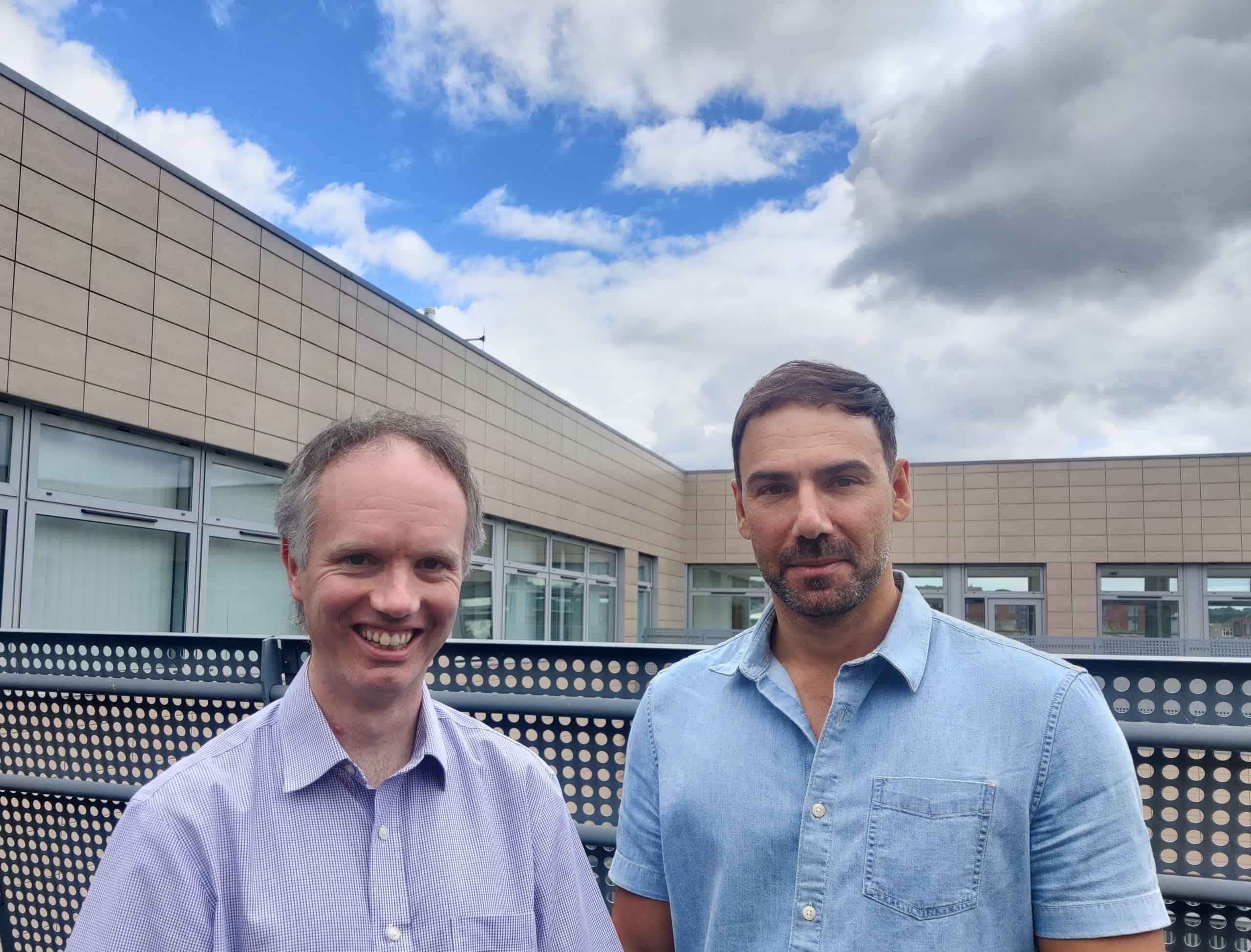
Daniel McGowan and Abolfazl Mehranian

The team developed three DL-TOF models (based on U-Net convolutional neural networks) to transform non-TOF PET data into corresponding TOF-like images. The models employed different levels of TOF strength (low, medium or high) to trade off contrast enhancement against noise reduction.

The researchers note that the neural network does not add TOF information to the PET coincidence data, but rather, it learns how TOF information alters image characteristics and then replicates these changes in non-TOF input images. “This is exactly the kind of task that deep learning algorithms do very well,” McGowan explains. “They can find patterns in the data and create the transformation that produces visually attractive and quantitatively accurate images that give high diagnostic confidence to the reporting radiologist or physician.”

Model evaluation

To train, validate and test the models, the team used PET data from 273 whole-body FDG-PET oncology exams performed at six clinical sites with TOF-capable PET/CT scanners. The PET data were reconstructed using the block-sequential-regularized-expectation–maximization (BSREM) algorithm, with and without TOF.

After training, the researchers evaluated the model performance using a testing set of 50 images. They examined standardized uptake values (SUVs) in 139 lesions and normal regions of liver and lungs, using up to five small lesions and five volumes-of-interest in the lungs and liver per subject.

Comparing the outputs of the three DL-TOF models with the input non-TOF images showed that the models improved the overall image quality, reducing noise and increasing lesion contrast. In the original non-TOF image, the lesion SUVmax differed from the target TOF image by −28%. Applying the DL-TOF low, medium and high models resulted in differences of −28%, −8% and 1.7%, respectively. The models also reduced differences in SUVmean from 7.7% to less than 2% in the lungs, and from 4.3% to below 1% in the liver.

Diagnostic application

In addition to the quantitative evaluation, three radiologists independently rated the testing set images in terms of lesion detectability, diagnostic confidence and image noise/quality. Images were assessed based on a Likert scale, which ranges from 0 (non-diagnostic) to 5 (excellent).

The DL-TOF high model significantly improved lesion detectability, achieving the highest score of the three models. In terms of diagnostic confidence, DL-TOF medium achieved the best score, while DL-TOF low scored the best for image noise/quality. In all cases, the top-performing model outscored the target TOF image. These results highlight how the DL-TOF model can be tailored to balance lesion detection versus noise reduction, according to the preference of the image reader.

“Overall, in terms of diagnostic confidence, the DL-TOF medium model provides a better trade-off in our test set, as a lower noise and improved detectability are desirable features for an image reconstruction or enhancement technique,” the team writes.

Finally, the researchers applied the DL-TOF models to 10 exams acquired on a non-TOF PET scanner, to illustrate the generalizability of the trained models. While there was no ground truth or target image for comparison, visual inspection showed that the images were free of obvious artefacts and exhibited the expected image enhancement. These findings suggest that the models may work on data from scanners that were not part of the algorithm training dataset.

McGowan notes that this initial work focused on whole-body FDG-PET for oncology as this is the main clinical application of PET today. “However, with the advent of new tracers and increased interest in organ-specific imaging, we are currently testing the existing algorithm in the context of these new applications, which were not represented in the training data, and deciding whether additional training is needed to achieve adequate performance for other indications,” he tells Physics World.

Sun NuclearAI in Medical Physics Week is supported by Sun Nuclear, a manufacturer of patient safety solutions for radiation therapy and diagnostic imaging centres. Visit www.sunnuclear.com to find out more.

Telescopes, accelerators and LIGO team up to probe neutron stars

Physicists have created a framework for better understanding the super-dense matter inside neutron stars by combining observations from gravitational-wave detectors and conventional telescopes with experimental results from particle accelerators.

The results, from a team led by Sabrina Huth of Technische Universität Darmstadt in Germany and Tsun Ho (Peter) Pang of Utrecht University in The Netherlands, indicate that many neutron stars experience greater degeneracy pressure in their interiors than predicted. As a consequence, some neutron stars have a larger-than-expected radius – a result that was previously hinted at in observations by the Neutron Star Interior Composition Explorer Mission (NICER) experiment on board the International Space Station.

Neutron stars are among the most extreme objects in the universe. They are the crushed cores of stars that have exploded as supernovae, and despite measuring only around 20 kilometres across, they pack in a mass up to 2.3 times that of the Sun. Inside them, the pressure is so great that negatively charged electrons and positively charged protons are crushed together, forming a body made almost entirely from neutrally charged neutrons.

The term “degeneracy pressure” refers to the inability of any two particles – in this case, neutrons – to inhabit the same energy level when crushed together. This inability produces a countervailing outward pressure that works to prevent neutron stars from being smushed any further. “Therefore, for high pressures, neutrons want to be farther apart, resulting in a larger neutron star,” Pang explains.

Equation of state

Knowing the radius of neutron stars will help astrophysicists constrain the stars’ so-called equation of state, which describes the properties of matter inside a neutron star and therefore dictates its radius. Since nobody knows exactly what the equation of state is, Huth and Pang’s team ran through 15 000 possible versions of it in their modelling, inputting data from several spinning neutron stars known as pulsars as well as gravitational-wave observations of two mergers between two neutron stars. These included the merger known as GW170817, which hit the headlines in 2017 when it was detected by the LIGO gravitational-wave detector and by telescopes observing at wavelengths across the electromagnetic spectrum. As such, it heralded the dawn of multimessenger astronomy.

The latest study took the multimessenger approach still further by incorporating information from collisions between gold ions accelerated to almost the speed of light. These collisions take place at high temperatures and low densities – unlike space, where temperatures are low but the density of objects like neutron stars is high. By combining the results of collisions at several particle accelerators (including the GSI Helmholtz Centre for Heavy Ion Research in Darmstadt as well as Lawrence Berkeley National Laboratory and Brookhaven National Laboratory in the US) with astrophysical observations, it is possible to begin bridging the gap in our understanding of matter in extreme environments.

“Because the data from heavy-ion collisions used in our analysis gives us information about the density region where nuclear theory and astrophysical observations are less sensitive, it provides us with a novel constraint [on the equation of state],” Pang says.

Afterglow consequences

The results also add to scientists’ understanding of what happens during a neutron star merger. In such events, two neutron stars orbiting in close proximity gradually spiral towards one another. As they begin to merge, gravity deforms their shape. This deformation shows up in the gravitational waves they emit during the merger, and is dependent upon the mass and radius of the neutron stars. A neutron star with a larger radius will be less compact and have weaker gravity, which can affect how much debris gets ejected by the merger. It is this glowing debris that is detectable in light as a “kilonova”, so the quantity of debris and its properties determines how visible a kilonova will be.

Nicolás Yunes, an astrophysicist at the University of Illinois at Urbana-Champaign, US, who was not involved in the latest research but who has previously used multi-messenger observations to discern properties of neutron-star matter, thinks we should anticipate more such results in the future. “Multimessenger astronomy is truly transformative and is already having an impact on our understanding of the state of matter at extreme densities and pressures,” he says.

With an upgraded version of LIGO (Advanced LIGO) expected to pick up gravitational waves from many more binary neutron-star mergers, at greater levels of sensitivity, and NICER continuing to provide independent measurements of the radii of pulsars, astrophysicists should soon be able to place even stronger bounds on the equation of state for neutron stars. “By combining information at high temperatures but low densities with information at low temperatures but high densities, we will begin to learn precisely how matter behaves in the universe,” Yunes concludes.

The research is published in Nature.

Continuous Bose–Einstein condensate opens the door to continuous-wave atom lasers

A continuous Bose–Einstein condensate (BEC) has been produced by researchers in the Netherlands. Claimed as a first, the achievement has been sought for years and could lead to continuous-wave atom lasers and a more fundamental understanding of the physics of condensed matter.

BECs form when a gas of bosonic atoms is cooled to ultracold temperatures. A large fraction of the atoms occupy the ground state of the system and the BEC behaves as a macroscopic system described by a single quantum wavefunction. BECs were first made in 1995 and their creators were rewarded with the 2001 Nobel Prize for Physics.

BECs are, strictly speaking, atom lasers, as quantum physicist Florian Schreck of the University of Amsterdam explains: “If you take the word laser as meaning light amplification by stimulated emission of radiation, and you translate all these words one by one to their atomic equivalents, then the process of making this macroscopically occupied mode is the same thing.”

A conventional laser beam is produced by drawing some of light out of an optical mode that exists within an optical cavity. To produce a continuous wave laser, one must pump energy into the cavity mode as fast as energy leaves via the laser beam and other loss processes.

Topping up atoms

One of the central goals of atom optics is to produce a continuous-wave atom laser – a system that delivers a continuous beam of coherent atoms. To achieve this, researchers would have to add new atoms to a BEC at least as fast as atoms in the beam left it.

Whereas photons are essentially non-interacting, ultracold atoms rapidly form molecules, which is usually the biggest cause of atomic loss in a BEC. To sustain a BEC continuously, therefore, physicists need to replenish these atoms rapidly and continuously. This alone has hitherto proved impossible, even without removing atoms to form a laser beam.

In 2013, Schreck and colleagues, then at Austria’s Institute for Quantum Optics and Quantum Information and the University if Innsbruck, created the first BEC by the laser cooling of atoms rather than evaporative cooling. Laser cooling is much faster and does not require most of the sample to be lost. They locked a laser to a narrow-linewidth atomic transition in strontium to cool a cloud of the atoms, while a second laser increased the trapping potential at the centre of the trap. This second laser made the centre transparent to the laser and allowed energy from these atoms, which heated up as their density increased, to diffuse away to the surrounding atoms.

Two-stage process

Laser cooling strontium atoms is a two-stage process, however: first the atoms are cooled from oven temperature using a broad, blue resonance, then a second, much narrower resonance cools the atoms from 1 mK to around 1 μK.

“Unfortunately, this trick that we used to protect the BEC from the laser cooling photons doesn’t work for the broad linewidth laser,” explains Schreck; “So we first cooled our sample using the blue light, then we switched that off.” This sequential approach could only produce a condensate intermittently, therefore.

In the new work, Schreck and colleagues designed a new machine with two separate vacuum chambers. This allowed them to guide a beam of atoms through both and replenish the BEC continuously.

“Instead of executing all these cooling transitions one after the other in time as people always did before, you’re executing them one after the other in space,” explains Schreck. The result was a condensate that was replaced faster than it decayed, allowing it to persist indefinitely.

Great progress

Several groups have previously attempted to implement sequential cooling stages using multiple techniques to cool a variety of atoms, Shreck says. “They made great progress, but they weren’t able to push it all the way through. Now the technology is just more mature, and strontium is just nice because it has this narrow linewidth cooling transition, which made it easier for us.”

The researchers’ main goal is continuous-wave atom lasers, which could find a host of uses in gravitational wave detection, dark energy searches, tests of the equivalence principle and elsewhere. Schreck says it is unclear precisely how much of the beam could be extracted at present as simulations are imprecise, but he is “absolutely confident it is more than 20%” and believes increasing the gain further should prove relatively simple.

Beyond atom laser beams, however, continuous BECs could answer important questions in condensed matter physics. “This is a driven, dissipative system and you can, have, in principle, novel quantum phases and quantum behaviour in dynamic systems where there’s drive and dissipation,” says Schreck, adding. “Theorists are quite interested in this”.

Jun Ye of JILA at the US National Institute for Standards and Technology and the University of Colorado is impressed. “Florian Schreck’s group has been working on a continuous source of ultracold strontium atoms for a number of years,” he says. “It is really satisfying to see that they have made a major breakthrough in combining this technology with a continuous Bose-stimulated scattering of thermal atoms into a Bose-Einstein condensate of strontium-84. This technology, once extended with a capability of continuous output, will have big impact for quantum sensors ranging from matter-wave interferometers to clocks.”

The research is described in Nature.

Climate change affects cherry blossoms, satisfying sizzle gives cooking temperature

Around the world from Kyoto to Washington DC, people enjoy the blossoming of cherry trees as a rite of spring. In some places – notably Japan and South Korea – blossom festivals are vital for local economies, so it is important that organizers get their timings right.

There has always been some year-to-year variation as to when peak blossoming occurs, but in 2021 it happened in Kyoto on 26 March 2021 – the earliest it has ever been since records began over 1200 years ago. More generally, the date of full blossoming has move steadily forward from mid to early April since the 1800s.

Scientists believe that this shift is caused by a combination of global warming and urbanization – the latter placing the trees in an urban heat island.

State-of-the-art climate models

Now, Yasuyuki Aono of Osaka Metropolitan University has joined forces with Nikolaos Christidis and Peter Stott of the UK’s Met Office to work out how global warming will affect the timing of future blossom events. Using historical data and 14 state-of-the-art climate models, the team calculated how blossoming times will change under various global-warming scenarios.

Under a medium greenhouse-gas emissions scenario, the trio reckon that full blossoming will be pushed ahead by nearly a week by the end of the century. That is on top of an 11 day shift forward that has already happened since the 1800s.

They also conclude that very early blossoming events like 2021 are 15 times more likely to happen now because of global warming and urbanization. Furthermore, they say that such events could become commonplace by 2100, happening once every few years. So, useful information for the long-term planning of blossom festivals.

The research is described in Environmental Research Letters.

Satisfying sizzle

When cooking, there is nothing as satisfying as the sizzle of food when it hits hot oil in a frying pan. But how do you know when the oil is hot enough to add your ingredients? In parts of Asia, cooks will put moist bamboo chopsticks into their pans and judge the temperature by watching the bubbles that form and listening to the sizzling sound they emit.

Now, an international team of scientists has looked at the physics underlying this clever test. “Many cookbooks teach this technique and it is widely used, but when we searched the academic literature, we couldn’t find any detailed scientific explanations,” says Zhao Pan of Canada’s University of Waterloo.

The team placed wet paper, moistened chopsticks and water droplets in hot oil and observed what happened using sensitive microphones and high-speed cameras. “We found three distinct types of bubble events in our experiments: an explosion cavity, an elongated cavity and an oscillating cavity,” explains Tadd Truscott at King Abdullah University of Science and Technology in Saudi Arabia.

Explosion cavities form when a water droplet enters hot oil and is vaporized to form a bubble that ruptures the surface of the oil. This is unlike elongated cavities, which explode without rupturing the surface. Oscillating cavities occur when a water droplet undergoes a multi-step explosion process and begins to oscillate before breaking up into numerous small bubbles.

The team found that these bubble events occur at oil temperatures that are favourable for cooking, explaining the chopstick test. Indeed, Pan says that the test can get the temperature right to within 5–10%.

The research is described in Physics of Fluids.

Advanced algorithm predicts outcome for patients with severe brain injury

A team of US-based researchers has created an innovative deep-learning model that analyses CT scans and clinical information to predict six-month outcomes for patients with severe traumatic brain injury (TBI). In addition to outperforming the predictions of neurosurgeons, the algorithm can also accurately steer TBI patients towards life-saving care.

Better clinical decisions

As part of the research, data scientists at the University of Pittsburgh School of Medicine worked with neurotrauma surgeons at the University of Pittsburgh Medical Center (UPMC) to create a novel artificial intelligence model that processes multiple head CT scans of severe TBI patients. The algorithm, described in Radiology, also analyses patients’ vital signs, blood tests and heart function, as well as estimates of coma severity.

In recognition of the fact that brain imaging techniques evolve over time, and that image quality can vary substantially from patient to patient, the team accounted for data irregularity by training the algorithm on a range of different imaging protocols.

The researchers, led by co-first authors Matthew Pease and Dooman Arefan, validated their model by testing it on two patient cohorts – one consisting of more than 500 severe TBI patients previously treated at UPMC and the other of 220 patients from 18 institutions across the country, through the TRACK-TBI consortium. They compared the model’s performance with that of the IMPACT model and the predictions of three neurosurgeons.

The developed model could accurately predict patients’ risk of death and unfavourable outcomes at six months following the traumatic incident. Importantly, the model maintained its ability when tested on an independent multi-institutional cohort from the TRACK-TBI consortium. The model was also shown to outperform the predictions made by three attending neurosurgeons.

Shandong Wu

As senior co-authors Shandong Wu and David Okonkwo explain, TBI is a disease that disrupts normal brain function and can lead to permanent neurological, emotional and occupational disability. When treating such injuries, physicians rely on prognostication to guide clinical therapy, yet struggle to accurately prognose outcomes in severe TBI. As such, Wu notes, there is a “great need and potential to leverage multimodal clinical information and machine learning to develop data-driven prediction models to improve outcome prediction for severe TBI patients”.

“We used deep-learning and curriculum-learning techniques to develop prediction models that process both head CT imaging data and other clinical variables of patients,” says Wu. “In practice, this model can provide an automated prediction to an individual patient’s recovery potential to better inform clinical decisions and patient care.”

Individualized predictions

Wu observes that, in recent years, machine learning and deep learning have transformed medical data analysis and improved performance in supporting computer-aided detection diagnosis and triage of medical diseases. Indeed, many machine learning-based models and tools are now under academic investigation and clinical evaluation.

In Wu’s view, the key advantage of the new model is that it is capable of effectively analysing multidimensional and multimodal data, such as images and non-imaging clinical data, in an automated manner. This means that machine learning can learn essential information from these complex data, which may be difficult for a human physician to digest and process.

“Our method can also provide individualized predictions compared with existing models such as the IMPACT model, which was designed to guide clinical trials and not prognose individual patients,” he says.

At present, the model is based on data acquired at a patient’s admission to the emergency room, but the project team plans to further enhance it by incorporating longitudinal data acquired during the course of the TBI patient’s care.

“We also plan to explore evaluation and identify potential barriers with regards to deploying such models in clinical workflow and settings,” adds Wu.

Sun NuclearAI in Medical Physics Week is supported by Sun Nuclear, a manufacturer of patient safety solutions for radiation therapy and diagnostic imaging centres. Visit www.sunnuclear.com to find out more.

Copyright © 2026 by IOP Publishing Ltd and individual contributors