Physics research is rarely a solo activity and physicists will often work in collaborations that can stretch around the world and across disciplines. In this episode of the Physics World Weekly podcast, Annette Bramley of the UK’s N8 Research Partnership talks about how to build successful research collaborations. Bramley has over 20 years’ experience fostering research excellence at UK funding agencies and is the is co-author with Liz Ogilvie of Research Collaboration: A step-by-step guide to success.
This year is the UN International Year of Glass and Physics World will be devoting a special issue to that amazing material later in 2022. Features editor Tushna Commissariat is on hand to give a sneak preview of what we have planned and to ask for your ideas about how we should cover the multiple intersections of physics and glass.
Colour in the above image represents bright radio emissions while fainter emissions are shown in greyscale. Running horizontal across the picture is the galactic plane, while the brightest object in the image is the galactic centre, which is home to a supermassive black hole that has a mass four million times that of the Sun.
The image also includes other sources of radio emissions such as supernova remnants, mysterious radio filaments and radio “bubbles” that span 1400 light-years across (seen as a broad vertical feature above).
The image was created from a mosaic of 20 separate observations using over 200 hours of telescope time. Radio waves can penetrate dust that permeates the region and obscures the view at other wavelengths such as optical.
MeerKAT, originally the Karoo Array Telescope, is a radio telescope inaugurated in 2018 and consists of 64 antennas spread over a diameter of 8 kilometres in the Northern Cape province of South Africa.
MeerKAT is the most sensitive telescope of its kind and is a precursor to the Square Kilometre Array radio telescope, which will be built in South Africa and Australia within the coming decade.
Quantum dots have many applications, but they would be even more useful if the light they emit wasn’t so blinking random. Thanks to researchers at the Massachusetts Institute of Technology (MIT), this goal is now within reach. By finding a means of keeping quantum dots from blinking on and off – a problem that has blighted these nanoscale chunks of semiconducting material since their discovery – the MIT team paved the way for new, precision applications, including single-photon sources for quantum cryptography and biological imaging.
Quantum dots behave much like atoms in the sense that the band gaps between their electron energy levels mean that they can absorb and emit light at discrete wavelengths. Shining ultraviolet light on a quantum dot, for example, excites it to a higher-energy state. When the dot subsequently drops back down to its ground state, it releases the excess energy by emitting a photon in the visible range of the spectrum, allowing the dot to glow with vivid colours. While this property allows quantum dots to be used in high-end display screens and televisions, the fact that their light is intermittent means they are not used for applications like single-photon emitters, which require more precise control.
Making the blinking stop
In the latest work, researchers led by MIT’s Moungi Bawendi demonstrated that they could achieve such control by firing a beam of mid-infrared laser light at the quantum dots for 200 ms. This technique, which builds on advances in ultrafast electric-field pulse technologies, forces the dots to stop blinking for a period five times longer than the duration of the laser pulse.
The technique works because the blinking effect is thought to stem from excess electrical charge that builds up on the quantum dots when extra electrons attach to their surfaces. These extra electrons change the dots’ surface properties such that the dots release energy via processes that do not involve the emission of a photon. When these extra charges are exposed to a flash of mid-infrared light, however, they get “knocked off” the dots’ surface, explains team member Keith Nelson. This allows the dots to emit light in a stable fashion rather than blinking.
Extended applications on the horizon
The newly stabilized quantum dots might be employed in applications like quantum information science, which require a reliable source of single photons without any intermittency. Biomedical research might benefit too, says team member Jiaojian Shi. “There are many biological processes that really require visualization with a steady photoluminescent tag, like tracking applications,” Shi explains. “For example, when we take medicines, you want to visualize how those drug molecules are being internalized in the cell, and where in the subcellular organelles it ends up.” This could lead to more efficient drug-discovery processes, he adds, “but if the quantum dots start blinking a lot, you basically lose track of where the molecule is”.
The team says the technique might also be used to stabilize other light emitters such as nitrogen-vacancy centres in diamond, which are employed in ultrahigh-resolution microscopy and as sources of single photons in optical quantum technologies. While producing mid-infrared laser light currently requires bulky and costly equipment, Nelson suggests the technique might not be limited to the mid-infrared range. If it could be extended to the terahertz (THz) range, he says, this could allow for much smaller and less expensive devices.
Following the full product launch in the spring of last year, the THALES 3D MR SCANNER has emerged as something of a game-changer in the radiation oncology clinic, providing a “gold-standard dose accuracy check” for medical physics teams tasked with overseeing the acceptance and ongoing verification of the new generation of MR-guided radiotherapy (MR/RT) systems.
Developed by laser and radiotherapy QA specialist LAP, the THALES 3D MR SCANNER provides the radiotherapy physicist with a 3D and MR-compatible motorized water phantom that’s tailored specifically for streamlined commissioning and QA of MR-Linac treatment machines. By necessity, the phantom is MR-conditional – i.e. all system components are made from non-ferromagnetic materials certified for use within the MRI scanner’s magnetic field – while the automated set-up (which takes under 15 minutes to prepare) and predefined measurement sequences are intended to help the medical physics team save time and simplify their test routines.
“It’s still early days, but we’re really encouraged by the clinical response and uptake of the THALES 3D MR SCANNER within the MR/RT user community,” explains Thierry Mertens, a physicist himself and LAP’s business development manager for healthcare.
Prioritizing workflow efficiency
Commercially, too, there’s no shortage of momentum for LAP’s new water phantom. Significant milestones ticked off in the first quarter of 2021 include 510(k) regulatory approval from the US Food and Drug Administration (FDA), while the subsequent CE mark has enabled LAP to ramp up installations with clinical customers in the European Economic Area (EEA). In both regions, the phantom comes with a yearly maintenance visit, software and hardware updates, and a configurable multiyear warranty.
Thierry Mertens: “It’s all about confidence and trust.” (Courtesy: LAP)
The traction with early-adopting clinical sites in the US and EEA is doubly important. After all, it’s feedback from these customers that will shape the development roadmap for the THALES 3D MR SCANNER in 2022 and beyond. Near term, it seems, product innovation is rooted in LAP’s relentless focus on workflow efficiency. “We are enhancing the phantom’s software functionality with this in mind – and specifically the introduction of a continuous scanning mode,” notes Mertens. “In this way, radiotherapy physicists will be able to spend less time on their machine QA so that they can focus on other important tasks.”
While the initial version of the phantom is optimized for the QA of ViewRay’s MRIdian MR/RT system, Mertens and colleagues are working on a custom water phantom to support Elekta’s Unity MR-Linac machine. The development work is already advanced, with LAP’s product development engineers in collective exchange with Elekta Unity clinical end-users. “The voice of the radiotherapy clinical user is fundamental to our requirements-gathering and for understanding – at a granular level – how the MR-Linac is being put to work in a clinical context,” Mertens adds.
Independent verification
At the clinical sharp-end, of course, it’s the medical physicist who is ultimately accountable for safe and effective MR/RT treatment delivery. This is where independent QA and verification tools – like the THALES 3D MR SCANNER – really come into their own, providing rigorous beam data and beam model visualizations to verify that the delivered radiation as it applies to the patient is indeed correct.
Made to measure: The reference detector is put in place with the help of the positioning plate, after which the phantom can be moved into the MR-Linac. (Courtesy: LAP)
Operationally, the THALES 3D MR SCANNER is being deployed alongside a portfolio of QA tools – some providing daily, weekly and monthly QA checks, with the water phantom reserved for system commissioning and ongoing verification of dose delivery after any major upgrades to the MR-Linac hardware or software (i.e. perhaps once or twice a year). “It’s all about confidence and trust,” adds Mertens. “The water phantom will give the medical physicist peace of mind, ensuring that their MR/RT system is calibrated accurately for the verification of delivered dose to the patient.”
Beyond initial QA applications for MR/RT systems, LAP has released a version of the water phantom for the commissioning and QA of traditional bore-type linacs – specifically targeting clinical users of Varian’s Halcyon image-guided radiotherapy system and ETHOS, the vendor’s AI-enabled adaptive radiotherapy machine. “Alongside the original THALES 3D MR SCANNER,” concludes Mertens, “we have now launched the THALES 3D SCANNER. In the long term, this ‘two in one concept’ will allow more clinics and treatment centres to use the water phantom.”
Workflow automation, intuitive software for data collection and analysis, plus minimal training overhead: these are just some of the recurring themes from LAP customers who have deployed the THALES 3D MR SCANNER over the past 12 months to support their MR/RT commissioning and machine QA efforts.
For Thierry Gevaert, professor and head of medical physics at UZ Brussel (UZB), Vrije Universiteit Brussel (VUB) in Belgium, and an early-adopter of the LAP THALES 3D MR SCANNER, the priorities when choosing a water phantom are ease of use, straightforward software to perform the measurements, and the ability to incorporate different types of ion chambers. “The reason for this is that our water phantom is only used extensively during commissioning and thereafter just a few times a year,” he explains.
In this regard, it seems the LAP THALES 3D MR SCANNER ticks all the boxes for the commissioning and QA of UZB’s ViewRay MRIdian system. “With only one day of training, we were able to use the phantom’s software to rapidly perform all the necessary data collection,” adds Gevaert. “Moreover, being fully automated, we gained time during the commissioning testing procedure versus our traditional manual phantom.”
That headline take is echoed by Randa El Gawhary, a medical physicist at San Pietro Hospital FBF in Rome, Italy, where the LAP THALES 3D MR SCANNER is also being used for the characterization and QA of the clinic’s ViewRay MRIdian system. “We have been impressed with the LAP phantom from the very beginning,” she explains. “It is a reliable QA system with high-quality electronics, while the software allows a very smooth workflow for fast and accurate measurements.”
Stretchable electronics are engineered with soft mechanical properties to enable conformal operation on complex biological surfaces. A typical skin-mounted device comprises thin semiconductor films, which act as the functional element, embedded into soft elastomeric layers. This layout enhances the overall stretchability of the device and protects the embedded rigid functional components from mechanical failure during flexing, twisting and bending.
The degree of protection and the device performance, however, are limited by the mechanical properties of the semiconductor layers. Replacing the rigid semiconductor films with skin-like elastomeric semiconductors that match the mechanics of soft tissues can increase the range of deformation. This could prove invaluable for future biomedical technologies that rely on photodetectors to convert light into electrical current.
Health monitoring: Canek Fuentes-Hernandez envisions applying the stretchable technology in wearable pulse oximeters. (Courtesy: Canek Fuentes-Hernandez)
With this aim, researchers from the Georgia Institute of Technology have developed an elastomeric bulk heterojunction (e-BHJ). The e-BHJ combines polymeric materials to create a photoactive layer with a Young’s modulus comparable to that of skin, and that can be stretched up to 189% before breaking. Writing in Science Advances, they describe the use of this e-BHJ in stretchable organic photodiodes (OPDs) that can detect ultralow light levels, even when stretched up to 60%.
“We have shown that you can build stretchability into semiconductors that retains the electrical performance needed to detect light levels that are around hundred million times fainter than produced by a light bulb used for indoor illumination,” explains corresponding author Canek Fuentes-Hernandez.
Organic materials as semiconductors
To create the e-BHJ semiconductor layer, the researchers combined the elastomer styrene-ethylene-butylene-styrene (SEBS) with a blend of synthetic polymers referred to as r-BHJ, which act as electron acceptor and donor materials and enable the photoactive properties.
Uniaxial tensile tests showed that the hyperelastic e-BHJ has a Young’s modulus between 2.4 and 6.9 MPa, comparable to that of human tissues. Further, the team found that the addition of SEBS to the r-BHJ increased the strain-at-break of the semiconductor layer from 6% to 189%, thereby making the e-BHJ an excellent candidate for photovoltaics applications using stretchable electronics.
The authors evaluated the electrical performance of the e-BHJ film using photodetector measurements of electronic noise in the dark. They also measured the specific detectivity, a common performance metric for photodetectors. The median RMS electronic noise was 72 fA at 0 V, at a measurement bandwidth of 1.5 Hz, which is comparable to industry-standard low-noise silicon photodiodes. The device showed a specific detectivity of 1010 Jones, high enough to detect weak light signals, at 653 nm.
Stretchable photodiodes
Next, the researchers fabricated elastomeric organic photodiodes (e-OPDs) by spin coating the e-BHJ film on top of a prestrained substrate, using a liquid metal alloy as the top electrode. The resulting e-OPD had an initial area of 0.09 cm2 and could be stretched up to 60% without significant degradation in performance. The device exhibited RMS electronic noise of 51 fA and an average specific detectivity of 2.3 x 10¹⁰ Jones at 653 nm.
Comparing the photoactive device before and after applying strain, the researchers found that the resistance of the electrode rapidly increased, causing the device to fail. They showed that increasing the prestrain in the substrate during fabrication increased the stretchability of the electrodes, enabling the e-OPDs to undergo strain of up to 100% without substantial degradation.
Stretchable semiconductor: Kyungjin Kim demonstrates a stretched elastomer film. (Courtesy: Kyungjin Kim)
Moving forward, the researchers envision applications of their technology in bioelectronic systems that interact directly with the body.
“The soft device also could be attractive for implantable electronics for bioelectronic applications since the interfaces comply with the dynamic motion of the soft biological tissues, reducing the foreign body reaction,” explains co-author Kyungjin Kim.
An important threshold in quantum error correction has been reached using silicon-based qubits. The feat was performed by three independent research groups, who used the spins of individual electrons or nuclei to create quantum logic gates, which carried out operations with over 99% fidelity. Silicon is widely used in the microelectronics industry, so this development could lead to quantum computers based on the large-scale integration of silicon devices.
Today’s nascent quantum computers are delicate devices that are constrained by the fact that their qubits can only maintain superpositions of multiple quantum states for a limited amount of time. Beyond this “coherence time”, qubits can lose their quantum information, introducing errors into calculations. To get around this problem, physicists are developing error correction algorithms to keep calculations on track. However, error corrections schemes require qubits that have at least a 99% fidelity – meaning that they only fail 1% or less of the time.
Such high-fidelity operations have been achieved in qubits made of superconducting circuits, trapped ions, and nitrogen-vacancy centres in diamond. However, these qubits are relatively large, which limits their practically when it comes to integrating large numbers of qubits in a single device.
Material of choice
Silicon is the material of choice when it comes to large-scale integration, and physicists have been developing qubits based on that material – often using the spins of individual electrons as qubits. To become competitive with existing approaches, operations involving these qubits would need to clear the 99% fidelity threshold, while ensuring that the spins are both accessible and easily controllable.
The Riken–QuTech and QuTech teams met these requirements by fabricating two nearby quantum dots within a silicon chip. The quantum dots behave much like atoms, each trapping and isolating an electron. Electric fields created by electrodes on the chip were then used to control the qubits.
These two teams used their qubits to implement quantum algorithms, which involved configuring the qubits to create single- and two-qubit logic gates. To do this, the researchers applied an electric field to act as a barrier between pairs of quantum dots. When this barrier is lowered, both electrons moved closer together until their wavefunctions overlapped – causing their quantum states to become entangled.
Implanted ions
The UNSW-led team took a different approach in creating its qubits. The researchers began with a silicon chip into which they implanted two phosphorus ions nearby to each other. Each ion has a nuclear spin, allowing them to be used as qubits. These ions are very well isolated within the silicon, so the qubits have long coherence times. The two phosphorus qubits are coupled using a nearby electron that is manipulated in a single-electron transistor on the chip. Microwave pulses are used to control the process.
In all three studies, quantum gate fidelities exceeding 99.5% were achieved, which is well above the widely accepted threshold for error correction. Each team took a different approach to demonstrate this advantage. The Riken–QuTech team implemented two quantum error correction algorithms. Here, the algorithms each achieved success rates as high as 97%, showing that silicon-based quantum computers can realistically perform highly accurate calculations.
To demonstrate the capabilities of its qubits, the QuTech team successfully calculated the dissociation energy of molecular hydrogen from first principles – which is a highly demanding task when done on a conventional computer. Finally, the UNSW-led team demonstrated entanglement between its two phosphorus qubits and the intermediary electron, thereby creating a three-qubit state. Since the electron spins in semiconductors can be further coupled to other electrons, or physically shuttled to other locations, this achievement presents realistic routes towards scalable quantum information processing, using a combination of nuclear and electron spins.
The results of all three studies now bring a silicon-based quantum computing platform a step closer to reality. However, much more work needs be done and the teams now aim to achieve larger arrays of two-qubit gates that remain above the 99% error correction threshold. If successful, large-scale quantum computers based on silicon may one day become a reality.
Biomedical engineers at Duke University have developed a new dual-axis optical coherence tomography (DA-OCT) system that increases the imaging depth by almost 50% compared with conventional OCT. This ability to reach deeper layers of tissue could enable the use of OCT to assess burn damage to skin, examine healing processes or help diagnose skin cancer.
OCT is widely used in ophthalmology, but its use in dermatology has been limited by the technique’s inability to obtain clear images beyond a depth of about 1 mm. The new DA-OCT system, described in Biomedical Optics Express, combines the inherently reduced scattering offered by a 1.3 µm light source with the depth enhancement afforded by a dual-axis geometry, The outcome: improved signal contrast with significantly increased penetration depth over previous DA-OCT approaches.
The new technique works by directing light onto the object being imaged at a slight angle. The detector is set up at an equal and opposite angle, creating a dual-axis configuration. This allows the detector to benefit from the slight scattering angle introduced by the object’s physical nature. “By tilting the light source and detector, the chances of collecting more of the light that is scattering off at odd angles from a tissue’s depths can be increased,” explains first author Evan Jelly.
Crossed beams: In dual-axis OCT, beams of light and their reflections enter and leave at an angle, capturing details from deeper than traditional techniques that focus straight in and straight out. (Courtesy: Modified from Matthews et. al Optica 1 105-111 (2014))
Jelly discovered that the depth of the focal point of light within the tissue makes a large difference to how well this dual-axis approach works in OCT. Because the axial field-of-view inherently decreases as the angle increases, he devised a method of scanning the focus of the narrower window through various tissue depths, and then combining the data into a single image. The technique uses a tunable lens to coordinate focal plane selection with image acquisition, enabling DA-OCT with an enhanced depth-of-focus (DOF).
The DA-OCT design utilizes a custom spectrometer design based on a low-cost architecture and incorporating a micro-electromechanical systems (MEMS) mirror for fast beam scanning. Graphics processing unit-assisted processing produces a frame rate of approximately 20 frames per second, with volumetric imaging performed in seconds. The team used a previously developed dual-window speckle reduction method to achieve real-time speckle reduction.
System characterization
To compare DA-OCT against conventional OCT, Jelly and colleagues used both methods to image a complex tissue phantom – mimicking several layers of skin, fat and fascia – containing an inserted 28-gauge needle. They also imaged laboratory mice with a needle inserted through the skin.
The researchers employed four imaging modes: conventional on-axis OCT; DA-OCT; on-axis OCT with enhanced DOF; and DA-OCT with enhanced DOF. In phantoms, they performed enhanced DOF imaging by scanning the optical focus from 500 to 1400 µm at 100 µm increments and stitching the images together during post-processing. For imaging in tissue, the researchers acquired repeated image frames with the focus of the tunable lens placed at 500 μm (3 frames), 750 μm (3 frames) and 1000 μm (4 frames). These images were compiled to create 10-frame averaged scans with an enhanced DOF.
Principal investigator Adam Wax reports that the dual-axis architecture provided improved penetration capability in both phantoms and mouse skin compared with conventional on-axis OCT. The dual-axis OCT was able to image the tip of a needle 2 mm beneath the murine skin surface, compared with 1.2 mm considered to be the traditional landmark depth of conventional OCT.
Duke researchers: Principal investigator Adam Wax (left) and first author Evan Jelly. (Courtesy: Adam Wax/Evan Jelly)
The resolution of the DA-OCT system was similar to that of a conventional OCT system when no scattering tissue was present, though resolution was degraded after multiple scattering events. However, the researchers showed that DA-OCT allows contrast from deep objects to be preserved when DOF enhancement is applied. They also note that while the focal zone of the DA-OCT system near 1–2 mm into the skin provides better image contrast than a co-axial OCT system, this enhancement depends on maintained overlap between illumination and detection beam foci.
Additionally, the researchers determined that the advantages of DA-OCT are only realised when anisotropy is high, as in many biological tissues. They note that while imaging at 1.3 µm inherently increases penetration, the reduction of tissue anisotropy at the 1.3 μm wavelength limits the additional benefit of DA-OCT.
“This research helps us better understand the underlying principles driving our technique and provides a roadmap for situations where dual-axis OCT may be applied for superior depth penetration,” Jelly explains.
The team has subsequently developed a portable device that can be administered at the point-of-care. “This involved engineering a robust handheld device that maintains extremely highly optical tolerances needed to keep the overlap between the two beams, which produce the DA-effect,” says Jelly. “Preliminary designs for the technology are currently in the early stages of development for dermatological and dental applications.”
Pending CAMPEP for approval of 1 MPCEC. Approved for 1 MDCB credit
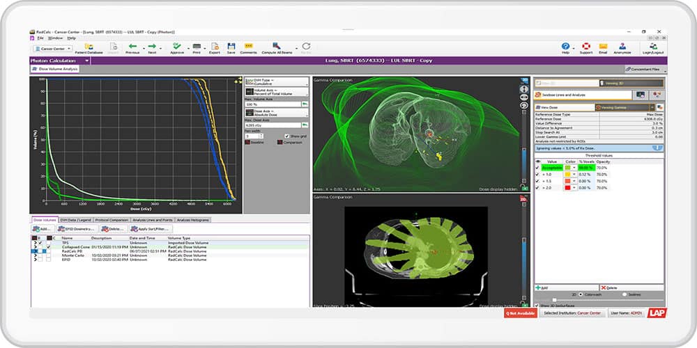
In this webinar, Jason Paisley will be sharing his experience with RadCalc’s 3D collapsed cone module. He will be covering his experience in setting up RadCalc’s dose engine, along with the hardware configuration he is utilizing within his hospital network. Jason will also be covering the setup and use of the automation that is included, since version 6.3, with RadCalcAIR. As well as any hurdles that he encountered in going clinical and points that TG 219 makes. Finally, Jason will be covering his experience using RadCalc’s 3D collapsed cone module clinically and fully automated, and why he chose to use a 3D second check solution.
Jason Paisley, MS, DABR is the chief physicist for Novant Health New Hanover Regional Medical Center in Wilmington, NC. Jason has been practicing medical physics for more than 10 years and is interested in treatment planning, machine QA and automation in medical physics.
The number one skill that I have developed in my career and that I use today is the scientific method – doing rigorous tests and analysis, checking again, duplicating the experiments. This is very important, so I try to teach my students that the scientific method is key. Serendipity is also an important factor when you use a rigorous method, so it’s essential to be curious about anything you come across and work hard to understand it.
Another important skill is to have an open mind and try to always learn, not only from your own area but also from different fields. If you want to explain something in your research, or discover something, you should not just look out for what you expect, but be open to seeing something that you were not expecting. This requires a kind-of multidisciplinary approach, and that’s what I have learned in my career. At the beginning, I did my Master’s in engineering, then a PhD in physics. When I moved to EPFL I moved into chemistry, and then went back to Italy as a professor of chemistry.
What do you like best and least about your job?
I think I’m very lucky as a scientist, because my job is also my passion, and I think most scientists would answer the same way. This makes a difference, not only for yourself, but also for your team, who can see that passion. Mentoring is another aspect I like a lot, and so is making new discoveries that can make a difference to the world, even if it’s a small one.
On the other hand, a research career is challenging, and for younger scientists it might be difficult to find a clear path towards their own independent work. I think there needs to be more attention and more action to guide younger researchers to develop their own independent thinking, rather than just doing whatever their supervisor is asking them to do. I’ve been very lucky because I met mentors, especially female mentors, who helped me navigate that. So finding a good mentor is very important.
What do you know today that you wish you knew when you were starting out in your career?
I think that it’s very important to go beyond your comfort zone and try to test yourself and your ability in an unexplored area. So for instance, my experiences abroad were extremely interesting for me and useful to develop my independent thinking. But maybe I could have looked for these experiences earlier.
Taking more opportunities to go and see different research approaches and labs, and to go to conferences, is really beneficial. Now with COVID, we are all in hybrid meetings, so it is very difficult for young researchers to engage with other scientists. I’m very lucky that I had those opportunities to travel and meet people, so I do not regret any of my steps.
The idea that particles can feel the influence of potentials even without being exposed to a force field may seem counterintuitive, but it has long been accepted in physics thanks to experimental demonstrations involving electromagnetic interactions. Now physicists in the US have shown that this so-called Aharonov–Bohm effect also holds true for a much weaker force: gravity. The physicists based their conclusion on the behaviour of freefalling atomic wave packets, and they say the result suggests a new way of measuring Newton’s gravitational constant with far greater precision than was previously possible.
Originally predicted by Werner Ehrenberg and Raymond Siday in 1949, the effect is named after Yakir Aharonov and David Bohm, who published an analysis of it a decade later. They showed that while classical potentials have no physical reality apart from the fields they represent, the same is not true in the quantum world. To make their case, the pair proposed a thought experiment in which an electron beam in a superposition of two wave packets is exposed to a time-varying electrical potential (but no field) when passing through a pair of metal tubes. They argued that the potential would introduce a phase difference between the wave packets and therefore lead to a measurable physical effect – a set of interference fringes – when the wave packets are recombined.
Seeking a gravitational counterpart
In the latest research, Mark Kasevich and colleagues at Stanford University show that the same effect also holds true for gravity. The platform for their experiment is an atom interferometer, which uses a series of laser pulses to split, guide and recombine atomic wave packets. The interference from these wave packets then reveals any change in the relative phase experienced along the two arms.
The Stanford team prepared a cloud of ultracold rubidium-87 atoms and used a pattern of overlapping laser beams (known as an optical lattice) to fire it up a 10 m-long vertical vacuum tube. A laser beam splitter then separated each atom’s wave packet into an upper and lower trajectory, with the former passing close to a semicircular ring of exceptionally pure (and therefore unmagnetic) tungsten weighing 1.25 kg and placed at the top of the tube.
The idea was to detect a tiny phase shift due to time dilation– the fact that two clocks at different heights in a gravitational potential will tick at slightly different rates. This phase shift is only measurable if the separation between wave packets is significantly larger than the distance between the closest interferometer arm and the tungsten source mass. As such, the researchers used beam splitters that transferred lots of momentum to the wave packets while spacing them as far apart as possible – up to 25 cm, compared with 7.5 cm for the wave packets that got closest to the tungsten.
Observing this effect, however, also required the physicists to account for the phase shift due to the gravitational tug of the source mass (the force field). They did this by also firing atomic clouds along interferometers with much more closely spaced arms, such that the wave packet separation, in this case 2 cm, was generally small compared to the distance of the tungsten mass, and therefore insensitive to the time dilation.
An extra effect
The researchers ran the experiment repeatedly, each time varying the minimum distance between the upper arm of the interferometer and the source mass. Plotting the variation of phase difference between the two arms with arm-mass distance, they found that the resulting curve for the interferometers with closely spaced arms matched expectations for shifts due solely to deflections of the wave packets by the gravitational field. But that wasn’t so for the interferometer with widely spaced arms. In this case, something other than the field itself had introduced phase shifts.
Kasevich and colleagues interpret this “something else” as relativistic time dilation, and therefore evidence of Aharonov–Bohm phase shifts. “These results show that gravity creates Aharonov–Bohm phase shifts analogous to those produced by electromagnetic interactions,” they write.
The researchers note that the phase shifts they observed are proportional to the mass of the atoms, in accord with predictions from theory. What’s more, these phase shifts depend on both Planck’s constant and Newton’s gravitational constant, G. As such, the researchers suggest that by precisely characterizing the source mass, this type of interferometer could be used to improve the measurement of G – the value of which is known far less accurately than any other fundamental constant.
In a piece written to accompany a paper in Science on the research, Albert Roura of the German Aerospace Centre in Ulm cautions that such experiments will have to overcome the unwanted effect of gradients in the gravitational field, as they make the phase shifts very sensitive to the atomic wave packets’ initial position and velocity. But he reckons that this problem can be overcome thanks to a technique he developed that gets around the fundamental limitations on position and momentum accuracy imposed by Heisenberg’s uncertainty principle. “The prospects for improved measurements of Newton’s gravitational constant based on atom interferometry are therefore very promising,” he concludes.
Guglielmo Maria Tino of the University of Florence in Italy is also upbeat about future research. He says that the latest results show “the potential of atomic quantum sensors as new tools to help us understand gravity and its relation to quantum physics”.
This article was amended on 28 January 2022 to clarify the roles of Aharonov and Bohm in describing the effect that now bears their names.