
A glimpse into the future world of hybrid imaging

© AuntMinnieEurope.com

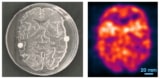
How can the clinical potential of PET/MRI be unlocked? What challenges lie ahead? Cancer imaging expert Vicky Goh took out her crystal ball and provided some insight into the future role of PET/MRI and how both modalities can be integrated effectively. She also touched on PET/CT.

Why is the UK lagging behind other countries when it comes to PET/MRI? That was one of several questions she was asked after her keynote lecture during the British Institute of Radiology (BIR) annual congress, held virtually on 4 and 5 November.

Scanner numbers and the stance on evidence-based practice play a major part in its progress in this area, according to Goh, who is professor and chair of cancer imaging and head of the cancer imaging department at King’s College London.

“We only have a handful of scanners in the country and the majority of those were funded through the Dementia Platform UK initiative. In terms of the neurology and neuro-oncology direction, we are probably further along, but we are still looking at PET/MRI as a clinical research tool,” she told attendees.

PET/MRI for staging cervical cancer

Goh, who received the Most Influential Radiology Researcher award in the inaugural EuroMinnies in 2019, said there needed to be changes in the “direction” of PET/MRI.

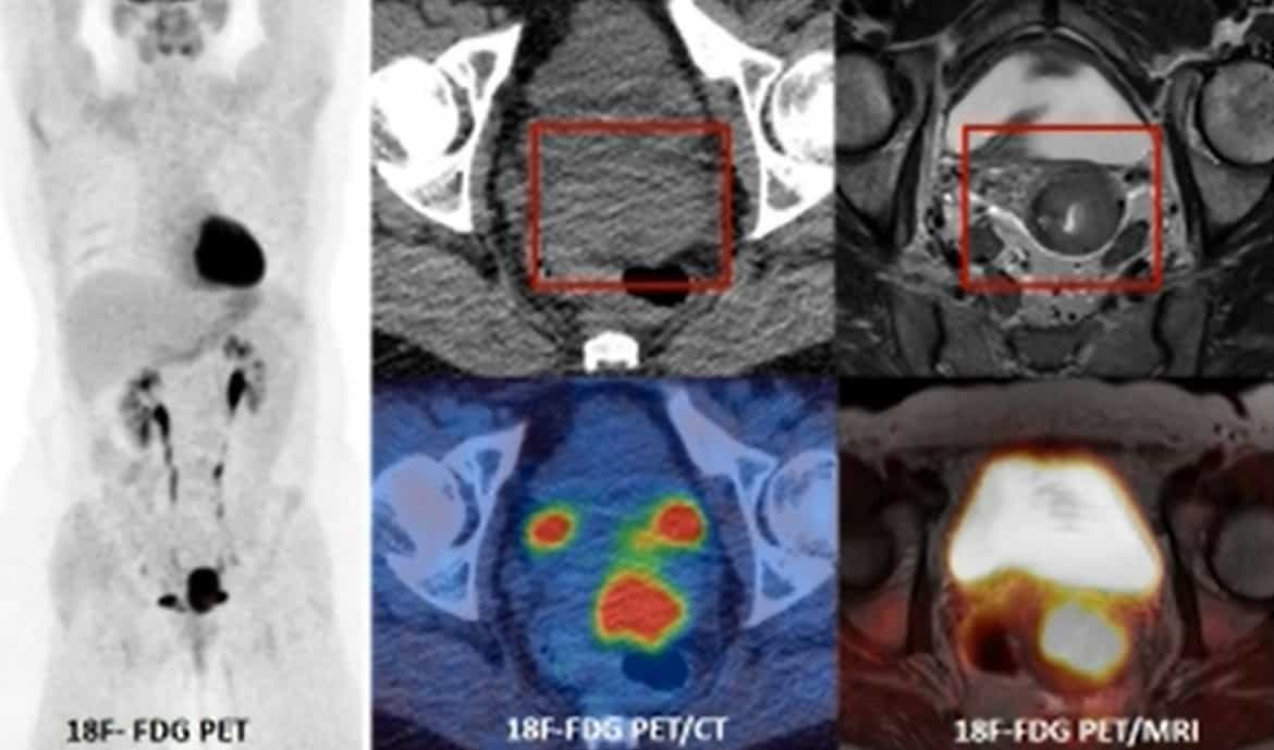
“We are very much an evidence-based practice and the evidence base is there for certain types of cancer. In prostate cancer you can see that prostate-specific membrane antigen (PSMA) PET/CT followed by targeted PET/MR would actually be of benefit to staging,” she explained.

“But the issues still remain: limited access, and limited dual-trained practices. In terms of outsourcing to other centres, a spoke and hub model will do quite well for referrals, but we need more scanners, we need more trained staff, and we need more evidence to push ahead in the UK with our practice.”

Evidence-based practice and AI

Given the need for evidence-backed protocols before wide-scale adoption and the lack of funding to provide PET/MRI in centres, Goh was asked by another online attendee how she envisaged the technique becoming widely adopted without significant delays.

Goh, who is also honorary consultant radiologist at Guy’s & St Thomas’ Hospital, noted that the only way was for institutions to work together and pool data and experience to get across the adoption gap in terms of data and protocols.

“Funding will always be an issue, but it’s one of those chicken-and-egg situations: the more data you have and the more utility you see, then the likelihood is that funding will increase,” she said. “It’s a case of ‘watch this space’, but in terms of cancer phenotyping and the type of radiologist that I want to be, there’s no doubt that this is one of the modalities I want to work with clinically.”

Goh was also asked if there is a role in the hybrid workflow for artificial intelligence (AI) systems.

She explained that in terms of improving prediction of prognostication, the data are still limited from research groups looking into this area, and larger datasets are needed. However, by improving noise and processing of the whole-body components there would be reduction in whole-body scan times, particularly for MRI diffusion scans.

“T1 and T2 scans are quick but it’s the diffusion we want to cut down to two or three minutes per station. It also means we can start looking at other things like segmentation, and quantitation in an automated fashion and again this is an area where we need to ‘watch this space’,” she said.

Clinical applications

Discussing myeloma treatment response with session chair and BIR president Sridhar Redla, Goh pointed to an exciting new role for PET/MRI. While MRI is the most sensitive modality for myeloma detection, there are, to date, more data for PET in the treatment response setting. PET-positive patients who show a reduction in PET uptake are more likely to do well and have a longer disease-free interval.

“What we really need is a modality that is both sensitive but also able to provide physiological information in the treatment response setting. And as a one-stop in this elderly population, we can offer them one examination [PET/MRI] rather than have them come back for three examinations (CT, whole-body MRI, then FDG-PET/CT) at multiple time points – in an elderly population this isn’t so viable. This is definitely one area where we can have a win,” she said.

Integrated PET/MRI

Goh also discussed the advantages of whole-body pseudo CT, whereby MRI is used to develop a CT scan with Hounsfield units equivalent to a CT that is acquired in clinical practice.

This can then be leveraged to improve standardized uptake value (SUV) quantitation from bone, which is one of the issues with attenuation correction, she noted, as well as for radiotherapy planning in which there is an issue of dosing. Although it’s early days, she pointed to great potential for this technique for prostate radiotherapy planning and gynaecological cancers.

In response to another question from an online viewer, Goh noted that for small-volume lung disease, chest CT remained the standard detection tool. However, she pointed to developments in the MR sequencing that might change this in future.

PET/CT versus PET/MRI

Another listener wanted to know if patients with a strongly suspected cancer based on chest X-ray should go directly to PET/CT rather than CT first and then PET/CT. For those who are going to have definitive therapy – for lung or oesophageal cancer, for example – PET/CT should be the way forward, Goh answered. This is because these patients often have metastatic disease presentation and PET/CT is the most sensitive way to pick up this disease.

What about colorectal cancer? Whole-body MRI can detect a higher number of metastases than just contrast-enhanced CT can, she continued. For colorectal cancer and metastatic disease in sites such as liver, brain and bone, FDG PET/MRI improves detection, she added.

But, does PET/MRI pick up more liver lesions than PET/CT? Responding to this question from Redla about neuroendocrine cancers and somatostatin receptor imaging, Goh noted that the answer wasn’t straightforward, and due to complex protocols.

“As we all know, diffusion is the most sensitive sequence for picking up lesions, and it’s the diffusion component of the MR that is really contributing here in whole-body PET/MRI. We’re also able to pick up discrepancies, lesions that are tracer positive versus those that are tracer negative, to provide a more realistic burden of disease and the proportion of it that is potentially going to respond,” she noted. Small lesions that are receptor-negative will probably not respond to therapy, she clarified.

So with more resources, will PET/MRI replace PET/CT? Goh’s short answer was “no”. She pointed to certain scenarios where PET/MR can either complement PET/CT, as in prostate cancer staging, and others where PET/MR would replace it, as in the myeloma setting. But PET/CT was far quicker to perform than PET/MR and would keep its place in the clinic.

“Think about scheduling and the number of PET/CTs we ask for. The number of patients examined in a day would be phenomenally different. So we can’t see this replacing PET/CT except in certain scenarios but we can definitely see it complementing it,” she said.

  • This article was originally published on AuntMinnieEurope.com ©2021 by AuntMinnieEurope.com. Any copying, republication or redistribution of AuntMinnieEurope.com content is expressly prohibited without the prior written consent of AuntMinnieEurope.com.

X-ray tomography breaks new world record

Researchers at the Helmholtz-Zentrum Berlin für Materialien und Energie (HZB) have developed a new X-ray tomography technique that’s capable of acquiring a record-breaking 1000 tomograms per second. The microscope could be used to monitor extremely fast processes in materials with high spatial resolution.

Computed tomography (CT) is a popular medical imaging tool in which a part of the body is X-rayed from all sides to produce 3D images of internal structures. The technique is also ideal for non-destructive analysis of materials. Here, intense synchrotron radiation is used to obtain micron-scale-spatial-resolution images in 3D and to monitor rapid processes and changes in a sample.

In 2019 a team of researchers led by HZB’s Francisco Garcia Moreno managed to record 200 tomograms per second using their technique, which they subsequently dubbed tomoscopy – in analogy to radioscopy. Indeed, the number of tomograms per second (tps) is the equivalent of the number of frames per second (fps) used to describe 2D X-ray image sequences.

High spatial and temporal resolution

In this latest work, Garcia Moreno and colleagues made use of the TOMCAT beamline X02DA of the Swiss Light Source at the Paul Scherrer Institute in Switzerland. The researchers placed their samples on a high-speed rotating table, developed in their lab. The angular speed of the table, which can reach 500 Hz, can be perfectly synchronized with the acquisition speed of the high-speed CMOS camera used to image each sample. They obtained the images by inserting the sample into a hollow, cylindrical boron nitride crucible in the rotation stage and heating it using two 150 W infrared lasers.

The technique, which achieves a spatial resolution of just 7.6 µm at 100 tps and 8.2 µm at 1000 tps, can take 40 2D projections of the sample in one millisecond. These projections are then stacked atop each other to create a tomogram of the sample.

To test their technique, Garcia Moreno and colleagues recorded the extremely rapid changes that occur as a sparkler is burnt (following ignition using the infrared lasers). This exothermal combustion process is technologically important and releases a huge amount of heat and a combustion wavefront moving as fast as 1–100 mm/s. This wavefront is difficult to image using conventional techniques.

The researchers also imaged dendrites forming in aluminium-germanium and aluminium-bismuth casting alloys as they solidify, and the growth and coalescence of bubbles in liquid aluminium-silicon-copper foams. This coalescence is an unwanted but unfortunately common process in such foams, which are interesting, lightweight materials from which future electric cars might be built.

Previous radioscopy and tomoscopy experiments revealed a film rupture time of less than 1 ms and a coalescence time (the time to form a new bubble) of 0.5–1.2 ms. Such time scales are now accessible with the temporal resolution of the new tomoscopy technique, say the researchers, and will allow for insights into the morphology, size and cross-linking of these bubbles – important factors when it comes to making mechanically strong and stiff components.

The research is detailed in Advanced Materials.

Physicists create discrete time crystals in a programmable quantum simulator

Time crystals are special quantum systems that exhibit periodicity in time, just as crystalline materials are periodic in space. Since 2012, when they were first proposed theoretically, several groups have built experimental systems that demonstrate key characteristics of time crystals, but these results lacked a method to generally stabilize the time crystalline phase. An international team of researchers has now gone beyond previous experiments by creating a type of time crystal known as a discrete time crystal (DTC) out of a chain of programmable spin quantum bits (qubits). The new system exhibits a phenomenon known as many-body localization that prevents the DTC from heating up and thermalizing and is considered a “smoking gun” that demonstrates its status as a genuine time crystal.

While time crystals are generally analogous to spatial crystals in terms of exhibiting periodicity across a dimension, the time periodicity of a DTC is subtly different. Specifically, a DTC’s behaviour repeats itself over an integer multiple (hence the name “discrete”) of the period of the force that drives it to change its quantum state.

To qualify as a time crystal, the system must meet two criteria: it must be robust to fluctuations (similar to normal crystals), and the energy that drives it must not cause it to heat up. This second requirement imposes special conditions on the system, because whereas most physical systems absorb energy to reach thermal equilibrium, for a time crystal such heating leads to the system’s destruction.

The way to avoid this is known as many-body localization (MBL), which essentially means creating a many-body system in which a certain amount of disorder is introduced. Although the particles inside such a system couple with different states, they do so with random variations, leading to destructive interference. The disorder therefore prevents the system from heating up, maintaining its stability.

A new spin

In the latest study, which is published in Science, researchers at QuTech (a collaboration between TU Delft and TNO in the Netherlands), the University of California, Berkeley, and the industrial diamonds firm Element Six created a DTC from nuclear spins in carbon-13 atoms located near a defect in diamond’s crystal lattice. These defects are known as nitrogen-vacancy (NV) centres, and their electronic spin can be controlled using optical techniques. This capability allows the nuclear spins in nearby carbon-13 atoms to be addressed as well, meaning that the nuclear spins act as well-isolated qubits with quantum states that are both controllable and detectable. To meet the criteria for MBL, the system should contain disorder. The diamond spin system achieves this because the position of the carbon-13 nuclear spins within the lattice is not ordered, which naturally causes variations in the coupling between spins.

Photo of Tim Taminiau, Mohamed Abobeih and Joe Randal with a model of the diamond they used in their experiment

To create the system’s initial state, the researchers set the all the nuclear spin polarizations to be the same. They then selectively altered these states by applying radiofrequency (RF) pulses. After one cycle, they retrieved the system’s initial state, meaning the cycle could start again. The period of this cycle was twice the period of the driving RF pulses – a characteristic of DTC behaviour – and the researchers found they could maintain the pattern over 800 cycles, for a total of eight seconds. The key DTC signature, however, came from altering the initial state of the many-body system and repeating the cycle. Regardless of system’s initial state, the experiment formed a stable DTC. This robustness was the key element the team was looking for to show that the time crystal was being stabilized by disorder in its internal interactions and not by prethermal effects that exponentially slow down the heating.

Perfect timing

Tim Taminiau, the QuTech physicist who led the research, says that the stability of time crystals is connected to fundamental questions in non-equilibrium statistical mechanics. “This work provides experimental input to such puzzles and helps lay the groundwork for understanding the scenarios in which time crystals appear and what their observable signatures are,” he says. He notes that another collaboration has reported a similar result on arXiv, using a quantum processor based on superconducting qubits. “It is exciting to see two experimental breakthroughs happen so shortly after another,” he says. “Both results are complementary: they used two times more qubits, but our time crystals lived ten times longer.”

As for future work, Taminiau points out that “a perfectly isolated time crystal can, in principle, live forever, so extending the lifetime is our next step”. To investigate the various settings in which time crystalline behaviour occurs, he and his colleagues want to study what happens in 3D systems (rather than the 1D case discussed here), as well as in other driven phases of matter such as topological phases. From a broader perspective, he adds that the group now has a powerful new quantum simulator with individual control over all the qubits, which can be used to investigate “a large variety of interesting problems in many-body physics and other fields”.

Peter Hannaford, a physicist at Swinburne University of Technology in Melbourne, Australia, who was not involved in the research, describes the result as being important for the field of time crystals, as the QuTech-led team unambiguously demonstrated a many-body localization DTC for the first time. He suggests that previously reported experimental results on DTCs based on spin systems were likely to be stabilized by prethermal effects, or else they did not employ disorder. As for future work, he notes that this study and the similar result currently on the arXiv both have two temporal lattice sites. “An exciting challenge for the future is to explore bigger DTCs with 100 or more temporal lattice sites,” he says. “Those can be ideal for studying (exotic) condensed-matter phenomena in the time dimension and might be realized, for example, from a Bose–Einstein condensate of ultracold atoms bouncing resonantly on an oscillating mirror.”

Merging neutron stars create more gold than collisions involving black holes

The amounts of heavy elements such as gold created when black holes merge with neutron stars have been calculated and compared with the amounts expected when pairs of neutron stars merge. The calculations were done by Hsin-Yu Chen and Salvatore Vitale at the Massachusetts Institute of Technology and Francois Foucart at the University of New Hampshire using advanced simulations and gravitational-wave observations made by the LIGO–Virgo collaboration. Their results suggest that merging pairs of neutron stars are likely to be responsible for more heavy elements in the universe than mergers of black holes with neutron stars.

Today, astrophysicists have an incomplete understanding of how elements heavier than iron are made. In this nucleosynthesis process, lighter nuclei must be able to capture neutrons from their surroundings. Astrophysicists believe this can happen in two ways, each producing about half of the heavy elements in the universe. These are the slow process (s-process) that occurs in large stars and the rapid process (r-process), which is believed to occur in extreme conditions such as the explosion of a star in a supernova. However, exactly where the r-process can take place is hotly debated.

One event that could support the r-process is the merger of a pair of neutron stars, which can result in a huge explosion called a kilonova. Indeed, such an event was seen by LIGO–Virgo in 2017, and simultaneous observations using light-based telescopes suggest that heavy elements were created in that event.

Gravitational disruption

Another possibility is that the r-process occurs just after the merger of a neutron star and a black hole. As the neutron star is disrupted by the huge gravitational field of the black hole, vast amounts of neutron-rich material could be blasted into space – providing an environment for the r-process. Astrophysicists believe this can happen when the black hole has a relatively low mass and is spinning at a relatively high rate. If the black hole is too heavy, the neutron star will be swallowed rapidly, and little neutron-rich material will escape.

Today, however, astrophysicists are unsure of the relative contributions of these two merger types to the universe’s overall abundance of heavy elements.

Ultimately, the amounts of heavy elements produced by these events depends on several factors: including the masses and spins of the merging bodies; the rate of occurrence of the merger types throughout the history of the universe; and the neutron star’s “equation of state”. The latter describes the mathematical relationship between the mass and radius of a neutron star. Over the years a variety of models have been developed to define these quantities.

Improved equation of state

In their study, Chen and colleagues have compared the contributions of both merger types for the first time. They began by studying LIGO–Virgo observations of the two different types of merger. Then, they used the latest simulations of ejections from these events – incorporating improved equation of state measurements, to test several models of how the r-process could proceed, which they deemed consistent with LIGO–Virgo’s observations.

In most simulation scenarios, the researchers found that binary neutron star mergers produced 2–100 times more heavy elements over the past 2.5 billion years than mergers between black holes and neutron stars. This outcome only changed when researchers assumed that black holes tend to have lower masses and faster spins than predicted by current theories.

Chen and colleagues now hope to improve their calculations using future observations from the upgraded LIGO and Virgo detectors – and the new KAGRA detector – which will all be back online in 2022. These efforts could ultimately improve astronomers’ estimates of the rates at which heavy elements are produced across the universe. In turn, this could help them to better determine the ages of distant galaxies, by measuring the abundances of heavy elements they contain.

The research is described in The Astrophysical Journal.

From geckos’ feet to Formula 1: how surface science underpins our world

I have to come clean about something. I thought I understood the science of surfaces. As a physicist working on nanomaterials for light-emitting diodes (LEDs), solar panels and sensors, I literally spend my days playing with polymers. The devices I build are multi-layered structures, where you have to really know what’s going to happen when you deposit one layer on top of another. For example, if the light-emitting layer of an LED doesn’t stick well to the electrodes below, then the pixels on our mobile phone and TV screens won’t light up. So, this being the field I work in, I thought I had a pretty good grasp of surface science. But within the first few pages of Sticky: the Secret Science of Surfaces by physicist and author Laurie Winkless, my confidence in my knowledge of surface science started to come unstuck.

It has been four years since Winkless’ first book, Science and the City, introduced the public to the science of the metropolis. While her enthusiasm and charm are apparent throughout both books, Sticky feels like the more grown-up sibling. Rather than providing a superficial overview of all the relevant scientific topics, Winkless takes the time to unpack those she is most interested in. The writing, including the technical descriptions, feels effortless, benefiting from her years of experience in science communication.

Unlike many popular-science books, Sticky is not just a disparate list of facts about forces. Every chapter, from “A gecko’s grip” to “Break the ice” is a story, masterfully assembled into an accessible, clear and highly engaging manuscript. Winkless’ writing is informed by discussions with a diverse group of scientists and engineers, and the unparalleled excitement of discovery is evident in every interaction. Alongside these interviews, the author provides an enticing (but not overwhelming) level of detail, introducing readers to the scientific method, peer review and even the politics of scientific theories. The science of the sport of curling, for example, is a hotly debated topic that has divided physicists for the past 30 years and fuelled “Broomgate” – the 2015 controversy over whether a new hi-tech broom was changing the fundamentals of the sport too much.

Winkless doesn’t shy away from complicated topics, such as fluid dynamics, organic chemistry and geophysics, but instead makes them relevant to her audience. The topic itself is ubiquitous by nature, so there’s something in the book to pique the interest of everyone from academic researchers to fans of Formula 1. For scientists, the properties of surfaces are an essential consideration. Double-sided Sellotape (of the appropriate stickiness) is required for nanoscale imaging using atomic force microscopes; carbon tape is crucial for mounting samples inside vacuum chambers; and Kapton tape is necessary for soldering. The “Scotch tape method”, by which tape is stuck to graphite and peeled off again, taking tiny fragments with it, paved the way for the graphene revolution. I’d even go as far as saying I’ve never met an experimental physicist who doesn’t rely on Blu Tack, whether that is to hold together pieces of equipment, secure a substrate to a stage or safely package samples to send to collaborators.

But adhesion principles also show up in everyday life and culture far beyond the lab, from the frescoes of Italian Renaissance art to non-stick frying pans and swimwear that helps you glide through the water more easily. So, while a truly brilliant read for scientists, I think Sticky appeals to a very broad audience. Irrespective of your level of expertise, Winkless’ language is not remotely patronising. The analogies are well thought through and the examples are appropriate. In fact, she somehow manages to make the mundane beautiful: “curling is a curious ballet of stones, brooms and ice”. For the super-keen, Winkless provides a comprehensive reading list (apparently just a handful of the 900+ references she read when researching the book) for further study.

My favourite chapter explores the gecko’s impressive ability to cling to even very smooth surfaces, which Winkless describes as the “smartest on-off adhesive in the world”. Little did I know that the attempt to unravel the mechanisms behind this creature’s grip has spanned two centuries, provided fuel for entire research careers and inspired a lot of questionable hypotheses.

Working out the hierarchical adhesive systems of geckos has presented a truly interdisciplinary research effort that is inspiring the design of futuristic robots and reusable adhesive tapes

The first theory – that geckos cling to surfaces through suction from the lamellae in their feet – was debunked a century after it was proposed, when it was shown that geckos maintain their grip even at vanishingly low air pressures. Next came the “climber’s boot” hypothesis: that densely packed hairs called setae on the gecko’s toes act as tiny hooks that grip onto bumps and grooves. This proposal was dismissed when experiments revealed that geckos can firmly attach to ultra-smooth surfaces with very low surface roughness. More theories followed, involving electrostatics (think sticking a balloon on a wall after rubbing it on your hair) and friction. It turns out that geckos stick to surfaces through a complicated interplay of many physical phenomena including Van der Waals forces and nanoscale networks of setae known as spatulae. Incredibly, they can detach their feet six times faster than you can blink. Working out the hierarchical adhesive systems of geckos has presented a truly interdisciplinary research effort that is inspiring the design of everything from futuristic robots to reusable adhesive tapes.

It will no doubt surprise fans of friction that there are still so many unknowns – such as how exactly curling works, or how we can quantify the slipperiness of swimmers – that continue to perplex scientists. So although this research field has existed for hundreds of years, it remains a lively and cutting-edge one. When I picked up Sticky, I wasn’t sure what I was in for, but I remained glued to every page.

  • 2021 Bloomsbury £16.99hb 336pp

Wireless implant uses optogenetics to control spinal cord activity in mice

Optogenetics – a technique that uses light to control the activity of neurons or other types of cell – has revolutionized our ability to manipulate and discern the mechanisms underlying brain function. Spinal cord activity underpins control of movement and several other basic physiological functions. But compared with the brain, optogenetics in the spinal cord presents a series of challenges that require the development of new light-delivery technologies.

For instance, implanting optical fibres is not a straightforward task, as the spine undergoes continuous displacement during natural movement. Therefore, one design challenge is the positioning of light-delivery sources over the very surface of the dura mater, the outer membrane that surrounds and protects the soft and dynamic spinal cord. Targeting deep intraspinal neurons presents additional obstacles. Light rapidly scatters when penetrating biological tissues. High irradiance, meanwhile, may result in local tissue heating due to light absorption, which can affect neuronal responses.

To overcome these challenges, and find a way to efficiently deliver light to the spinal depth of interest, researchers at the Swiss Federal Institute of Technology (EPFL) have developed a compliant, wireless-controlled optoelectronic implant customized for optogenetic studies of the spinal cord. They describe this novel implantable system in Nature Biotechnology.

NeuroRestore group

Technology features

The new implant technology encapsulates miniaturized LEDs that can be switched on and off on the surface of a mouse’s spinal cord. A miniature head-mounted, wireless platform powers these micro-LEDs and performs customized on-chip processing to control light stimulation trains. This enables modulation of light pulses in real time, for instance, in response to the animal’s muscular activity or routine physiological signals. The wireless electronic circuit can control the duration and intensity of the emitted light with high precision.

In contrast to wire-based systems usually used for this type of research, wireless optoelectronic systems allow for unrestricted photostimulation of target neurons in the spinal cord of mice under untethered and ecological conditions.

In one key aspect of this work, the researchers, led by Grégoire Courtine and Stéphanie Lacour, used the micro-LED arrays to take advantage of the evolving library of experimental opsins (light-sensitive proteins activated by specific wavelengths) to target a broad range of cellular mechanisms.

They designed the micro-LED array to shift the emitted blue light toward a desired wavelength, enabling photostimulation using a broad spectrum of light, including red-shifted wavelengths that penetrate further into the spinal cord.

To optimize LED positioning and avoid tissue heating, the team ran simulations and performed in vivo recordings to quantify heating within the spinal cord for different levels of irradiance and cycles of photostimulation.

The researchers designed the new soft optoelectronic implant to be resilient and adaptable for long-term implantation and to meet the demanding mechanical dynamics of the spinal cord in freely behaving adult mice. Using a hybrid approach combining stiff LEDs and elastic interconnects, they created miniaturized implants that wrap around the surface and move along with the spinal cord.

The team’s research has potential to pave the way for the development of therapeutic optogenetic applications. The ability to control the activity of spinal cord neurons with light could allow doctors to reduce pain and improve autonomic function.

Even though it may still be some time until these implants can be used clinically, Courtine finds optogenetics “revolutionary” and is eagerly anticipating further developments in biointegrated optoelectronic implants.

Photonic band structure created in electrically driven OLED microcavities

A photonic crystal emitter has been fabricated by stacking multiple organic light emitting diode (OLED) microcavities into a single structure. It was created by a team led by Matthew White at the University of Vermont in the US, that was able to demonstrate that the degree of interaction between the cavities depends strongly on the different layers employed, meaning that both the colour of light emission and the band structure of the devices are highly tunable. This paves the way towards electrically driven photonic crystals that allow further control over the emission of OLEDs. The full study is described in Nature Communications.

OLEDs have received a large amount of attention in recent years due to their numerous applications in display screen technologies and efficient low-cost lighting. They are also easy to integrate into other technologies and structures.

White and colleagues have placed semi-transparent mirrors on either side of an OLED to form a microcavity – that is, a cavity with a length at the order of a micron. By varying the distance between the two mirrors, the length of the cavity and therefore the emission wavelength (colour) of the OLED can be finely tuned, even without changing the organic material.

Communicating OLEDs

The team then created a stack of N microcavities separated by semi-transparent mirrors to make a device with a photonic band structure analogous to that in a photonic crystal. If the mirrors are much thicker than the penetration depth of light, no communication between the cavities is possible and they act as N separate cavities. Conversely, if the mirrors are infinitely thin, the N cavities will act as one cavity with a total length Nd, where d is the length of each individual cavity.

Blue emission from an OLED microcavity

If the mirror thickness lies between these two extremes, the system acts similarly to that of the extended cavity, but with small perturbations in the electric field caused by the internal mirrors. This results in a hybridization of the energy states, forming a photonic band structure that is similar to a photonic crystal or hybridized orbitals in molecules. This hybridization alters the energy of the states and therefore affects the OLED emission.

The researchers have shown that the band structure is heavily dependent on the properties of the stack. For example, the internal mirrors used in the stack alternate between silver and aluminium. This symmetry doubles the unit cell size so that it consists of two cavities with a semi-transparent internal mirror and total length equal to 2d.

Tuning the band

As aluminium has a shorter penetration depth and higher losses than silver, the aluminium mirrors primarily control the length of the unit cell and therefore the energy separation of the hybridized states. The researchers have shown, however, that the presence of the silver mirror perturbs the states, decreasing the energy gap between the two sub-bands. Therefore, by altering the thicknesses of the different mirrors, it is possible to control the size of the band gap, the separation of the energy states and the total bandwidth of the OLED stack.

“The effect is the result of structural colours so we can use green organic emitters, which are known to be more stable, to produce any desired colour combination”, says White. In this way, it will be possible to “tune the emission from OLEDs to include broadband white light, narrow bandwidth single peaks, or multiple peaks forming a low-fidelity frequency comb”.

Help me if you can: founding a business doesn’t have to be lonely

In 1978 the Silicon Valley entrepreneur Nolan Bushnell was fizzing with ideas for new businesses. He had just left Atari, where he had masterminded the creation of Pong – one of the earliest and most successful video games – and many other pioneering tech products. But one of the biggest problems with start-up companies, Bushnell realized, was the sheer amount of “bullshit housekeeping stuff” involved.

His solution was to create Catalyst – believed to be the world’s first hi-tech business incubator. “My idea was that I would fund [the businesses] with a key,” Bushnell told Fast Company in 2017. “And the key would fit a lock in a building. In the building would be a desk and chair. They would sign their name 35 times and the company would be incorporated. So in 15 minutes, they would be in business working on the project.”

The idea was not entirely new. The first formal business incubation service that I can find details of dates back to 1959 in the US, but Catalyst was geared specifically to the demands of hi-tech companies. So as well as providing fledgling firms with office space, IT systems and legal and accounting services, Catalyst offered laboratory space and most importantly money and support – in fact, Bushnell was often chief investor and chairperson of each firm’s board.

Catalyst was designed to help those companies get through the initial challenges of starting up. Within a year, it had funded 10 businesses, many of which were very much ahead of their time. They included Etak, which released an in-car computer navigation system in 1985 – fully four years before the GPS network was available for civilian use. Parts of the press ridiculed the product (it didn’t help that the symbol of the car on the driver’s screen looked uncannily like the ship from Atari’s Asteroids game) but sat-navs are now a feature of almost all modern cars.

Catalyst folded but the blueprint for business incubation had been set

James McKenzie

Catalyst created around 20 companies, but it unfortunately over-invested in some of them, which therefore took a lot longer than expected to generate a return. Eventually, in 1986, Catalyst folded but the blueprint had been set and by 2006 there were more than 1400 incubators of all kinds in North America, up from only a dozen in 1980. A report that year from the US National Business Incubation Association indicated that US incubation programmes had helped more than 27,000 companies, together employing over 100,000 people and generating annual revenues of $17bn.

Global growth

The idea quickly spread across the world as more and more start-ups saw the benefits of lab space, help, advice and access to capital. In 1997 there were around 25 incubation environments in the UK, but by 2017 that number had risen to almost 400, according to a report from the Department for Business, Energy and Industrial Strategy.

With hi-tech incubators and accelerators usually needing specialist facilities, many are located at (or linked to) universities and other national labs in areas where there are lots of similar companies with a local talent pool. The Institute of Physics also has a business accelerator at its new headquarters, giving small firms access to affordable office space in central London and letting them hook into the IOP business and member networks.

As well as many business incubators, the UK now in addition has the Catapult Network of technology innovation centres. These have been set up over the last decade in the wake of a 2010 government report into technical innovation. Written by Hermann Hauser – the physicist-turned-entrepreneur and honorary fellow of the IOP – he reckoned that the UK has a science capability second only to that of the US, but was falling short on translating scientific ideas into leading positions in new industries. “Technology and Innovation Centres can help solve this shortcoming and give Britain the lead in some of the most promising technologies leading to new industries with transformational economic impact,” he wrote.

The Catapult Network, which is part of Innovate UK, now has nine technology and innovation centres spanning more than 40 locations across the country. These centres cover everything from compound semiconductors, renewable energy and high-value manufacturing to satellites, energy systems and digital technology. They provide high-quality facilities for hi-tech businesses of all sizes and have a support programme for start-ups and small-to-medium-sized enterprises (SMEs). Since 2013, more than 8000 start-ups and SMEs have been supported. Innovate UK also offers grants to support its industrial strategy, with various competitions running throughout the year.

So whether you are an early-stage firm, a pre-seed innovator, or a more established company, there are many networks and resources to support your innovation journey. A good place to start is the Knowledge Transfer Network, which has experts in many areas on tap. There is also Innovate UK Edge – part of UK Research and Innovation – which has business advisers who can provide advice and introductions. In addition, it can carry out a grant-funded audit of your firm’s intellectual property. It even has an “investment readiness” programme as well as a showcase called Pitchfest to help you improve your pitch to investors.

There is now more help than ever for new hi-tech start-ups

James McKenzie

I know from personal experience that starting a new hi-tech firm is not easy and many physicists have great ideas but are unwilling to take the plunge. But there is more help on offer than ever before: through business incubation, catapult networks, grants and business advice. So why not have a go? Whether it’s energy, medicine, quantum tech or new materials, there are plenty of opportunities.

From whole-organ to cellular resolution: synchrotron X-ray images reveal COVID-19 lung damage

The ability to image human tissue at scales from an entire intact organ down to individual cells is key to advancing our understanding of health and disease. To meet this challenge, a European research collaboration has developed a new imaging technology known as hierarchical phase-contrast tomography (HiP-CT), using X-rays from the Extremely Brilliant Source (EBS) at the European Synchrotron Radiation Facility (ESRF).

The EBS is the first high-energy (6 GeV), fourth-generation synchrotron source. It provides the brightest source of X-rays in the world, with the spatial coherence required to resolve faint density contrasts at high resolution. Using the EBS, HiP-CT can perform non-destructive 3D scanning of an entire human organ and then zoom down to the cellular level.

“The ability to see organs across scales like this will really be revolutionary for medical imaging,” says first author Claire Walsh from University College London. “As we start to link our HiP-CT images to clinical images through AI techniques, we will – for the first time – be able to highly accurately validate ambiguous findings in clinical images. For understanding human anatomy, this is also a very exciting technique, being able to see tiny organ structures in 3D in their correct spatial context is key to understanding how our bodies are structured and therefore how they function.”

Walsh and colleagues have used the new synchrotron X-ray tomography technique to scan donated human organs, including lungs from a COVID-19 donor, reporting their findings in Nature Methods.

High-resolution scanning

Using the EBS test beamline, BM05, the researchers developed the specialized sample-preparation, scanning and reconstruction approaches required for HiP-CT. They designed a scanning geometry that reduces sample dose (to avoid tissue damage), optimizes the detector’s dynamic range, reduces artefacts and suppresses beam hardening.

For imaging, organs are fixed, partially dehydrated and stabilized in agar–ethanol in a jar. The team recorded reference scans of a jar of agar–ethanol mounted on top of the sample to provide a background that can be removed during image reconstruction. This process eliminates low-frequency background variations and enables extreme off-axis local reconstructions.

Synchrotron X-ray imaging of human organs

The HiP-CT scans are performed hierarchically, typically starting at 25 μm/voxel over the whole organ, then followed by magnified imaging of selected volumes-of-interest (VOIs) at 6.5 and 1.3–2.5 μm/voxel. They team assessed the performance of the HiP-CT technique by scanning an intact human lung. The estimated image resolutions were 72±3.4, 18.3±0.6 and 10.4±0.17 μm, for images recorded at 25, 6.5 and 2.5 μm/voxel, respectively.

To assess the consistency of scans at various depths and distances from the organ centre, the researchers analysed images of two high-resolution VOIs. They found minimal differences in mean intensity or image quality between the two, suggesting that HiP-CT can achieve high-resolution scanning in any region of the lung with consistent quality.

Next, they imaged five intact donated human organs – brain, lung, heart, kidney and spleen – performing HiP-CT at 25 μm/voxel to provide a structural overview of each organ, followed by multiple higher-resolution scans of selected VOIs. The 25 μm/voxel scans clearly identified macroscopic features such as individual lobules in the lung, for example, and the four chambers of the heart and associated coronary arteries.

The higher-resolution scans successfully visualized functional units in the organs, as well as imaging certain specialized cells. In the brain, for example, HiP-CT revealed layers of the cerebellum and individual Purkinje cells. Lung images showed the intralobular septa and septal veins, as well as terminal bronchi and bright cell-sized objects identified as pneumocytes and/or alveolar macrophages. Images of the heart showed bundles of cardiac muscle fibres comprising individual cardiomyocytes, while epithelial tubules were evident in the kidney, and red and white pulp was seen in the spleen.

COVID-19-related lung damage

The team also used HiP-CT to investigate structural changes in the lung tissue of a patient who died from COVID-19-related acute respiratory distress syndrome. Lung slices imaged at 25 μm/voxel contained high-intensity regions in the lung periphery, consistent with clinical radiology findings.

COVID-19-related lung damage

Scanning VOIs at 6 μm/voxel revealed heterogeneous parenchymal damage, with some secondary pulmonary lobules displaying greater deterioration than others. The team performed higher magnification images of a more affected lobule at 2 μm/per voxel. The scans revealed how severe COVID-19 infection remodels the finest blood vessels in the lungs, causing blood to shunt between the capillaries that supply oxygen to the entire body and those that feed the lung tissue itself. This cross-linking prevents the patient’s blood from being properly oxygenated – a process that was previously hypothesized but not proven.

“By combining our molecular methods with the HiP-CT multiscale imaging in lungs affected by COVID-19 pneumonia, we gained a new understanding of how shunting between blood vessels in a lung’s two vascular systems occurs in COVID-19 injured lungs, and the impact it has on oxygen levels in the circulatory system,” explains Danny Jonigk from Hannover Medical School.

The researchers emphasize that HiP-CT will continue to evolve alongside advances in synchrotron technology. Completion of a new ESRF beamline next year is anticipated to provide several key advances.

“The beam [will be] much larger and higher energy, hopefully enabling scanning of a whole torso at 20 micron voxels, while still being able to zoom locally to one micron. The increased beam size enables increased speed for the same sample size,” project lead Peter Lee tells Physics World. “We hope to form a hub to help us populate the Human Organ Atlas with a statistically significant number of organs in health and disease.”

  • All image data from this study are publicly available in an online Human Organ Atlas.

RadCalc adds in vivo dosimetric verification, intelligent automation for independent QA

Customer-driven innovation and continuous improvement are once again front-and-centre in the latest release of RadCalc QA secondary check software, a suite of widely deployed quality-assurance (QA) tools that provides medical physicists and dosimetrists with fully automated and independent dosimetric verification of their radiotherapy treatment planning systems (TPS). Top billing in RadCalc v7.2 is the commercial introduction of 3D EPID-based functionality to underpin measurement-based patient QA and in vivo dosimetric verification. Meanwhile, speed and workflow efficiency remain ongoing priorities for the RadCalc development team following the integration of a range of customization features for intelligent automation into RadCalc AIR, the software’s control centre for automated data import and reporting.

With the release of v7.2, RadCalc’s portfolio of calculations now encompasses secondary dose checks, with the use of point-dose analysis, 3D Monte Carlo or 3D Collapsed-Cone Convolution Superposition algorithms to identify clinically relevant deviations within the entire patient volume; EPID for pre-​treatment dosimetry, in which the software reconstructs the dose from the delivered pre-treatment plan on the patient’s original planning CT (giving a direct comparison with both the intended dose from the TPS and RadCalc’s 3D dose second check); and EPID for in vivo dosimetry to reconstruct the dose delivered during treatment, yielding a direct comparison on the actual delivered dose reconstructed on the original planning CT to evaluate intrafraction changes in the patient.

“We are not aware of any other product on the market able to match RadCalc’s in vivo 3D capability using EPID-based dosimetry,” claims Craig Laughton, CTO and co-founder of the RadCalc software portfolio, part of LAP’s growing QA product line in radiotherapy. “In the clinic,” he adds, “RadCalc 7.2 enables the medical physics team to compare the whole dose volume versus the original treatment plan, measuring what is actually being delivered in vivo to the patient during radiation therapy.”

Dynamic systems

Such insights are especially powerful given that patients are dynamic systems, always in flux rather than steady-state. Between treatment sessions, for example, patients can gain and lose weight; their stomach, bladder and bowel contents change; their organs may shift, rotate or deform; and their tumours may shrink, move or rotate.

Those changes can be problematic for traditional radiotherapy regimes that rely on a single CT snapshot of the patient at the start of treatment, with most clinics currently limited in their ability to track geometric deformations in patient anatomy over time. Put another way: a treatment plan attuned to the initial simulation can quickly become suboptimal as radiotherapy progresses over the course of many fractions typically spread across a month or more.

Craig Laughton

“Our EPID-based module is going to pick up any changes in dose delivery over a course of multiple fractions, triggering a conversation between the clinical physicist and radiation oncologist to understand what’s happening inside the patient,” explains Laughton. “That dialogue could ultimately mean reimaging and replanning of the patient – another important step towards a more personalized approach to radiation therapy.”

What’s more, RadCalc’s 3D EPID module also has an enabling role to play in supporting the latest hypofractionated and ultrahypofractionated radiotherapy schemes, in which an increased dose per fraction results in significantly fewer overall treatments over a compressed timeframe. The goal, as always, is enhanced targeting accuracy and enhanced dose distribution accuracy – minimizing collateral damage to adjacent organs at risk and critical structures – over a course of treatment running to just one or a few high-dose fractions.

“With a hypofractionated treatment scheme,” notes Laughton, “you need to know if something’s not right straight away – for example, in the case of a machine error or incorrect patient set-up. You can’t afford to wait a week, because in a week the treatment’s done and it’s too late.”

Automate to accumulate

More broadly, intelligent automation remains a long-term fixture on the RadCalc development roadmap, giving medical physicists the power to optimize processes versus their own clinical requirements – customizing which DICOM tags to trigger actions, for example, and new layouts for cleaner workflows. “While we see user-defined automation as a key differentiator,” notes Carlos Bohorquez, RadCalc product manager, “automation is ultimately all about patient safety and minimizing human errors and the burden on clinical resources associated with manual QA tasks.”

Carlos Bohorquez

According to Bohorquez, the v7.2 release incorporates significant customizations to the RadCalc AIR import and reporting tool to streamline workflows in the clinical environment (e.g. the import/export of clinical protocols; also specific tolerance criteria for each treatment plan based on any DICOM tag the user’s TPS is able to export). “It’s an ever-evolving process to provide clinicians with true and faithful automation for existing and emerging radiotherapy technologies,” he explains.

That’s certainly true for RadCalc’s new 3D EPID module, which imports the necessary EPID data/image files for processing before sending to the Collapsed-Cone dose engine to calculate the dose. “The EPID-based solution needs further automation so that the physics team can use it on a routine off-line adaptive basis, eliminating manual interventions within the RadCalc UI,” explains Bohorquez. “That work will be the focus of our next release in a few months’ time, increasing the clinical and operational impact of this functionality.”

Another priority for the RadCalc development team is to tap into the scripting features of leading TPS offerings. “In effect,” Bohorquez concludes, “these are changes that we will implement outside of RadCalc to make the software’s automation tools even more powerful to the end-user in the clinic.”

Copyright © 2026 by IOP Publishing Ltd and individual contributors