The slow kinetics of the oxygen evolution reaction (OER) limits the overall efficiency of water electrolysis for hydrogen production. As spin-dependent kinetics exist in triplet oxygen production, the spin alignment in active OER catalysts is critical for reducing the kinetic barriers in OER.
In this presentation, recent progress in investigating the OER on magnetic oxides is introduced as well as the finding of the spin pinning effect to make the spins in the most active oxyhydroxides more aligned for higher intrinsic OER activity. The control experiments were conducted to confirm the enhancement of OER under an external magnetic field. Some other factors that may affect the observed phenomenon are also discussed. The spin pinning effect is found at the interface of oxideFM/oxyhydroxide. The spin pinning effect can promote spin selective electron transfer on OER intermediates to generate oxygens with parallel spin alignment, which facilitates the production of triplet oxygen and increases the intrinsic activity of oxyhydroxide by ~1 order of magnitude. The spin polarization process in OER is sensitive to the existence of active oxygen ligand (O(-)) in oxyhydroxide. When the O(-) is created in the first deprotonation step under high pH, the spin polarization of ligand oxygens will be facilitated, which reduces the barrier for subsequent O-O coupling and promotes the triplet O2 turnover. Combining the above understanding and the magnetic domain evolution, why an external magnetic field can promote OER can be answered.
Zhichuan Xu is a professor in the School of Materials Science and Engineering, Nanyang Technological University (NTU). There he holds leadership appointments as deputy director of the CNYang Scholars Programme and Cluster Director of Materials for Energy and Catalysis, ERI@N. Prof. Xu’s research focuses on electrocatalysis and related materials. He serves as president of the ECS Singapore Section, and is a fellow of the Royal Society of Chemistry (FRSC) and member of the International Society of Electrochemistry ISE). Named a Clarivate Analytics Top 1% Global Highly Cited Researcher for the last four years (2018–2021), Prof. Xu was awarded the 2019 ISE Zhaowu Tian Prize for Energy Electrochemistry. He received his PhD from Lanzhou University, Institute of Physics at the Chinese Academy of Sciences in 2008 with a period as an exchange student in materials chemistry at Brown University (2005–2007). Prof. Xu worked as a research associate at the State University of New York at Binghamton from 2007–2009, followed by a postdoc at the Massachusetts Institute of Technology (2009–2012).
A new ultrathin quantum dot light-emitting diode (QLED) that bends and creases like a piece of origami paper could be ideal for next-generation displays and foldable mobile phones. The device was made using laser etching, and researchers at the Institute of Basic Sciences in Korea say that the same technique could also be used to create QLED structures with complex 3D shapes that stand up to repeated folding.
Flexible light-emitting devices based on quantum dots and organic luminophores are at the heart of modern display technologies. Some devices made from these electroluminescent materials are now so thin – QLEDs, in particular, can be thinner than 5 microns – that they continue to operate despite being bent, folded or even rolled up. Researchers are now looking to go beyond these technologies and develop displays that can transform from 2D to 3D or vice versa. Such displays could be used in next-generation large-scale screens or miniaturized for use in mobile phones.
Origami technique cuts and folds
Origami is a simple and reliable way to convert 2D materials into 3D structures, and it has recently been applied in electronics to make a 3D photodetector array out of molybdenum diselenide. In that case, researchers converted an ultrathin 2D photodetector into a dome-shaped 3D pop-up structure.
In the latest work, which is described in Nature Electronics, a team led by Dae-Hyeong Kim and Taeghwan Hyeon of the Center for Nanoparticle Research at the IBS used a new type of laser patterning technique to form folding and cutting lines in conventional planar QLEDS. In the team’s fabrication process, the light-emitting layers of the material are protected from over-etching by a silver-based “etch-stop” layer deposited on the QLED surface. A pulsed, power-controlled carbon dioxide laser enables researchers to precisely control the depth of etching. Since the laser-etched part of the device is thinner than the surrounding area, the device folds easily along these etched lines.
Protected from external strain
Kim, Hyeon and colleagues succeeded in creating QLED architectures with bending radii of just 0.047 mm. Accordingly, the fold lines on the devices resemble sharp edges with no visible curves, allowing the devices to be folded into 3D structures in which the fold lines take on most of the applied deformation. The finished devices are thus protected from external strain and continue to emit light even after they are repeatedly folded 500 times.
The researchers used their foldable QLEDs to build a star-shaped passive matrix array displaying letters and numbers, as well as other 3D structures shaped like a pyramid, a cube, an aeroplane and even a butterfly. Kim notes that while the IBS team fabricated QLED arrays composed of 64 individual pixels, the same technique could be used to make more complex displays in the future.
The researchers now plan to develop deformable QLEDs that can take on various form factors for next-generation displays. In the longer term, Hyeon suggests that the new technique could be used to create electronic paper that folds as easily as the traditional kind. “Who knows when the day will come when electronic paper with a display unit can replace real paper?” he says.
When Syukuro Manabe, Klaus Hasselmann and Giorgio Parisi received the time-honoured phone call from Stockholm last month telling them they had won the 2021 Nobel Prize for Physics, the trio surely knew their lives were going to change forever. Newer, rival awards might offer more money, but the Nobel prize is still the accolade that every physicist dreams of winning. Conferring prestige, kudos and honour, a Nobel immediately places the recipients in the pantheon of great physicists of the past.
The ability and confidence to venture into new territory and question the status quo is often what led these scientists to do their Nobel-prize-winning work in the first place
The prize also gives the winners new freedoms. Unfettered by the need to “prove” themselves or continue on the treadmill of bringing in grants, equipment and students, Nobel laureates can branch out into new research directions. But tackling novel topics is usually second nature for Nobel laureates. In fact, the ability and confidence to venture into new territory and question the status quo is often what led them to their Nobel-prize-winning work in the first place. You don’t, after all, win a Nobel prize by playing safe.
For Andrea Ghez, who shared one half of the 2020 Nobel Prize for Physics with Reinhard Genzel for discovering a huge black hole lurking in the middle of the Milky Way, the award has already opened new doors. “I’m really excited to take on a more ambitious and riskier research agenda that would not have been possible otherwise,” Ghez told Physics World. She wants to explore how gravity works near supermassive black holes and how these exotic, but poorly understood, objects regulate the formation and evolution of galaxies.
No doubt, Ghez will do more great studies in astrophysics – the field in which she made her name. But there are lots of Nobel-prize-winners of the past who’ve gained notoriety for work beyond their Nobel endeavours. Some changed direction even before winning the prize, while for others the switch has been forced on them due to personal circumstances.
In the run-up to this year’s Nobel prize announcement, Physics World editors profiled Ghez and four more Nobel laureates – finding out what motivated these physicists to strike out in new directions. You can read all their profiles online:
Researchers in the US have developed a new technique that allows wood to be shaped into complex 3D structures. Shaoliang Xiao, Bing Hu and colleagues at the University of Maryland, have shown how useful components can be made by breaking down the molecular structures of wood cell walls, and then moulding the material into desirable shapes. The approach could allow the manufacture of components that are normally made from plastics and metals, but with far lower environmental impacts.
Plastics and metals can be easily processed into lightweight structural components, with widely varying shapes and sizes. This property makes these materials particularly valuable for use in vehicles and buildings, where weight-saving measures are often vital for reducing costs and improving performance. Yet due to the environmental costs of producing metals and plastics, there is now a growing need for more sustainable alternatives.
As a mechanically strong, lightweight, and widely available resource, wood is now being studied as a potential replacement material. Since it is completely renewable, its production can be far more environmentally friendly than metals and plastics, provided it comes from sustainable sources.
Lignin inconvenience
But compared to these materials, wood is also far more difficult to mould into complex shapes. This inconvenience stems from lignin. This is a biopolymer that is a key component of the walls of wood cells, which typically have long, slender shapes running parallel to each other. Although lignin is essential to wood’s mechanical strength, it cannot easily change shape without breaking.
In their study, the researchers showed how the cell walls of wood can be engineered to overcome the challenges posed by lignin’s rigidity. To transport water and nutrients from their roots to their leaves, trees use several different types of wood cell: including “vessels”, which are around 100 micron in diameter, and narrower “fibres”. In the first part of their process, the researchers closed off these transport passages by removing around 55% of the lignin from their cell walls, and then air drying the material to remove any water.
Afterwards, the team re-swelled the wood using a “rapid water-shock” process – which selectively re-opened the vessels, while keeping the fibres closed. This technique produced distinctly wrinkled structures in the cell walls – with enough empty space to allow for compression, while also being able to support high levels of strain. This meant that the wood could be easily folded and moulded into different shapes, and then dried to fix its shape.
To demonstrate this, the researchers shaped flat sheets of hardwood into versatile 3D structures: including a honeycomb composite material, which they made by joining specially corrugated sheets. This structure was about six times stronger than the original wood – giving it a similar tensile strength to aluminium alloys, but with a far lower density. Based on their success, the team hopes their technique could soon expand the use of wood as an environmentally sustainable structural material.
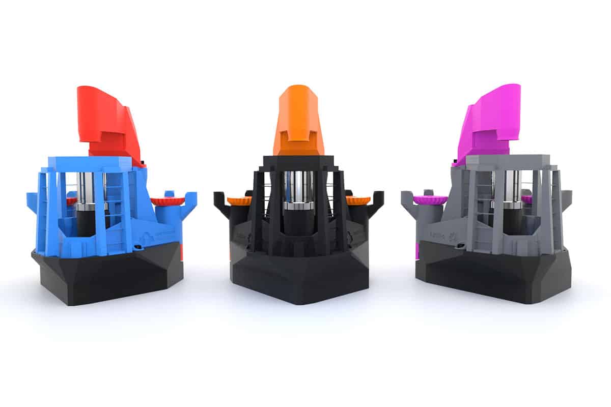
Six years ago, on a Friday afternoon, I (Richard Bowman) made a microscope focusing stage with the £400 3D printer in our lab. What started as a moment of idle curiosity quickly snowballed and pushed aside my “day job” research, to grow into so much more. Today, the OpenFlexure Project – developing a 3D-printed laboratory-grade motorized microscope to analyse samples and detect diseases – has a global community of users and developers that spans hobbyists, research scientists, entrepreneurs and clinical researchers.
All of them are supported by a core team spread around the globe – at the universities of Bath and Cambridge in the UK, along with the Tanzanian engineering company BTech (Bongo Tech & Research Labs, formerly STICLab) and the Ifakara Health Institute. We are currently working towards medical certification in Tanzania, and exploring commercialization options in various parts of the world. This growth has largely come about because of our commitment to openness and reproducibility – and the OpenFlexure project is just the beginning of what open-source hardware enables.
Complementary to open-source software, the open-source hardware movement aims to allow people all over the world to make, modify and share hardware for scientific use. Such technology could be particularly appealing for education and training purposes, where expensive professional equipment is particularly prohibitive. More essentially though, for researchers and medical professionals in the Global South, open-source hardware built and specified by themselves could be a game-changer.
Optical microscopes are an essential tool, both to detect disease in clinics, and for scientific research in general. But commercial high-performance microscopes are expensive – usually selling for tens of thousands of pounds – and are hard to maintain. Our most basic 3D-printed microscope, in contrast, can be built for as little as £15 (the cost of the printed plastic, a camera and some fastening hardware). Our top-end version – including a microscope objective and an embedded Raspberry Pi computer – would cost a couple of hundred pounds. If you visit openflexure.org, you can download, print and assemble the latest version of the OpenFlexure microscope. The fully automated microscope is highly customizable, with a number of options readily available for optics, camera and control, including motorized sample positioning and focus control.
Setting the stage
A key feature for any microscope is the ability to precisely position samples and probes – so it must have a precise and stable translation stage to focus the microscope, and move to the right area of the sample. This is a ubiquitous challenge when designing apparatus, and is one reason why research microscopes cost so much. Indeed, machining a fine translation stage that doesn’t wobble or stick requires smooth surfaces, hard materials and precise dimensions.
We found that simply 3D printing the stage designs used in most microscopes results in poor performance. That’s because printed plastic is soft, the surface finish is usually rough, and the printed object often doesn’t match its nominal dimensions exactly. Instead, our design exploits the flexibility of the plastic by using a deformable mechanism to move the sample. The “flexure hinges” that give the project its name are used in many precision instruments, but are usually made of metal and have a limited range of motion. The greater deformability of plastic, and the ability of 3D printers to create intricate shapes, mean our flexure stage can move a relatively long way: 12 × 12 × 4 mm.
The initial publication of the microscope in 2016 focused on its mechanics (Review of Scientific Instruments87 025104). At that time, there were a number of low-cost and/or open-source microscope projects that provided a good optical solution, but there were far fewer easily manufactured solutions for the mechanics of such a microscope. We characterized our microscope’s drift over time; how repeatable the stage was when moved by stepper motors; and how linear the motion was. By all those metrics, our microscope compared well to others costing orders of magnitude more.
Open and sustainable 3D rendering of the OpenFlexure microscope using an all open source toolchain (OpenSCAD and Blender). (CC-BY 4.0 Joel Collins)
By the time we’d completed this work, the OpenFlexure microscope was already starting to be noticed by others. A team in Cambridge picked up the OpenFlexure design and turned it into OpenScope – a programmable open-source microscope. Also, the ability to do low-cost timelapse imaging meant the microscope was the perfect base for the first generation of water-testing technology used by WaterScope – a company Richard co-founded in 2015 to provide low-cost methods for testing water quality in the Global South.
Now that we had active users, they started requesting features and improvements. In particular, we improved our optics options, which used conventional microscope objectives in addition to the inverted webcam lens that enabled most of the early work. The OpenFlexure microscope was then a capable lab prototype, able to take good quality images and even move the stage automatically. However, it was very much a lab prototype. The interface involved obscure keyboard commands with little documentation; there were loose wires and circuit boards; and the whole thing was usually packed inside a shoebox padded out with blue kitchen roll.
Building equitable partnerships
In 2018 we were funded by the UK’s Engineering and Physical Sciences Research Council and the National Institute for Health Research to develop and evaluate the microscope for malaria diagnostics – a project run in collaboration with Ifakara Health Institute and BTech in Tanzania. A central theme of this project is local manufacturing, rather than shipping microscopes from the UK to Tanzania, so that the microscopes used at the Institute can be built a couple of hours’ drive away, at BTech.
We believe this local manufacturing is crucial, as the World Health Organization estimates that nearly 70% of donated medical equipment in sub-Saharan Africa is out of service or not in use usually because authorized service engineers, proprietary consumables and spare parts are not available locally (Med. Biol. Eng. Comput.49 719). Our approach means BTech can provide this missing infrastructure, thereby making a sustainable impact as well as creating skilled jobs. Ultimately, releasing the hardware under an open-source licence is a way to share ownership and control of the project, which we feel is key to building equitable partnerships between groups with very different economic contexts.
Working in collaboration, we improved the design to make it more acceptable in a hospital lab – for example, adding a better interface and enclosing the electronics. Together with BTech, we made it easier to manufacture too, adding 3D-printed tools for tricky steps, simplifying the bill of materials, and taking more care over the specification of the components. In particular, our initial prototype was very hard to reproduce as it required specific components that are expensive to order or re-order and are typically only found in well-funded labs. The current version of our microscope (Biomedical Optics Express11 2447) is a capable lab microscope with options for high-resolution bright field, fluorescence and reflection imaging.
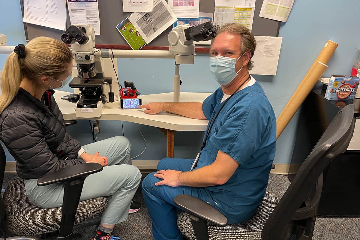
Educational value Pathologist Daniel Rosen, at the University of Houston, using the OpenFlexure microscope for teaching. (Courtesy: Daniel Rosen)
Over the three and a half years that our malaria project has run so far, we have collected terabytes of images of blood smears, to evaluate the microscope and train machine-learning algorithms to understand the images. We have also identified and fixed many more issues that only came to light when we had multiple microscopes running long-term in realistic scenarios, in the field. When we scan a blood smear by taking a grid of images spread over the sample, for example, those images can now be evaluated on the fly, so that we can repeat any measurements that are out of focus. This approach of making the instrument smarter, so it can self-correct and become more reliable as well as more capable, is a key theme in the OpenFlexure project.
Open-science test drive
In tandem with our funded research focused on malaria, we have worked to build up a community around the microscope. Most project communication was initially done on the software-development platform GitLab. While GitLab is still where all our designs are managed, last year we set up an online forum that opened up the project to many more people. The forum is a much easier place for users and contributors to engage with the project without having to navigate the complicated, software-focused interface of GitLab.
Our community now includes scientists and engineers from all over the globe – from both physical and life science backgrounds – as well as hobbyists, teachers, members of community groups, and even companies using parts of the hardware or software in their own products. Our best estimate is that there are hundreds of microscopes that have been manufactured “in the wild” without our direct involvement. That means the OpenFlexure project is a useful testbed for many aspects of how to create and share a piece of open scientific hardware.
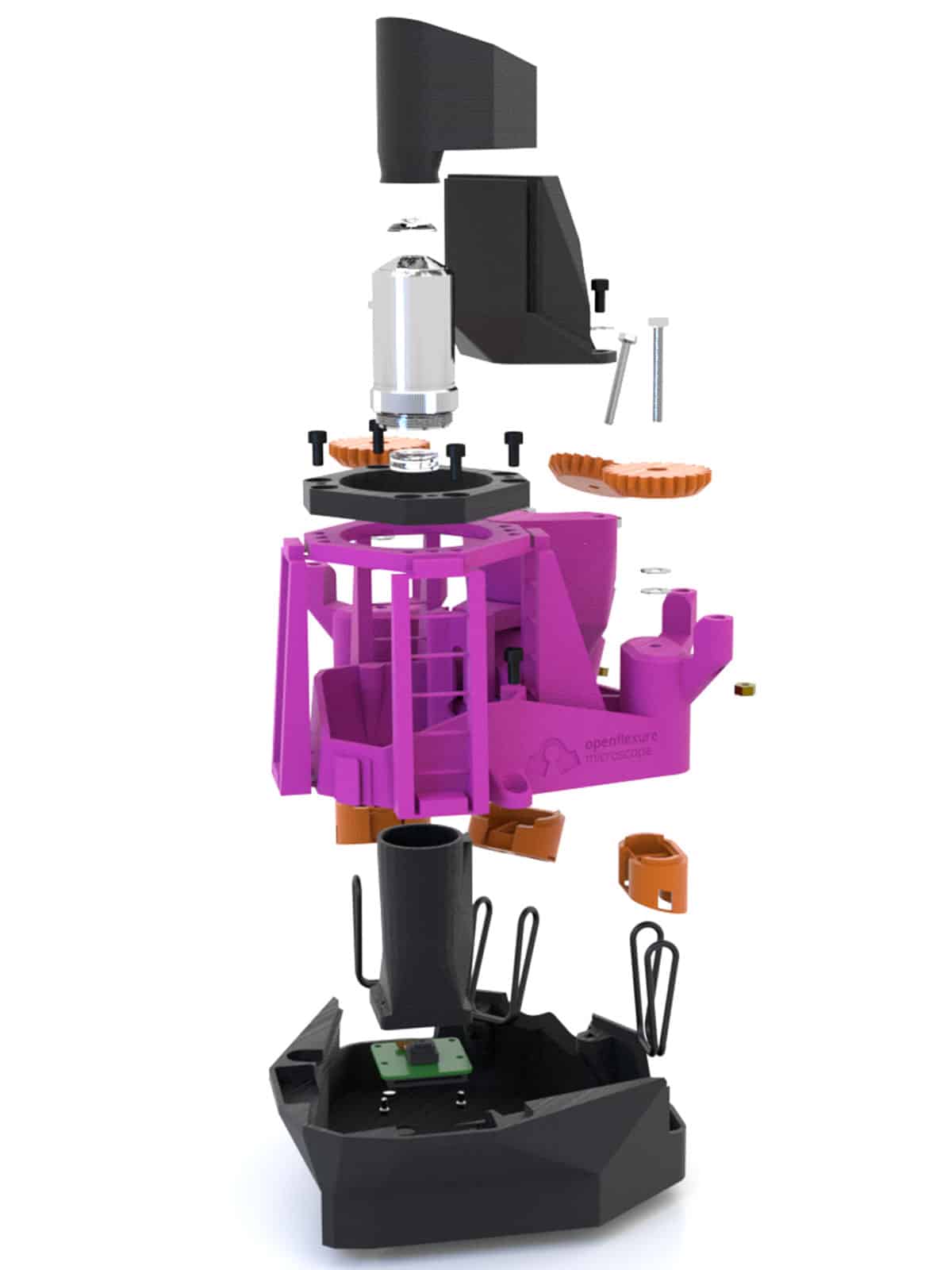
Build your own An exploded render of the OpenFlexure microscope showing all components is part of the build instructions. (CC-BY 4.0 Joel Collins)
The single most important thing we have learned about how to allow a piece of lab apparatus to be replicated exactly is the importance of good documentation. Our build instructions have gone through numerous complete rewrites and lots of minor updates – and there is still a long way to go. It’s not uncommon for replications of scientific experiments, even ones that intend to be fully open, to rely on direct communication between research teams. While this is less work for both teams the first time a project is replicated, it doesn’t scale well. Relying on word of mouth also means that crucial know-how never gets written down, which means the scientific record isn’t enough to reproduce an experiment once the researchers involved have moved on.
The growth of the OpenFlexure community has forced us to work in ways that scale better. Well-honed instructions are a good starting point, but moving all the questions and answers about the project from spoken or e-mailed conversations into an open, searchable forum means that not only the core instructions, but also a great deal of associated know-how is now available for others to learn from. It’s great to see more and more researchers adopting a similar approach, and we try very hard to make sure that what we’ve learned from this project is made available so that others can learn from our experience.
On the open record
One aspect of sharing a project openly that’s not often talked about is the emotional journey of opening up. The decision to release a project openly might seem simple, but there are many worries that often stop academics from fully sharing their work. Many scientists, for example, feel unable to share details before their work is published, in case they are “scooped”. We too did not initially share the first version of the OpenFlexure microscope online – only doing so when the preprint of our first paper was uploaded to arXiv – but we have since moved to sharing projects much earlier. Doing so eliminates worries about who we share ideas with, and more fully realizes the benefits of open working (The Design Journal 10.1080/14606925.2020.1859168). Sharing designs as they evolve actually makes us less concerned about others “stealing” our work, as there is a verifiable, public record of who did what in our repositories. This makes it much harder for someone to claim the work as their own, than if we kept all of our work a secret until we’re ready to publish.
One aspect of sharing a project openly that’s not often talked about is the emotional journey of opening up
The OpenFlexure microscope adapted for the WaterScope project. (Courtesy: University of Bath)
The deeper worry, however, is usually that releasing the project openly will diminish the creators’ ownership and control of it; meaning they don’t receive full credit for the work – be it financial, or through authorship and citation of published articles. This is a reasonable concern, but our experience is that the benefits far outweigh the downsides. By and large, people who make use of open designs are happy to give credit where it’s due. After all, citing the design properly is a small effort to repay something useful, and the scientific community is increasingly paying attention to good practice around open science.
While there may be people and projects who have not credited their use of our work properly, this is more than made up for by the large network of collaborators who do recognize its value. That network is far bigger than it would be if we had taken a less open approach, and indeed it is much larger than we could support if we had to deal with requests individually rather than through open, reusable platforms like the forum.
Experimental ecosystem
As scientific instruments go, the OpenFlexure microscope is more of a workhorse tool than a state-of-the-art microscope existing in only a few labs. However, it has been extended to do super-resolution and phase imaging, and can be connected to another open-source toolbox – the UC2 modular optics system – for even more customizable use.
Valerian Sanga adjusting the illumination of an OpenFlexure microscope. (CC-BY 4.0 Gathering for Open Science Hardware)
One of the reasons it has been extended so frequently is its ease of replication: it is simple and inexpensive enough for other labs to replicate on a whim, without feeling the need to justify their efforts with a new publication or apply for a research grant to pay for it. A big part of our vision for the project is that it can support an ecosystem of experimental techniques that can be fully replicated at moderate cost. Our hope is that this will lead to experimental science becoming more transparent, being repeated more often, and ultimately improving the quality and trustworthiness of our experiments.
The tools and working practices we develop to support the sharing of instrument designs need active, frequently replicated projects like ours for their development, but are intended to scale well to more expensive and specialist projects. This is reflected in CERN’s White Rabbit Project – a multi-laboratory, multi-company collaboration to develop new technology for control and data-acquisition systems that covers hardware, gateware and software; or the mesoSPIM initiative, which focuses on open-source light-sheet microscopes (Nature Methods16 1105).
Support and funding in the long run
Looking to the future, one of the biggest obstacles to achieving a long-term, sustainable impact with a project such as ours is supporting the team over a number of years. We are fortunate to have been funded for five years through the Global Challenges Research Fund (GCRF) – part of the UK’s aid budget that funds partnerships between UK scientists and their counterparts in low and middle-income countries. Building the trust that is needed for a productive, equitable partnership takes a long time, as does learning to work around the many differences in culture and working practices between different countries and institutions.
The project so far has been a great success, but recent cuts to the UK aid budget mean that future funding for our work, as well as hundreds of other projects, is now scarce and the phase 2 funding for our pan-African network was cancelled earlier this year. The GCRF represents a significant investment from the UK, and if we are serious about achieving a meaningful impact with this kind of work we must make sure that we have a plan in place to support its translation out of academia. In the UK, this support often comes from companies that will commercialize the work, supported by government grants.
If we are serious about achieving a meaningful impact with this kind of work we must make sure that we have a plan in place to support its translation out of academia
In the spirit of “sustainable development”, we want to avoid leaving our African partners dependent on a UK company, as this would undermine many of the key benefits of an open, distributed project. However, start-up capital and support for small businesses is much harder to come by in countries with fewer economic resources. Transferring work from academia into charitable organizations is also harder, and much less well supported, than simply patenting and licensing technologies in a way that entrenches the inequalities already present in the world’s economic system.
On an individual level, this project could not have succeeded without postdoctoral researchers on short-term contracts who have devoted several years to building relationships, sharing knowledge and documenting projects to get ready for real-world use – often at the expense of chasing prestigious publications that would further their career. The recent aid cuts, and the consequent slashing of the GCRF programme, means that these incredibly valuable and committed people will now have to find jobs elsewhere. This comes at a huge personal cost to the people involved, but it also means that their skills, knowledge and partnerships are lost.
There’s a great deal of anger from the GCRF research community that has flourished in recent years, and problems will continue even if the UK does reinstate aid funding to meet its legal aid budget commitment of 0.7% of gross national income. We are pursuing other ways that our project and others like it can continue to make progress, for example by creating a foundation that can help open projects move towards medical certification, and are keen to collaborate with others to bring this about. We believe that open-source hardware truly has the potential to revolutionize the way we do science – from building instruments and developing educational tools, to launching clinical applications and growing global partnerships.
Many medical imaging techniques rely on a mathematical process called tomography, which reconstructs data recorded in one or two dimensions into three-dimensional image volumes. Positron emission tomography (PET) – which images the body using an injected radiotracer that emits positrons as it decays – offers the unique possibility of reconstruction-free 3D imaging. This is because PET can also localize the signal source by exploiting the time difference between detection of the two photons created when the positrons annihilate with electrons in the body.
Today’s state-of-the-art PET systems have a timing resolution of around 210 ps that, based on the speed of light, translates to a spatial localization of 3.15 cm. As such, image reconstruction is still required to create accurate images. But if this timing resolution could be improved, the tomography step could be eliminated completely.
A collaborative team from the University of California, Davis and Hamamatsu Photonics has now developed radiation detectors with an average coincidence timing resolution of 32 ps. The researchers – also from University of Fukui and Kitasato University – have used these detectors to create the first experimental cross-sectional medical image that doesn’t require tomography, a technique they call direct positron emission imaging (dPEI). They report their findings in Nature Photonics.
Ultrafast detection
First author Sun Il Kwon and colleagues achieved dPEI by combining three technology innovations. Firstly, they exploited the Cherenkov luminescence emitted when annihilation photons interact in the detector. If the detector material has a high refractive index and high atomic number, this interaction creates electrons with sufficient energy to produce Cherenkov photons.
To convert this prompt optical signal into an electronic signal, the researchers developed a photosensor based on microchannel plate photomultiplier tubes (MCP-PMTs), which have extremely high single-photon time resolution (SPTR), approaching 20 ps. They integrated the Cherenkov radiator (lead glass) with the photocathode in the MCP-PMT to eliminate any optical boundaries and increase the chances of detecting the small number of Cherenkov photons. Finally, they employed convolutional neural networks (CNNs) to predict the timing information for detected events from the detector waveforms.
To assess the performance of the new detector, the team measured the signal from a 22Na point source placed between two Cherenkov-radiator-integrated MCP-PMTs. The set-up detected Cherenkov photons with an average timing resolution of 32 ps, allowing annihilation events to be localized with a spatial precision of 4.8 mm.
Experimental set-up: The brain phantom in the imaging system. (Courtesy: Sun Il Kwon)
The researchers then tested the system using three test objects filled with the PET radiotracer 18F-FDG: an image quality phantom; a spatial resolution phantom; and a 2D Hoffman brain phantom representing 18F-FDG distribution in a slice of human brain. To capture the signals, the detector pair was translated linearly over the width of each object. The system then created images directly from the measured data, without using any tomographic reconstruction algorithm.
The image quality phantom has a uniform background activity with two 8-mm diameter voids filled with air and with water, all of which could be clearly visualized. The dPEI image of the resolution phantom resolved 3 mm rods, demonstrating a spatial resolution of 4–5 mm. The dPEI image of the larger 18.4-cm diameter brain phantom faithfully captured its detailed structure, with a spatial resolution of around 4.8 mm, suggesting that the method could be scaled for human imaging.
The two-detector set-up, with its average timing resolution of 32 ps, produced images with a similar spatial resolution to that achieved by diagnostic PET scanners. The team note that these initial experiments used long acquisition times and high levels of radioactivity. The signal collection efficiency could be increased, however, by replacing the lead glass radiator in the MCP-PMTs with a higher atomic-number radiator, increasing the radiator thickness, and tiling multichannel detectors to increase geometric coverage.
Such an upgraded system would enable shorter acquisition times and/or lower radiation doses. For example, these three changes could reduce the data acquisition time for the brain phantom image from 24 h to roughly 1 min. In addition, multidetector configurations that cover the entire imaging volume-of-interest would remove the need for detector translation and allow dynamic radiotracer imaging.
The team is already working to implement some of those advances. “We are testing new detectors that have the scintillator bismuth germanate (BGO) integrated in the MCP-PMT, rather than lead glass,” senior author Simon Cherry tells Physics World. “This leads to a significant efficiency improvement because BGO has a higher atomic number; it also produces more Cherenkov light because of its higher refractive index.”
The greatest number of fast radio bursts (FRBs) ever to be observed from a single source has been reported by an international team of astronomers. Led by Di Li and Pei Wang at the Chinese Academy of Sciences, the group used the Five-hundred metre Aperture Spherical radio Telescope (FAST) in China to detect more than 1600 radio bursts from an object called FRB 121102 within a span of just two months. The observations allowed them team to carry out the first detailed statistical studies of the FRB phenomena – shedding new light on their astrophysical origins.
In 2007, two astronomers at West Virginia University detected an extremely rapid burst of radio waves, just under 5 ms in duration. Since this discovery, hundreds of similar FRBs have been detected in separate studies. Most FRBs are detected as single events, but in a small fraction of cases, FRBs have been shown to repeat. This has allowed astronomers to roughly identify the galaxies where these events originated.
Compelling mystery
Although they are relatively weak once they reach Earth, astronomers know that the bursts involve the release of large amounts of energy – a year’s worth of the Sun’s total energy output over the span of just a few milliseconds, for example. A variety of theories have been put forward to explain FRBs, including merging black holes, magnetized neutron stars, and exotic defects in the early structure of the universe. So far, however, no one theory has emerged as being more plausible than any other, and today, the astrophysical sources of FRBs remain a compelling mystery.
Over 47 days between August and October 2019, Li and Wang’s team detected a record-breaking 1652 FRBs originating from FRB 121102, which appears to be located in a dwarf galaxy about three billion light-years from Earth. This is the first known repeating FRB and it was discovered in 2021.
High activity
The FAST tally of 1652 events is greater than all previous observations of FRB events combined. At one point of particularly high activity, the astronomers recorded as many as 122 bursts in a single hour.
Such an extensive sample size allowed the team to do a detailed mathematical analysis of the properties of the bursts. This revealed key differences between lower- and higher-energy FRBs. Contrary to some theories, the analysis of FASTS’s data revealed little repetition in the bursts over timespans ranging from just 1 ms to 1000 s. This lack of periodicity challenges the idea that FRBs may originate from single rotating objects, such as neutron stars.
In addition, the fast rate of bursts within the event implies that FRBs can be generated extremely efficiently – discrediting the idea that their triggering mechanisms requiring large amounts of energy. Altogether, the team’s discoveries place tighter bounds on existing FRB theories and could bring astronomers a step closer to uncovering their enigmatic origins.
Due to breathing motion, lung tumours move during stereotactic body radiation therapy (SBRT). Scatter imaging, which collects photons scattered out of the radiation therapy beam, is a potential technique for real-time tracking of lung tumours.
In this webinar, presented by Kevin Jones, the scatter imaging method is characterized through simulation, phantom experiments and analysis of clinical patient scatter images.
Kevin Jones is a medical physicist in the department of radiation oncology at Rush University Medical Center in Chicago, Illinois, USA. He is board certified by the American Board of Radiology. His research focuses on developing novel imaging techniques to guide radiation therapy.
For the first time ever, researchers have delivered an antibody therapy drug directly into the brain to target breast cancer metastases. A team at Sunnybrook Health Sciences Centre in Toronto used MR-guided focused ultrasound (MRgFUS) to non-invasively and temporarily open the blood–brain barrier (BBB), enabling the monoclonal antibody trastuzumab to reach specific areas of the brain. Writing in Science Translational Medicine, the researchers describe the first visual confirmation that focused ultrasound can improve delivery of targeted antibody therapy across the BBB.
The BBB consists of a thin layer of cells that protect the brain from toxins, viruses and bacteria, but which also blocks the delivery of therapeutic drugs. Trastuzumab, an antibody therapy that helps the immune system fight cancer cells, is used in conjunction with chemotherapy and radiation therapy to treat breast cancer metastases in the brain. However, it is 100 times larger than typical compounds able to enter the brain across the BBB.
This first-in-human trial suggests that MRgFUS provides a safe and effective method to deliver drugs across the BBB. It sets the stage for the possibility of delivering both established and novel therapies for numerous brain conditions that otherwise cannot gain access to the brain.
Early findings
Principal investigator Nir Lipsman, director of Sunnybrook Research Institute’s Harquail Centre for Neuromodulation, and colleagues report the results from the first four patients participating in the ongoing Phase I clinical trial. All patients had Her2-positive breast cancer with brain metastases, and had previously received whole-brain radiation and/or stereotactic radiosurgery, plus chemotherapy, to treat progressive intracranial and stable systemic disease.
To perform BBB opening, the team used Insightec’s ExAblate, a helmet-like focused ultrasound device containing over 1000 transducers that converge ultrasound waves onto discrete points in the brain. The pulsed FUS was delivered in conjunction with a continuous intravenous infusion of microbubble ultrasound contrast agent and trastuzumab-based therapy.
Drug delivery: Left: baseline MR/SPECT images obtained prior to treatment; tumours are indicated with a white arrow. Right: images obtained 48 hr after BBB opening, indicating a significant increase in trastuzumab within the tumours. (Courtesy: Sunnybrook Health Sciences Centre)
The trastuzumab used in the study, developed by Raymond Reilly from the University of Toronto and colleagues, was radiolabelled with 111In. Use of this radiotracer-infused trastuzumab allows direct visualization of trastuzumab distribution in single photon emission computed tomography (SPECT) images.
The team delivered a total of 20 outpatient treatments combining transcranial MRgFUS with standard of-care trastuzumab injection, with an average treatment time of 138 min. Patients had a SPECT scan prior to treatment, 4 hr after treatment and 48 hr later. The researchers performed additional SPECT scans 30 and 90 days following the final treatment and continue to monitor the patients.
The SPECT images showed trastuzumab precisely targeting the tumours. After BBB opening, the researchers could directly visualize increased SPECT signal within the sonication volume for all lesions treated. MR images revealed that the patient’s brain tumours decreased in size by between 7% and 31%, at up to four months after treatment. The procedure caused no serious adverse effects. Lipsman reports that three of the patients remain stable, and that one patient died from progressive, non-intracranial systemic disease.
The researchers note that one potential advantage of MRgFUS over other physical and biological approaches to brain drug delivery is the high degree of spatial and temporal control that it provides. MRgFUS enables selective targeting of single or multiple brain lesions, which can be located at the extremes of the brain or in deep eloquent central regions of the cortex that control bodily function.
“This study adds the brainstem, cranial nerve nuclei and cerebellum to the list of regions that can be safely and precisely targeted with MRgFUS, all areas in which radiation and surgery may be limited,” the authors write. Also, because MRgFUS can be combined with diverse therapeutic agents, therapies with less side effects can be used, improving treatment tolerability and patient safety.
Future outlook
This study is continuing to enrol patients, with the goal of performing a 10-patient trial. “Completing our current trial is an important first step,” Lipsman tells Physics World. “It will help inform what the design of a larger trial would look like, what kinds of outcomes we should be measuring, and how to optimize a study for safety and feasibility. The goal is to determine what role focused ultrasound will play in the management of brain cancer patients. For that, creating partnership and collaborations with other institutions will be key.”
Sunnybrook is a Focused Ultrasound Center of Excellence, the only Canadian site recognized as such by the Focused Ultrasound Foundation, which is helping to fund this study. Lipsman says the team is investigating several aspects of using focused ultrasound in brain cancer.
“We want to determine whether peripheral biomarkers, or so-called liquid biopsy, can be enhanced following BBB opening, to help with less invasive approaches to diagnosis and treatment monitoring,” he explains. “We also will conduct more comprehensive tests looking at the safety of BBB opening, including what impact, if any, we are having on important inflammatory markers and other features of the BBB.”
“Importantly, we are interested also in streamlining the procedures, making them faster, more efficient and more comfortable for patients,” he adds. “Enhancements in imaging technology will aid in targeting, and improvements in hardware and software will give us even better control of microbubbles and ultrasound transducers, to control just how much of the BBB is open and hence how much of a therapeutic can be delivered. All of these advancements are underway and will be rolled out in future trials that are now under active development.”
Researchers in China and Russia have discovered superconductivity in two new phases of a hydride material at pressures much lower than those needed to stabilize other recently-discovered hydride superconductors. The work will aid the search for lower-pressure and potentially room-temperature superconductors.
Superconductors are materials that conduct electricity without any resistance. Many materials display superconductivity when they are cooled to low temperatures, and the phenomenon was first observed in 1911 in solid mercury, which has a superconducting transition temperature Tc of 4.2 K. The search for superconductors that operate at warmer temperatures – perhaps even room temperature – has been on ever since. A material that remains superconducting at ambient temperatures would hugely increase the efficiency of electrical generators and transmission lines, as well as simplifying existing applications such as superconducting magnets in particle accelerators and MRI scanners.
Step closer to holy grail
Physicists came a step closer to this holy grail in the 1980s and 1990s with the discovery of high-temperature superconducting copper oxides, which have Tcs between 30–133 K. It wasn’t until 2015, however, that the maximum critical temperature took a major leap forward with the discovery that hydrogen sulphide has a Tc of 203 K when compressed to pressures of 150 GPa.
While materials that only superconduct at such extreme pressures have few (if any) practical applications, studying them may offer a path to discovering new compounds that superconduct at milder temperatures and pressures. For this reason, the hydrogen sulphide result sparked a flurry of interest in solid materials containing hydrogen atoms bonded to other elements. Since then, several other hydrogen-rich superconductors have been made in the laboratory. In 2019, researchers reported breaking hydrogen sulphide’s record with lanthanum decahydride (LaH10), which they found to have a Tc of 250–260 K. And in 2020, another group reported observing a Tc of 288 K in the C-S-H system at around 275 GPa.
Superconductivity in cerium hydrides
The team, led by Tian Cui, Xiaoli Huang and Wuhao Chen from Jilin University in China together with Artem Oganov and Dmitrii Semenok of Russia’s Skolkovo Institute of Science and Technology (Skoltech), took a slightly different approach. In 2019, members of the team synthesized a new cerium hydride with the formula P63/mmc-CeH9. Each cerium atom in this material is enclosed in a H29 cage in the atomic hydrogen sublattice, and it had previously only been studied theoretically. This same team has now found superconductivity in novel phases of both CeH9 and another newly-synthesized material, CeH10.
The researchers note that cerium hydrides are remarkable materials, as they are stable and display high-temperature superconductivity at lower pressures (about 0.8 million atmospheres) than other so-called “superhydrides”. As such, they say that these compounds “serve as an ideal starting point to further study the mechanism of superconductivity in these fascinating compounds, and design other superconductors, stable at even lower pressures”.
In their previous work, the Skoltech-Jilin researchers found a close relationship between the location of elements within the periodic table and the superconductivity of hydrides made from these compounds. They now believe that this relationship may also apply to materials other than hydrides. “Take La and Ce, for example – they are neighbours in the periodic table and indeed both form high-temperature superconductors,” Oganov explains. “However, there are differences: LaH10 superconducts at higher temperatures, while CeH10 is stable at lower pressures.”
Now that the binary hydrides have been well-studied, the Skoltech-Jilin team says that it will focus on achieving higher-temperature superconductivity at lower pressures in ternary hydrides. “We know which elements lead to higher-temperature superconductivity and have begun to learn which ones lead to stability at lower pressures,” Semenok says. “These are the main notes, but it takes imagination to combine them into a melody.”


 Due to breathing motion, lung tumours move during stereotactic body radiation therapy (SBRT). Scatter imaging, which collects photons scattered out of the radiation therapy beam, is a potential technique for real-time tracking of lung tumours.
 Kevin Jones is a medical physicist in the department of radiation oncology at Rush University Medical Center in Chicago, Illinois, USA. He is board certified by the American Board of Radiology. His research focuses on developing novel imaging techniques to guide radiation therapy.