Magdalena Bazalova-Carter and Nolan Esplen with the X-ray tube. (Courtesy: Magdalena Bazalova-Carter)
The curative potential of radiation therapy is limited by normal tissue toxicity, which restricts the dose that can be delivered to nearby non-target tissue. Recently, interest in ultrahigh dose rate radiotherapy has been rekindled, following preclinical studies showing increased normal tissue tolerance at high dose rates. This approach – known as FLASH radiotherapy – employs dose rates exceeding 40 Gy/s and can improve the therapeutic ratio by increasing the differential response between normal and tumour tissues.
The expectation is that FLASH could one day provide tumour ablation in a single sub-second treatment, while substantially reducing radiation-induced side-effects. The underlying mechanism, however, is not yet understood and requires further research. And to date, FLASH dose rates typically require specialized electron sources or substantial modifications to clinical linacs.
“Access to FLASH beamlines is limited and the progress on understanding of the FLASH mechanism is quite slow,” explains Magdalena Bazalova-Carter from the University of Victoria’s XCITE Lab. To remove this constraint, Bazalova-Carter and PhD student Nolan Esplen are investigating the feasibility of using a conventional high-powered X-ray tube for FLASH radiotherapy (Med. Phys. 10.1002/mp.13858).
“Our results will hopefully inspire researchers without access to high-power electron sources or proton beamlines to perform FLASH in vitro, and possibly some limited in vivo experiments, with a standard X-ray tube,” says Bazalova-Carter.”
The researchers used Monte Carlo (MC) modelling to evaluate the maximum dose rates achievable by two conventional X-ray tubes: the 3 kW MXR-160/22 (which was being validated in the XCITE lab) and the 6 kW MXR-165, which benefits from a short distance from the focal spot to the tube surface. For both tubes operating at maximum power, they simulated the output of an unfiltered 160 kV beam and calculated the dose deposited in a water phantom placed against the tube surface. They then converted the MC-calculated dose to dose rate.
The simulations revealed that both X-ray tubes were FLASH-capable, with maximum phantom surface dose-rates of 114.3 and 160.0 Gy/s, for the MXR-160/22 and MXR-165, respectively. Dose non-uniformity due to the heel effect – an inherent directional variation in the X-ray intensity emitted by the anode – was seen in both cases. For a 1 cm diameter region-of-interest within the high-dose region, dose rates were 110.6 Gy/s for the MXR-160/22 and 151.9 Gy/s for the MXR-165.
Plotting dose rate versus depth revealed a rapid fall-off for both 160 kV X-ray beams. At 2 mm depth, for example, dose rates decreased to 23% and 28% of that at the surface, for the MXR-160/22 and MXR-165, respectively. The dose rate remained FLASH-capable at depths of up to 1.4 and 2.1 mm, for the MXR-160/22 and MXR-165, respectively.
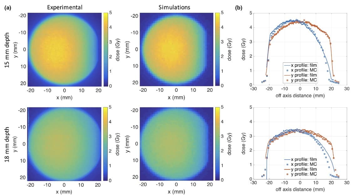
To validate their MC models, Bazalova-Carter and Esplen measured the dose in a plastic water phantom irradiated with a 120 kV beam from the MXR-160/22. They placed Gafchromic EBT3 films at 15 and 18 mm depth in the phantom and compared the measured 2D dose profiles with those from MC simulations of the 120 kV beam.
Experimental and simulated 2D dose distributions in the solid water phantom (a). Comparison of dose profiles at the two depths (b). (Courtesy: Med. Phys. 10.1002/mp.13858)
In the region not affected by the heel effect, the simulations agreed well with the film measurements. The mean X-profile differences between experiments and simulations were 1.5% and 3.2%, at 15 and 18 mm, respectively; the mean y-profile differences were 1.5% and 3.5%, at 15 and 18 mm. Agreement in the heel-effect region was poorer, however, with a mean difference of up to 17.8% along the X-profile.
This validation experiment demonstrates the ability of conventional X-ray tubes to deliver FLASH therapy. The researchers suggest that these particular tubes could be suitable for FLASH skin irradiations, in vitro experiments or testing dose-rate dependence of small-field dosimeters.
They are now working to further tailor the X-ray tubes for FLASH applications. “We are currently building a shutter mechanism that will inset in the X-ray tube, which will further increase the dose rate,” Bazalova-Carter tells Physics World. “We are also designing experiments to test cell survival for FLASH irradiations with and without gold nanoparticles.”
The human brain undergoes significant functional and structural changes during the first decades of life, as the fundamental building blocks of human cognition are established. However, relatively little is known about maturation of brain function during these critical times. Non-invasive imaging techniques can provide information on brain structure and function, but brain scanners tend to be optimized for adult head-sizes. Traditional fixed scanners also require patients to stay completely still, which can be highly challenging for children.
A UK research collaboration aims to solve these problems by creating a wearable magnetoencephalography (MEG) system that allows natural movement during scanning. They have now used the wearable MEG for the first time in a study with young children (Nature Commun. 10.1038/s41467-019-12486-x).
The researchers, from the University of Nottingham, the University of Oxford and University College London, developed a lightweight ‘bike helmet’ style MEG scanner and used it to measure brain activity in children performing everyday activities. As well as enabling studies of neurodevelopment in childhood, this system should allow investigation of neurological and mental health conditions in children, such as epilepsy and autism, for example.
MEG measures the small magnetic fields generated at the scalp by neural current flow, allowing direct imaging of brain activity with high spatiotemporal precision. Traditional MEG systems use an array of cryogenically-cooled sensors in a one-size-fits-all helmet. Such systems are bulky and highly sensitive to any head movement.
To address these issues, the team is using optically pumped magnetometers (OPMs) to measure the magnetic fields generated by the brain. These small, lightweight sensors can be positioned on a 500 g helmet that can adapt to any head shape or size. The OPMs can also be placed far closer to the head than conventional sensors, increasing their sensitivity. The researchers also employed an array of electromagnetic coils to null the residual static magnetic field inside the magnetically shielded room, allowing individuals to be scanned whilst they move freely.
“The initial prototype scanner was a 3D printed helmet that was bespoke – in other words only one person could use it. It was very heavy and quite scary to look at,” explains PhD researcher Ryan Hill who led this latest study. “Here, we wanted to adapt it for use with children, which meant we had to design something much lighter and more comfortable but that still allowed good enough contact with the quantum sensors to pick up signals from the brain.”
The researchers designed and built the new bike helmet style scanner and used it to successfully analyse the brain activity of a two-year old (typically the hardest age to scan without sedation) and a five-year watching TV whilst their hands were being stroked by their mother. The children were able to move around and act naturally throughout.
To show that the MEG system is equally applicable to older children, the researchers also used it with a larger helmet to scan a teenager playing a computer game. Finally, they used the new scanner to examine brain activity in an adult learning to play a sequence of chords on a ukulele. Despite the substantial head and arm movement required to complete this task, clear electrophysiological responses were observed.
“This study is a hugely important step towards getting MEG closer to being used in a clinical setting, showing it has real potential for use in children,” says Matthew Brookes, who leads the MEG research at the University of Nottingham. “The challenge now is to expand this further, realising the theoretical benefits such as high sensitivity and spatial resolution, and refining the system design and fabrication, taking it away from the laboratory and towards a commercial product.”
The researchers conclude that their study demonstrates that the OPM-based MEG system can generate high quality data, even in a 2-year-old child, and can be used to measure brain activity during naturalistic motor paradigms. “OPM-MEG, with generic helmet design, paves the way for a new approach to neurodevelopmental research,” they write.
When taking a deep breath you draw a range of gases into your lungs from oxygen and nitrogen to carbon dioxide and argon along with traces of water vapour. But that same breath could also contain microscopic amounts of copper, iron, zinc and even chromium. Despite making up a tiny fraction of the pollutants in the air, transition metals have some of the most damaging impacts on our health.
A study has now assessed the abundance of various airborne transition metals in urban areas across the US. While the overall particulate pollution has decreased over the past two decades, particularly in urban areas, some cities have, however, seen a rise in the amount of air-borne transition metals. By studying the trends, researchers are beginning to pinpoint what the likely sources of various metals are and how their emissions can be better controlled.
Diluting concentrations
Clean air is a staggeringly good investment. Since 1990, the US has spent an estimated $65bn on implementing the 1990 Clean Air Act but gained $2 trillion in benefits. According to the Environmental Protection Agency, this year alone the act has prevented around 230 000 early deaths, avoided 120 000 emergency room visits, and stopped 5.4 million sick-days in schools and 17 million sick-days at work.
Particulate pollution is responsible for some of the worst health and economic impacts of air pollution with transition metals believed to be more damaging than other compounds. That is because the metals act as a catalyst and help to produce oxidants, which can lead to oxidative stress. “Oxidative stress has been linked with the genesis and progression of many different diseases – it’s why there is so much research and marketing for foods that contain antioxidants,” says Christopher Hennigan, an environmental engineer at the University of Maryland, Baltimore County.
There is a strong body of scientific research that shows that a transition to more sustainable energy sources will have co-benefits in air quality
Christopher Hennigan
Hennigan and colleagues analysed seven different transition metals over the period 2001 to 2016, across ten different urban areas — Atlanta, Baltimore, Chicago, Dallas, Denver, Los Angeles, New York City, Seattle, St Louis and Phoenix. They found that around a decade ago concentrations of nickel and vanadium in port cities were around five times higher than non-port cities but that the difference has now all but disappeared. “The reductions in port-cities were most likely from regulations on marine fuel sulphur content,” explains Hennigan.
The strong downward trend in vanadium across all urban areas clearly matched the introduction of diesel-fuel regulations in 2006. Yet copper, meanwhile, has stayed stubbornly constant in most areas. “Our results suggest that vehicle brake-lining dust is a major source of copper,” says Hennigan. The team also found higher concentrations of iron in western cities (by around a factor of two) than cities in the east, most likely because soil and dust are major sources of iron and prevailing winds cross more land and carry more dust to western cities.
One puzzle, however, was chromium, which increased in cities in the east and midwest, with a distinct spike in 2013. “We don’t have a good explanation for this which indicates a gap in our understanding of chromium sources and their magnitude,” says Hennigan.
The findings confirm how beneficial air-quality legislation has been for the US. It also makes a strong case for continuing to improve air quality with Hennigan believing there are still big gains to be make. “There is a strong body of scientific research that shows that a transition to more sustainable energy sources will have co-benefits in air quality,” he says.
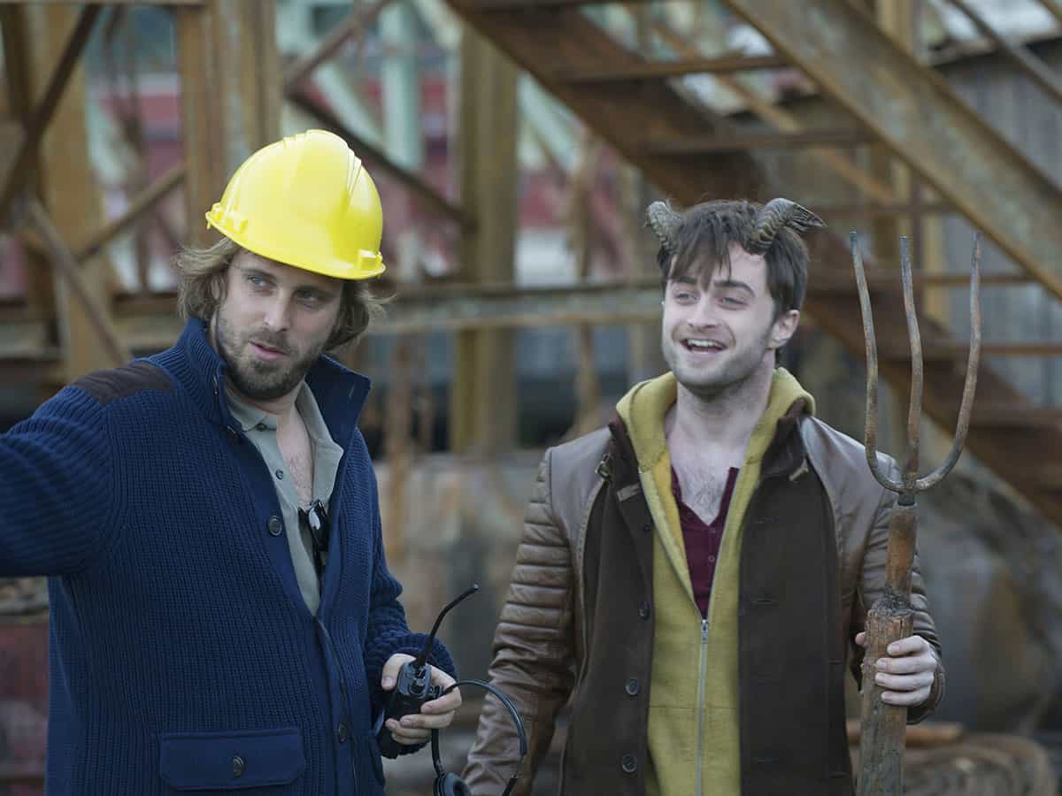
Fantastic beast: Daniel Radcliffe (right) on the set of Horns with director Alexandre Aja. This film used prosthetics and practical effects as well as VFX. (Courtesy: Red Granite/Mandalay/Kobal/Shutterstock)
Jess Wade: You have been in a bunch of films that use VFX in the most progressive and creative ways. What was it like starting your acting career with the extraordinary VFX in the Harry Potter films [2001–2011]?
Daniel Radcliffe: For some of the experienced actors on Potter, it was their first time working with VFX on that kind of scale. It was different for us kids. Telling us that “the dragon is this tennis ball on the end of the stick” is a little different from giving an older actor that instruction – we’d never known anything different. And we were all kids, so using our imagination was something that we were doing a lot anyway.
JW: Has VFX changed how you act?
DR: I don’t think so – it’s always been a big part of my career. I enjoy the challenge of it. I think I’m weirdly good at following numbered cues now. I remember when they shot all the audience reactions during the Tri-Wizard Tournament [in the fourth film, Harry Potter and the Goblet of Fire (2005)], and there would basically be a bunch of the cast and background artists on a big stand – sometimes on a green screen, depending on what the backdrop was. Assistant directors would hang big numbers around the studio and just say, for example, “1” so everyone would turn to the same eye line at the same time.
JW: The Harry Potter films ended eight years ago, and you’ve done some really exciting things with VFX since then. Has it changed a lot?
DR: Potter came at a time when people were leaning heavily toward visual effects and away from “practical” make-up or special effects. Even though, of course, we had plenty of them too. In the last couple of years, we’ve reached a nice balance – where big franchises like Star Wars and Mad Max use a lot of practical stuff in creature effects and stunt work. People see the value of having practical, on-set effects, but VFX are so good. It can also make stunt work safer because you don’t have to put a human being through what you can get VFX to do.
But certainly, VFX is improving at an extraordinary rate. If you were to look at the difference between the first and last Potter films [in 2001 and 2011] – they get exponentially better over time.
JW: How does working with all that VFX compare to stage acting?
DR: I think that’s the joy of my job – I’ll do some films where there’s almost no VFX whatsoever, then I’ll do films like Swiss Army Man[2016] where it’s a crazy mix of VFX and old-school practical stuff such as camera tricks. There was one scene in that film where my character gets punched in the mouth, then swallows the hand that punches him… and punches himself in the stomach to make the hand that’s in his mouth get forced back out. I wondered “how are we going to do that?”. There was no VFX involved – it was entirely clever camera angles and a bit of make-up on the arm to make it look like it was covered in spit. It’s wonderful to be able to flit between those things – the very low-fi and the highly sophisticated ways of solving problems on film.
JW: Do you ever get involved with VFX? Do you go and see what they’re doing?
DR: The closest you get on set is when the film’s big enough to do previs [previsualization] sequences – like an animated storyboard that no-one else ever sees. For example, when there was a big quidditch sequence on Potter, they’d have that all mapped out on a visual storyboard first, and we’d try and stick to that when we filmed. But the majority of the time, the VFX is in post-production, when the actors aren’t around.
JW: But sometimes you go in to do that funny thing – what’s it called – ADR?
DR: Yeah, ADR – additional dialogue recording. At that point you might see some sequences with half-finished VFX – and that’s always cool; it’s always fun to see it in a primitive phase. For someone who is interested in how films get put together it’s kind of fascinating. In this rough cut of the film there will be shots like, if you did a driving sequence on a green screen, they’ll just show the shot on a green screen with a little caption saying “VFX needed”.
When films started using huge sets that were just entirely blue screen and VFX, I think actors were a bit whiney about it – there’s something about being on a bright blue or green screen that can drive you slightly insane. At first it was something to be remarked upon, but now it is so much part of the industry – I don’t think anyone sees it as a novel thing anymore.
JW: What’s your favourite example of VFX that you’ve worked with?
DR: That’s really hard. There are some amazing sequences in Potter – there is some really beautiful stuff. The Hall of Prophecy in [the fifth film, Harry Potter and the] Order of the Phoenix [2007] was almost entirely green screen if I remember rightly.
And then in Horns [2013], when my on-screen brother took some hallucinogenic drugs and had this really visual trip – that’s a really good mix of practical prosthetics, VFX and tricks the designers built into the sets.
There’s also the other side of VFX, which is less glamorous but even more useful. Like driving sequences – when you’re filming in a place where you can’t shut down roads, you have to do it on green screens. Then there’s patching up a prosthetic. Sometimes things look fantastic when they’ve been put on at 9 a.m., but when you’ve been wearing it for 10 or 11 hours, visual effects can be helpful for polishing up that stuff.
JW: What has been the most ridiculous thing that you had to work with?
DR: None of it feels too ridiculous at the time. The hippogriff [a magical creature that’s part eagle, part horse] in [the third film, Harry Potter and the] Prisoner of Azkaban [2004] – the reality of the hippogriff and the flight of it was quite funny. If you imagine a limbless, headless bucking bronco…
JW: [descends into laughter] Like…a mechanical thing?
DR: Yeah, a mechanical bucking bronco on hydraulics. Just a grey torso with no texture, filmed on a blue screen and a green screen with a motion control camera.
JW: [can’t stop laughing] But you were all kids! I imagine when one 14-year-old starts laughing, everyone starts laughing.
DR: Sure, there would be an element of that. Thankfully, for the hippogriff sequence I was on my own at the start – so I’d got used to it. Of course, it also feels slightly strange when you mark it through for the first time if you’re acting alongside something like a tennis ball, but you get used to it.
JW: Is it weird to watch yourself after you’ve been VFX-d?
DR: It’s not weird so much as it is cool! It’s satisfying and really fascinating to see the finished product all put together, after having seen it at its most basic stages.
JW: Have you had experience with any cool VFX technologies?
DR: On Potter there was something called cyber-scanning. You’d stand in the middle of around 30 cameras and a computer would make a 3D map of you. And you know, as a kid, I had to be very still for a long time. They also had to keep doing it for every film because us kids were growing up.
JW: What did they use that for?
DR: If there’s a scene where you’re being thrown around in a crazy way – or you’re falling from a broom or something – and they didn’t want to do it with a stunt man. They use the cyber scan to recreate a digital version of you.
JW: It’s kind of cool but also intimidating. I think I’d hate to have 30 cameras pointing at me from all different angles.
DR: Yeah, for sure, it’s weird. You don’t just sit there either – you sometimes have to make expressions. There will be six or seven “first do a neutral face, then do smiling, then smiling with teeth, then surprised, then scared…” – so you have to make slightly caricatured versions of facial expressions. It’s one of the weirder parts of my job – but I enjoy all of those parts of my job!
JW: Does it feel like there’s a movement in the film industry to go back to more old-school techniques, away from VFX?
DR: Maybe a little bit. If you go to one of J J Abrams’ sets for the new Star Wars films there are lots of practical prosthetics, make-up effects and creatures – it’s really cool. It’s one of the things people love about the films that he has made.
The directors of Swiss Army Man, Daniel Kwan and Daniel Scheinert, love doing stuff practically. There are sequences in the film where we’re attacked by a bear, and there is no safe or practical way of doing that really, and we didn’t have the money that The Revenant [2015] had to do a bear attack. But Dan Kwan has a VFX/animation background and knew how to film things to make the VFX easy – there are tricks.
People used to say they didn’t want movies to look like video games – but video games look incredible at this point in time, so it’s not really a valid criticism anyway anymore. I don’t think we’ll ever get to a point where we completely do away with human actors and have entirely VFX movies – though there is a place for those movies right now, and they’re awesome.
You see how people respond to films like Mad Max: Fury Road [2015], which had a lot of practical stunts, the crazy cars – that was all real. But it was coupled with a tonne of VFX – removing wires, stunt harnesses. I think the industry has got to a point where we realize the value of both and find a compromise between the two.
JW: When you think about your career – of course you think about acting, but increasingly producing and directing – do you see yourself getting more involved with VFX?
DR: Depending on what level of VFX is in the film, VFX teams work very closely with the director. I think it’s really important to work with people you get on with and who understand the vision of the film. I cannot overstate how important that relationship is – the VFX team can really bail you out of stuff. On Guns Akimbo [2019] there was a lot of VFX, and we had a very chill, cool VFX co-ordinator called Tony [Kock] – and whenever there was a problem on set we’d say, “Hey Tony, can you fix that?” and he’d be like, “Yeah, that’s fine.”
JW: When you find someone like that do you not just want to ask them a tonne of questions about the technical parts of it?
DR: I do, but it’s like when I ask you about physics – I can only understand so much.
JW: Talking of physics, it’s not often we have a film star in Physics World. If you played a physicist who would you be?
DR: I will reverse the question: who would you cast me as?
JW: Paul Dirac would be great. Remember we read that great book about him [Graham Farmelo’s The Strangest Man]. But I want to know more about whether you like physics?
DR: I was always excited by space but there was way too much maths in it for me to ever feel truly at home. I’m interested in it now though – absolutely. You know I always watch science shows and listen to podcasts. I guess I’d say I’m an enthusiast but I’m not informed. Maybe I got it from my teachers at school and my tutors on set. Even though I wasn’t great, they got me interested. But I think pretty much across the board, every subject I didn’t think I was good at when I was at school, I’m fascinated by now. I’m fascinated by mathematics. I don’t understand anything about mathematics, but I love hearing people talk about it. It blows my mind.
By mimicking how a tiny fish clings to rocks and other objects, researchers in California have made suction cups that adhere to rough surfaces in air and underwater. The team also showed how a robotic arm fitted with such a suction device can manipulate delicate objects such as a strawberry and a raw egg. They hope that their design could be used by deep-sea remotely operated vehicles (ROVs) for the recovery of fragile archaeological specimens and brittle marine samples.
Suction cups work well on smooth surfaces such as car windscreens, where the pressure difference they rely on can be maintained for a very long time. Rough surfaces are much more challenging because creating an effective seal is difficult.
Evolution has solved this problem for sea creatures that use natural suction cups to cling onto rugged rocks both above and beneath the waves. Trying to mimic these natural structures has long been an active area of research in soft robotics — however, scientists have only recently begun investigating the passive mechanism by which the northern clingfish avoids being tossed about by intertidal surges.
Michael Tolley’s soft robotics group at the University of California, San Diego, began to look at clingfish adhesion when PhD student and ROV pilot Jessica Sandoval shared her frustrations of gathering objects underwater.
Softness built in
“[ROVs] have rigid manipulators that don’t have much fine tune control,” said Tolley. “It started us thinking, can we do manipulation underwater with some softness built into the system to delicately handle things?”
Tolley struck up a collaboration with Dimitri Deheyn, a marine biologist at the Scripps Institution of Oceanography in nearby La Jolla. With Deheyn’s guidance, Sandoval dissected clingfish specimens from the Scripps’ extensive fish collection, and some fresh specimens collected along the San Diego coastline. They examined the structure of the clingfish’s suction cup using various optical techniques, identifying four core features likely to be involved in the adhesive strategies employed by the clingfish.
The team then studied how these features — slits, a soft sealing layer, microfibrils and the shape of the cup — impacted adhesion. They fabricated 25 mm diameter silicone suction cups, with different combinations of these features and tested them in different scenarios.
Secret to suction
The tests involved applying a relatively small force to attach a cup onto surfaces of varying roughness, both underwater and in the air. Then the force needed to remove the cup was recorded.
“The commercial suction cup always did better on flat surfaces, but with any sort of roughness our prototypes did much better. It’s an exciting start, but we’ve not yet reached the actual performance of the clingfish suction disc,” explains Tolley.
Different combinations of shape and slits performed better in water and air, but the soft sealing layer is essential for adhesive performance on rough surfaces below and above water.
Another feature thought to help clingfish maintain a seal on challenging rough surfaces, is the dense bed of microfibrils or “micropapillae”, which are tiny soft protuberances that line the cup perimeter. The team mimicked these micropapillae by adding silicone micropillars to their cups.
Somewhat surprised
“We were somewhat surprised the microstructures weren’t an improvement ……on the soft sealing layer,” said Tolley. “But we looked only at structure and material properties, while the clingfish has other features, like mucus secretion that could affect papillae adhesion.”
The team followed up these investigations by examining the impact of curved surfaces and analysing how slits in the suction cup enabled it to conform to concave surfaces.
The researchers then turned their attention to demonstrating what a suction cup could do by attaching it to the end of a robotic arm handling a variety of delicate fresh foods such as tomatoes, strawberries in the air. Tests were also done underwater, where the arm picked up several objects including a crab and a knobbly vase. Finally, with her pilot hat on, Sandoval used a ROV arm with a suction cup attachment to handle a raw egg without breaking it.
High pressure
Nicola Pugno at Italy’s University of Trento, who was not involved in the study, praised the team’s extensive investigations into suction cup performance in different scenarios. Pugno adds that he is intrigued to see how the suction cup, which relies on establishing a pressure differential for suction, would perform when subjected to the high pressures ROVs experience on the ocean floor.
The team is keen to perform further underwater tests and plans to study live clingfish to find out how suction cups are actively altered according to the environment.
“I see these types of adhesive components as being a very specific piece of the puzzle that fits into a larger soft robotic system,” said Tolley, who hopes to combine adhesion with other work in his team on pneumatics and smart muscles, to create robots with greater utility.
Tumour spheroids irradiated with 50.0, 50.25 or 50.4 keV X-rays for 20 min. Fluorescence images reveal the energy dependence of spheroid destruction in spheroids containing Gd-loaded nanoparticles. (Courtesy: CC BY 4.0/Sci. Rep. 10.1038/s41598-019-49978-1)
The combination of gadolinium-loaded nanoparticles and monochromatic X-rays completely destroyed tumour spheroids within three days after 20 to 60 minutes of irradiation in a laboratory setting in Japan. The technique, which selectively amplifies the effect of radiation delivered to a tumour site, could eventually pave the way for a new type of cancer radiotherapy, according to researchers from Kyoto University’s Institute for Integrated Cell-Material Sciences (Sci. Rep. 10.1038/s41598-019-49978-1).
The research was conducted at Kyoto University and SPring-8, the largest third-generation synchrotron radiation facility in the world. The facility creates synchrotron radiation consisting of narrow, powerful monochromatic X-ray beams. These X-ray beams can be precisely tuned to target the K-shell of high-Z atoms, such as gadolinium, which causes ejection of inner K-shell electrons (K-edge activation) and triggers a series of events that releases Auger electrons. This approach, called photon activation therapy, has been shown to enhance DNA damage that can kill cells.
Fuyuhiko Tamanoi.
Principal investigator Fuyuhiko Tamanoi and colleagues hypothesized that nanoparticles loaded with high-Z atoms located close to the nuclei of cancer cells could improve this photon activation therapy. They selected gadolinium as the high-Z material because it can generate Auger electrons and cause DNA damage. After loading gadolinium into mesoporous silica nanoparticles, they added the nanoparticles into a culture media of human ovarian cancer cells and confirmed that the particles could enter the cells without causing toxicity.
The researchers next prepared tumour spheroids from ovarian cancer cells that express green fluorescence protein. Fluorescence imaging confirmed that the nanoparticles were uniformly distributed in the spheroids. They then irradiated tumour spheroids without and with varying levels of gadolinium using monochromatic X-rays at 50.0, 50.25 and 50.4 keV.
Tumour spheroids containing 50 ng of gadolinium-loaded nanoparticles and irradiated with 50.25 keV X-rays broke up into pieces 72 hours after a 10 minute exposure. After 60 minute exposure, these spheroids were completely destroyed. Spheroids irradiated with 50.4 keV X-rays showed slightly less levels of destruction, while 50.0 keV X-rays caused almost no spheroid damage.
Tumour spheroids containing 10 or 20 ng of nanoparticles were only partially destroyed, and there was no damage at all when the nanoparticles did not contain gadolinium.
“Destruction of the tumour spheroids was exposure time dependent and was also dependent on the amount of gadolinium loaded to spheroids,” the researchers write. “The dramatic difference between the effect of 50.25 and 50.0 keV X-rays is consistent with the idea that the Auger electrons are exerting cellular effect.”
As for the slight difference in destruction efficiency between 50.25 and 50.4 keV, noting that similar levels of energy are likely to be absorbed, the researchers speculate that the energy release processes may have differing degrees of efficiency and/or that the energies of electrons released from the inner shell differ. “It is also interesting that tumour spheroids were broken into pieces after irradiation, which may suggest that the treatment has some effect on cell adhesion,” they write.
The researchers are hopeful that a compact X-ray generator capable of producing monochromatic X-ray beams in a clinical treatment facility will be developed for experimental and clinical use. They are now planning studies using animal model systems, Tamanoi tells Physics World. After this research is successfully completed, the nanoparticles will need to be approved for use in human clinical trials.
“My guess is that it will take more than five years to be able to use this technology in a clinic,” Tamanoi says. “But I would like to emphasize that our work opens up a possibility to develop a new type of radiation therapy. This could have a major impact on how radiation therapy is carried out.”
Scientists in the US have shown that plant growth under extreme-warming conditions could be boosted thanks to more nitrogen in the soil. While plant growth is limited by the low level of nitrogen in the soil during modest warming conditions, the study shows that this is not the case in hotter temperatures due a surge of soil microbes that act to boost nitrogen supply. The researchers add, however, that this increase could be curtailed by the greater amount of carbon dioxide in the atmosphere.
Previous studies have shown that elevated carbon dioxide can boost plant growth, whilst increased temperature may have the opposite effect. But few studies have looked at the combined effects of increased carbon dioxide and temperature. Current projections suggest that both atmospheric carbon-dioxide levels and average temperature will increase over the coming decades. To understand what kind of impact this will have on ecosystems, Genevieve Noyce from the Smithsonian Environmental Research Center, and colleagues manipulated growing conditions at Kirkpatrick Marsh in Chesapeake Bay — a tidal marsh environment on the east coast of the USA.
Shoots and leaves
Using infra-red heaters, soil-heating pins and carbon-dioxide chambers the researchers carefully controlled the conditions on several different plots and measured both root and shoot growth of sedge plants over two growing seasons. The ratio of root-to-shoot growth indicates how much nitrogen is available to the plant as shoots put on more weight when nitrogen is readily available.
Under modest warming conditions — 1.7 °C above present day — they found that root growth outpaced shoots, indicating that plant demand for nitrogen outstripped supply. But under more extreme warming (5.1 °C above present day) shoots outpaced roots indicating that there was surplus nitrogen available in the soil. “Microbes generally become more active under warmer conditions, so as the soil warms up, the rate of microbial mineralization increases, which leads to more plant-available nitrogen being added to the soil,” says Noyce.
It is likely that our results apply to other unmanaged ecosystems including grasslands and forests
Genevieve Noyce
However, when elevated levels of carbon dioxide were added to the warming treatment, the response changed, with a swing back towards greater root growth. “As the temperature rises, soil nitrogen supply increases, but as carbon dioxide rises, plant demand for nitrogen also increases, so the net result is going to be the balance between the two,” says Noyce, whose findings are published in Proceedings of the National Academy of Sciences. “It is likely that our results apply to other unmanaged ecosystems including grasslands and forests, provided the soils contain adequate soil organic matter to be broken down by microbes to yield plant-available nitrogen.”
This response to climate change demonstrates the complex interaction between plants, which increase growth even at low levels of warming, and soil microbes that don’t increase their activity until it is significantly warmer. In recent decades rising atmospheric carbon dioxide has boosted plant growth and helped trap more carbon in land sinks. Levels of nitrogen in the soil have usually been the limiting factor to growth. But as temperatures continue to rise microbial activity looks set to boost nitrogen supply, such that it is no longer a limiting factor for plant growth.
The liquid film that develops as an object glides across ice is as viscous as oil and much thinner than expected, say a team of researchers who have developed a way of probing the ice-water interface much more precisely than was previously possible.
Ice and snow have exceptionally low friction coefficients, making them good for skiing, skating and sledging, but dangerous for drivers on icy winter roads. Although these materials have been studied for more than 150 years, scientists still do not understand why they are so slippery.
Unanswered questions
Some have attributed the low friction coefficients to the formation of a thin layer of liquid water between the ice and the sliding object – caused, paradoxically, by frictional heating slightly melting the ice. However, this hypothesis raises many unanswered questions, says Lydéric Bocquet of the Physics Laboratory at the Ecole Normale Supérieure (ENS) in Paris. Water is a bad lubricant compared to oil, and the thickness and properties of the proposed interfacial water layer have not been measured. Indeed, its very existence has been under debate.
Now, however, a team led by Bocquet and his ENS colleague Alessandro Siria have used a new instrument – dubbed a stroke-probe tribometer – to measure the properties of this interfacial water layer. Their work shows that the liquid film does indeed exist, but it is just a few hundreds of nanometres to a micron in depth, and its viscoelastic properties resemble those of polymers or polyelectrolytes rather than simple water.
Tuning fork technique
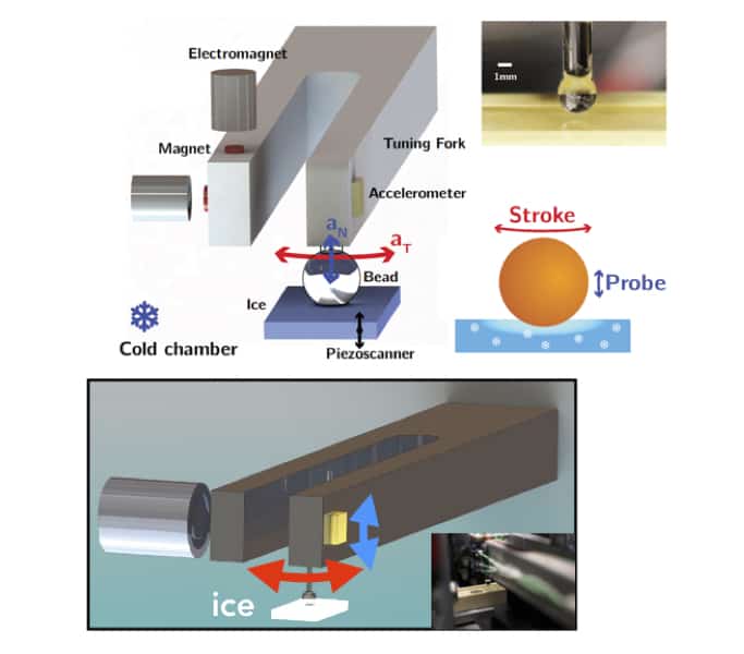
Bocquet, Siria and colleagues studied “interfacial water” using a modified double-mode Tuning Fork Atomic Force Microscope (TF-AFM). The instrument they developed comprises a millimetre-sized probe ball glued to a macroscopic tuning fork. Although the fork is very similar to a piano tuning fork, it can be excited by a very low frequency vibration, typically several hundred Hertz. The system can be accurately modelled as a stiff mass-spring resonator with a quality factor of around 2500.
Schematic of the experimental setup. Courtesy: L Bocquet
When the researchers bring the vibrating ball at the end of the fork in contact with the surface of a centimetre-sized block of ice (using a piezo element with an integrated motion sensor of nanometric resolution), the lateral stroke of the ball slides across the ice with a fixed amplitude and velocity. The frequency of the system then changes, but so does its quality factor.
The ENS team use the frequency offset to measure the elastic properties of the contact surface, and the change in quality factor to evaluate the dissipation processes occurring there. The two measurements together give the layer’s interfacial viscosity.
“Listening” to forces
The researchers say the instrument allows them to “listen” to the forces between the probe and the ice with remarkable precision. Indeed, despite being centimetres in size, the instrument’s sensitivity is such that it is possible to probe ice contact and friction properties at the nanometre scale. “The system allows us to access several vibration frequencies, offering us the possibility to simultaneously probe the tribology of the contact (‘how it rubs’) by moving the ball in a lateral direction and its rheology (‘how it flows’) by moving the ball in a perpendicular direction,” Bocquet explains.
The experiments confirm the super-slippery nature of the interfacial ice, but they also – for the first time – confirm that friction generates a film of liquid water when the probe ball is set in motion. This film is, however, much thinner than previous theoretical calculations have suggested, and it is also as viscous as oil, with a viscosity of up to hundreds of mPa-s – two orders of magnitude larger than the viscosity of water. The researchers also showed that the film’s viscosity depends on the shear velocity – a behaviour known as shear thinning.
Crushed ice and water state
According to Bocquet, one interpretation for this unexpected behaviour is that surface ice does not completely transform into liquid water when an object glides across it. Instead, it may enter a mixed “granité-like” (crushed ice and water) state. This mixed film, he suggests, lubricates the contact between the solid ice and the ball and prevents any direct contact between the two surfaces.
Separate experiments by the ENS team show that making the probe hydrophobic reduces friction even further by modifying the interfacial viscosity. This “waxing” process is practiced empirically by skiers, but the reason why it made skis glide better was not previously understood.
Towards a new theory for interfacial ice
The team’s result means that existing theoretical descriptions for interfacial ice need an overhaul, Bocquet tells Physics World. A new theory would provide a better understanding of sliding on ice, which would come in useful in developing winter sports equipment or self-healing, ultra-low-friction lubricants for industrial applications. It might also help, conversely, to find ways of increasing friction, which is essential to avoid slipping on icy roads.
Angelos Michaelides of University College London, UK, who was not involved in the research, says that the ENS study is very exciting. “I am not aware of such a nice and elegant set of measurements on the friction of the quasi-liquid layer and think it is an extremely interesting new perspective on this age-old story,” he comments.
Spectacular connection: Fireworks have long linked science with the public and the state. (Courtesy: Jan Bogumil Plersz (1732–1817))
Fireworks are essential to many of today’s celebrations – from national holidays and sporting events to musical concerts and the gatherings held on Bonfire Night (5 November) in Britain each year. Once upon a time, though, fireworks were serious scientific business. Designing the rocket and preparing the propellant and coloured fire required, after all, a detailed knowledge of chemistry and physics (see “Whizz-bang science” by Pierre Thebault, December 2018). Mounting effective firework displays required other skills too, including architecture, artillery, ballistics and even poetry.
Fireworks, it turns out, also played a critical role in the complex and evolving relations between science, the public and the state. That, at least, is the intriguing argument in a book by Simon Werrett, a science historian at University College London, entitled Fireworks: Pyrotechnic Arts and Sciences in European History (University of Chicago Press 2010). Previous histories of fireworks had ignored this connection. As Werrett put it, the true history of fireworks has “gone up in smoke”. His book brings it back.
Up in smoke
Fireworks originated in China, where by the 12th century they were routinely used in public spectacles. Werrett, though, focuses on the European story, which started in around the 14th century, when gunners began to develop a new genre of spectacle – “artificial fireworks” – for a general audience. The spectacles were called “artificial” because they were specially crafted for non-military purposes, and “fireworks” because they used gunpowder to produce fiery effects. The people who made the fireworks, meanwhile, were known as “artificers” and worked in spaces called “laboratories” (a name also used by alchemists) well before the modern scientific use of the term.
The first grand firework display over the Thames took place in 1613. Indeed, in his novel New Atlantis, published in 1620, one of the crucial tasks that the philosopher and statesman Francis Bacon assigned the scientists in his utopian world was to produce fireworks. Fireworks were on the way to becoming an important undertaking of nations, not so much because they demonstrated knowledge of military capital such as explosives and rockets, but because they symoblized power and authority.
“In a world without electric light,” Werrett writes, “fire was a powerful medium, a source of light and heat whose divine and magical connotations were strong”. Indeed, the ability to control, tame and exploit fire in spectacular and artistic displays seemed to demonstrate an ability to bring the divine and celestial down to Earth and under human control.
The ability to control, tame and exploit fire in spectacular and artistic displays seemed to demonstrate an ability to bring the divine and celestial down to Earth and under human control
By the end of the 17th century, fireworks had become an important element in public displays and extravaganzas in several European states. Monarchs gave resources to those who could manufacture them and stage their displays, and supported the institutions where they worked. Fireworks makers were encouraged to invent new and more dramatic effects, fostering a culture of innovation. Power and prestige came to those who could successfully innovate.
One of Werrett’s unusual stories involves the quest to create green fireworks. While artificers could produce most colours, green was difficult and in the early 18th century the ability to produce it became the subject of quests at Imperial courts – rather like the modern hunt for blue light-emitting diodes. Scientists at the St Petersburg Academy of Sciences eventually succeeded, and for a time were able to keep their knowledge a trade secret. The Russians, typically, attributed the discovery of green fireworks to Peter the Great himself. But the key breakthrough occurred at the St Petersburg Academy, when its scientists began treating fireworks as based on a chemical rather than a mechanical process.
In the 17th and 18th centuries, Werrett writes, Britain, France, Italy, Russia and other nations sought to outdo each other in the grandeur and scale of the fireworks displays they staged, with the manufacture of fireworks serving to promote science. How precisely this occurred depended on local conditions. At the time of the restoration of the monarchy in England, for instance, fireworks were sometimes associated with Catholic plotting and religious zeal, provoking a counter-reaction – but English philosophers and natural scientists also debated the significance of fireworks for understanding nature. In Russia fireworks appealed chiefly to the Imperial Court’s thirst for spectacle, which fostered its support for the country’s first generation of Western-style scientists.
In Russia fireworks appealed chiefly to the Imperial Court’s thirst for spectacle, which fostered its support for the country’s first generation of Western-style scientists
“With no scientific tradition in Russia,” Werrett writes, “academicians found that experimental lectures failed to interest the Russian nobility, whose support was critical to the survival of the academy. Simultaneously, academicians learned that the design or ‘invention’ of allegorical fireworks could improve their fortunes as spectacles appealing to the Russian court.” Werrett’s book opens, for instance, with a description of a firework display intended to symbolize the incremental but inexorable growth of the power and prosperity of the Russian state.
In the 1750s, seeking to exploit competition amongst their academicians, the St Petersburg Academy commissioned two of its prominent scientists – Mikhail Lomonosov and Jacob Stählin – to work separately on fireworks displays, with the intention of choosing whoever was better. Lomonosov was offended when Stählin’s was chosen, and announced that he was giving up firework-making. Fireworks were not only an important activity of the young academy, but also elevated its position and prestige, as well as of Russian science itself.
The critical point
The lesson I draw from Werrett’s book is that producing fireworks was not a hobby or side occupation that scientists tacked on to their “real” work. Scientists who produced fireworks were simply carrying on the practice of science, not trying to promote themselves or curry favour. A modern-day equivalent would be researchers consulting on governmental projects. Such activity is not only an integral part of the work of science, but it also bolsters the confidence of legislators and the public in science and their awareness of its value.
The spacecraft Voyager 2 left the heliosphere and travelled into interstellar space over the course of a day in November 2018, according to a suite of papers published today by scientists working on the mission.
The spacecraft was launched in 1977 along with its twin Voyager 1, which crossed-over into interstellar space seven years ago. Scientists analysing data from Voyager 2 have found both similarities and differences to the crossing of Voyager 1.
The Sun is surrounded by a huge bubble called the heliosphere that is inflated by the supersonic solar wind of charged particles emitted by the Sun. The edge of this bubble is called the heliopause, which is where the outgoing solar wind is halted by the interstellar wind of charged particles.
Different crossings
Both Voyager missions crossed the heliopause on the windward side of the bubble but at different locations. Voyager 1 left the northern hemisphere of the heliosphere and Voyager left the southern hemisphere at locations separated by about 160 au (1 au is the distance from Earth to the Sun).
Voyager 1’s departure point was about 122 au from the Sun, while Voyager 2 exited at 119 au from the Sun. According to scientists working on the Voyager 2 mission, these slightly different distances could be a result of the exit events occurring a different times in the 11-year solar cycle. This cycle changes involves changes in the intensity of the solar wind that could make the size of the heliosphere fluctuate.
One big difference between the two spacecraft is that all five instruments onboard Voyager 2 are still functioning, whereas the plasma instrument that measures the solar (and then interstellar) wind was damaged on Voyager 1 in 1980. This meant that Voyager 1 was unable to measure the transition from the hot, low-density solar wind to the cold, high-density interstellar wind.
Thinner and smoother
Analysis of the Voyager 2 data suggest that the heliopause it encountered was thinner and smoother than the boundary crossed by Voyager 1. Indeed, Voyager 2 made the crossing in less than one day. The Voyager 2 data also suggested that the interstellar medium that the spacecraft first encountered is hotter than had been expected.
Voyager 2 also discovered a region between the heliopause and interstellar space where the solar and interstellar winds interact. This layer was not detected by Voyager 1.
Both spacecraft found little change in the direction and magnitude of magnetic fields across the heliopause. This is surprising because scientists had expected an abrupt transition between solar and interstellar magnetic fields to occur at the interface.
Gaining a better picture of the heliopause and heliosphere could provide important clues about how life emerged on Earth – and how it could emerge on exoplanets orbiting distant stars that would also be surrounded by bubbles. That is because the heliosphere shields Earth from many cosmic rays impinging on it – radiation that is harmful to life.