
FLASH radiotherapy – it’s the technique that’s got everyone talking. The idea is that delivering radiation at ultrahigh dose rates, roughly 50 Gy/s and above, will vastly reduce normal tissue toxicity while preserving anti-tumour activity.
The premise has been demonstrated in preclinical studies by several research groups and, as delegates at the recent AAPM Annual Meeting heard, the first human treatment has just taken place. A dedicated symposium examining the promise of FLASH saw a ballroom packed full of delegates keen to find out more about this cutting-edge treatment.
A giant leap
The first speaker was Julianne Pollard-Larkin from MD Anderson Cancer Center, who presented a talk entitled “FLASH Photon: one small step for physics, one huge leap for cancer therapy”.
Pollard-Larkin began by citing a 2018 study highlighting that mice receiving thoracic irradiation at 3600 Gy/min had 70% less pulmonary fibrosis than those treated with conventional radiotherapy at 1.8 Gy/min. “Ultrahigh dose rates showed a marked reduction in fibrosis at every time point investigated,” she told the audience. “I could stop my talk now…”
But she didn’t. Instead, she presented a brief history of the FLASH effect, which was first noted in some form as far back as 1959. In 1971, researchers showed that high dose rate electron beams induced hypoxia. And in 2014, a report revealed the differential response to high dose rates between normal and tumour tissue in mice. “In 2014, it was reborn and rebranded as FLASH radiotherapy,” said Pollard-Larkin.
She described several studies demonstrating the advantages of FLASH over conventional radiotherapy. One early investigation of mice with lung tumours showed that FLASH spared normal lung and was toxic to tumours, with “almost no double-edged sword”, while another showed memory sparing in mice after whole-brain irradiation. In larger mammals, FLASH increased progression-free survival of six cats with naturally occurring nasal tumours, and conferred a protective effect on irradiated skin of mini-pigs.
So how is FLASH actually delivered? Several of the preclinical studies used the Oriatron eRT6 from PMB-Alcen, an experimental high dose‐per‐pulse linac that delivers an electron beam with dose rates of up to 200 Gy/s.
But not everyone has an Oriatron, so Pollard-Larkin described how she adapted a redundant linac to deliver FLASH. “We had an old decommissioned linac and they allowed me to work with this,” she explained. Adaptation included removing safety systems, tuning the 20 MeV board and creating a holder for a miniature ion chamber and film. The measured output ranged between 40 and 60 Gy/s over time. “Get a linac that nobody cares about and you can do it too,” she told the audience.
As for how FLASH actually works, the biology is inevitably complex and various ideas have been proposed. One possibility is that the ultrahigh dose rate irradiation converts the endogenous oxygen in all tissues into reactive organic species, which normal tissues can remove more effectively than tumours. Another suggestion is that FLASH causes transient hypoxia and preferentially spares normal tissues due to differential oxygen tensions between tumour and normal tissues.
And while researchers strive to unravel the exact mechanisms underlying FLASH, work continues alongside to implement clinical translation. One challenge is the lack of available systems to perform FLASH. Pollard-Larkin noted that treatment of deep tumours will require very-high-energy electrons, or X-ray or proton FLASH. “We also need to come up with a comprehensive dose monitoring system,” she added. “Safety is really critical when you are delivering such high dose rates.”
Pollard-Larkin concluded by emphasizing FLASH’s potential, particularly its 30–80% protective effect. “FLASH treatment times are economical and beneficial to clinics, especially those in low- and middle-income countries,” she explained. “And the reduced number of fractions necessary could provide better quality-of-life for patients. I see FLASH as one of the cancer breakthroughs of 2020 that we need to work on as a community and push through.”
FLASH for protons
Lei Dong from the University of Pennsylvania followed with a look at FLASH using proton beams, which could provide an option for treating deep-seated tumours. He pointed out that in pencil-beam scanning proton therapy, the dose is packed into a tight spot and thus already has a relatively high dose rate.
He examined the requirements to achieve ultrahigh dose rates required for such a proton therapy system. For a typical 200 MeV pencil beam spot of 1 cm diameter, he estimated that 22 nA at the nozzle would create a dose rate of 100 Gy/s. However, the need for beam propagation and creating a 5 x 5 cm field, for example, ups this requirement to about 600 nA at the nozzle to create 100 Gy/s.
Dong described the current status of proton FLASH research at various proton therapy facilities. Researchers at Institut Curie in France, for example, have adapted a clinical system to perform proton FLASH irradiation of small animals. They optimized a single scattering system with a ridge filter and a high current monitoring system. For a 12 × 12 mm field, the set-up achieved dose rates exceeding 40 Gy/s at energies between 138 and 198 MeV.
At Penn, Dong and colleagues are using an image-guided small-animal X-ray irradiation coupled to a proton beamline to study proton FLASH with mice. They performed whole-body irradiation at 1 or 75 Gy/s, delivering a 7.5 Gy dose. “We see that FLASH produces better survival compared with conventional irradiation – an indicator of the normal tissue sparing,” Dong explained. He notes that, due to the high dose rates of their set-up, dosimetry with a parallel plate chamber is preferable to a cylindrical ionization chamber.
A team at the University of Maryland performed a similar study comparing 1 and 40 Gy/s thoracic irradiation in mice. They saw a 30% reduction in lung fibrosis, reduced skin dermatitis and an increase in overall survival.
Dong also shared a slide showing proton FLASH outcomes in human studies. This one was blank… for now. So how could proton FLASH be implemented clinically? He pointed out that, for starters, extra room shielding might be needed – with high-energy beams to maximize ultrahigh dose rates, the protons may not stop inside the patient. On the plus side, this eliminates the issue of range uncertainty. “In proton therapy we would never have imagined having no need to worry about range uncertainty!” he said.
Dong concluded by reiterating that proton FLASH demonstrates significant normal tissue sparing in animal studies, though the biological effects are still unclear. Dose rate will become an important quality assurance issue, while treatment planning will need to incorporate machine delivery information, he explained.
Into the clinic?
Finally, Billy W Loo Jr from Stanford University School of Medicine took a look at the clinical translation of FLASH. He first described some of the preclinical work performed at Stanford, in which the team customized a clinical linac to use the electron beam at high currents and without a photon conversion target.
They employed the reconfigured system, which could deliver dose rates of 0.1–300 Gy/s, to perform whole-brain irradiation on mice and examine cognitive sparing after FLASH. “Like other groups, we found that FLASH produces a similar neurocognitive outcome to unirradiated controls, while a clear detriment was seen with conventional irradiation,” Loo explained.
He also described work investigating total abdomen irradiation with 16 MeV FLASH electrons. “In normal mice, we saw a loss of gastro-intestinal function using a conventional dose rate; but this function was preserved after FLASH.” In tumour-bearing mice, both dose rates produced similar levels of tumour reduction. As for late effects, one year after abdominal irradiation using a non-lethal dose, two conventionally treated mice developed radiation-induced cancers, while none of the FLASH group did.
In a further study, the team noted a decreased incidence of spontaneous lung metastases after FLASH irradiation of subcutaneous tumours, suggesting a possible immune effect at distant sites and leading Loo to propose the possibility of a “FLASHscopal” effect.
Next, Loo moved onto the headline act: the first case report of FLASH radiotherapy in a human patient. Published by researchers at Lausanne University Hospital a few days before the AAPM meeting, the paper described treatment of a 75-year-old man with widely spread cutaneous T-cell lymphoma. The patient had undergone over 110 localized radiotherapy courses for different lesions over the past decade, with good tumour control but severe skin toxicity.
In this study, the team treated a 3.5-cm wide ulcerated lesion, using an Oriatron linac to deliver 15 Gy in 90 ms. Notably, the patient felt no sensation during treatment. At day 15, the tumour was healing; optical coherence tomography showed some thickening at the skin surface in the treated region, but no breakdown at the dermis–epidermis interface. Five months later, the patient’s tumour was completely healed with no toxicity.
“These results are very intriguing,” said Loo. “This is suggestive, though not conclusive, of a FLASH effect in a human patient and certainly a demonstration of clinical feasibility of delivering FLASH.”
The study also demonstrated the technical feasibility of delivering FLASH to patients using an existing preclinical system. Loo noted that while electron beams can treat superficial targets and proton pencil beams can treat small volumes, “I argue that new technology is needed for FLASH to target general cancer patients.”
Loo pointed out that the fastest radiotherapy treatment today takes just over three minutes to deliver 25 Gy, which is extremely fast, but still a long time compared with the motion of a tumour. “The ultimate motion management strategy is to freeze all motion,” he said.

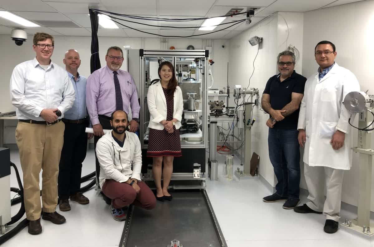
With this aim, Loo and colleagues are developing the PHASER linac, a compact, power efficient radiation delivery system that can treat in a fraction of a second – three hundred times faster than SABR and fast enough to freeze motion. “This would be a platform for translating FLASH to the clinic”, he said. “It’s the first fundamentally new linac design in 60 years.”


