
Where the energy goes

Particle physicists have been using calorimeters of one type or another for around 70 years. The basic principle of these workhorse instruments is straightforward: the idea is to measure the energies of elementary particles such as the electron, proton and neutron, as well as artificially produced ones such as pions and kaons, by sending them into a dense medium where they interact. Each interaction produces more particles at lower energies, which also interact with the medium, and the process continues until the original particle’s energy is fully exhausted. By interspersing the interaction medium with charge-sensing detectors and summing up the signals recorded, we can obtain a measurement of the initial particle’s total energy.

A particle “shower” induced by a high-energy electron is shown in the image above. Here, the charged particles of the shower (electrons and anti-electrons, or positrons) are made visible via a cloud chamber: a classic type of calorimeter called a sampling calorimeter. The development of these particle showers is highly random. The number of particles generated in the shower, N, is a direct measure of the energy, E, of the initiating particle. N follows a Poisson distribution so the random fluctuations in N are equal to √N and, therefore, the relative precision in N, which is the energy resolution, is √N/N. The energy resolution is a simple formula, σE/E ≈ k/√E. This cloud chamber calorimeter has k ≈ 85% when E is expressed in GeV units (for reference, the rest mass energy of a proton is about 1 GeV); for a modern electromagnetic sampling calorimeter, k is typically 10% or better.

The hadron challenge

Electron energies are easy to measure in calorimeters because these particles interact via the electromagnetic force, with only two simple allowed interactions. In contrast, particles subject to the strong nuclear force (such as protons, neutrons, pions and kaons – collectively known as hadrons) interact through a multitude of widely fluctuating mechanisms, with additional complications associated with breaking up nuclei
and the energy expended for nuclear binding energies.

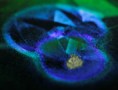
The tremendous complexity of a shower of hadrons is illustrated in figure 1, which shows the results of a simulation (created using the CERN code GEANT4) of a 500 GeV proton entering a copper absorber. Charged hadrons are shown in blue while electrons and positrons are in red. Visually, the intensity of the colour indicates the amount of energy lost by the particles and represents the signal generated by the calorimeter.

The particle interactions in showers such as this are a complex but highly important area of research in particle physics, and in the CERN R&D project RD52 we are studying them using a new type of calorimeter (full results from the project are available at www.phys.ttu.edu/~dream, where one can view a collection of hadronic showers induced by protons in a copper absorber). These “dual-readout” instruments are made from copper or lead and interspersed with two types of optical fibres: scintillating fibres that sense all charged particles, and clear fibres in which Cherenkov light is generated predominantly by the electrons and positrons of the shower. These two very different signals from a shower are used in combination to extract a highly accurate measurement of hadron energies – including, most importantly, the energies of “jets” of particles resulting from the fragmentation of a quark or gluon produced in fundamental interactions such as those studied at CERN’s Large Hadron Collider and other facilities around the world.

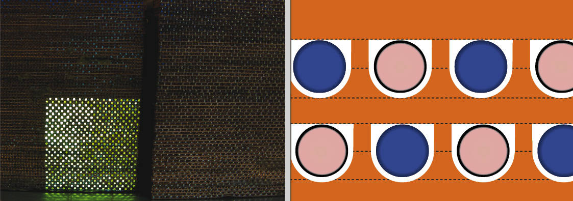
A calorimeter specifically designed to measure these jets of particles is shown in figure 2. It consists of 1 mm-diameter fibres on 1.5 mm centres uniformly interspersed in a copper absorber. The size of this absorber is dictated by the characteristic interaction distances of electrons and hadrons. The distance over which an electron will interact and produce more particles is called the radiation length, and is around a centimetre for most metals (including copper). The corresponding distance over which a hadron will interact is known as the nuclear interaction length. This is considerably longer, typically 20–30 cm, and it takes several nuclear interaction lengths to fully absorb a hadronic shower. The difference is evident in the simulated shower in figure 1, which shows the blue charged hadrons travelling longer distances before interacting. The red-coloured electrons and positrons clearly interact on a much shorter scale. The salt-and-pepper red dots evident throughout the volume are electrons from Compton scattering of low energy (about 1 MeV) photons which have a minimum cross section to interact at this energy and therefore spread out spatially in the calorimeter.

The variation and complexity in the development of showers are challenging for calorimeters. Getting an accurate measure of the total energy requires us to measure each fluctuating component of a shower. The electromagnetic part consists of electrons and positrons and is measured by the Cherenkov light generated in the clear fibres. The charged hadrons are measured by the scintillation light generated in the scintillating fibres, and the neutrons from nuclear break-up are measured by the late-developing recoil protons from neutron-proton elastic scattering in the scintillating fibres.

This dual-readout approach has been thoroughly tested and yields energy resolutions near σE/E ≈ 30% /√E in both simulations and data. This means that the energy of 100 GeV jets can be measured with 3% precision, which is much better than achievable with currently available calorimeters. The main difficulty in constructing a calorimeter of this type is forming the copper absorber to the required precision: about 10 µm over a length of about 2.5 m. This spatial precision is needed in order to maintain a highly uniform (better than 1% when averaged over a cigar-shaped volume) distribution of the two fibre types relative to the absorber. Since the energy measurement depends critically on the light generated in the fibres, if a region of the calorimeter has 1% higher fibre density, then showers developing here will yield 1% more light. An ensemble of showers of the same energy will have a 1% wider signal distribution.

We have tested various methods of creating these copper absorbers, including rolling copper sheets, skiving, extruding, water-jet grooving, chemical etching and cutting with blades. Only the cutting procedure was successful, but it was also difficult and expensive. An industrial procedure involving rolling – perhaps with multiple rollings – might succeed, but in all our tests, rolling copper always resulted in highly warped sheets due to work hardening. Before we can really take advantage of our calorimeter’s improved performance, we need a better means to manufacture the copper sheets for large-scale tests.

Cellular building blocks create life-like constructs

One of the main challenges in tissue engineering today is to create a complete network of blood vessels and capillaries throughout an artificial tissue. A promising solution, now demonstrated by researchers in Belgium, is to fabricate micron-sized cellular building blocks that already incorporate a capillary network, which can then be 3D bioprinted to form a large tissue structure. As well as helping to engineer large-area tissues or organs, the technique would also be useful for making in vitro structures that can be used in cancer research, drug testing and disease modelling.

Tissue engineered constructs are useful in many applications, including as in vitro models for injury, disease, drug-screening, or to repair, regenerate or replace dysfunctional tissues or organs. Much progress is being made in the field, but it is easier to engineer thin tissues with a low metabolism, such as skin or cartilage, than to make thick 3D tissues. The problem is that cells cannot diffuse more than around 100 to 200 µm in space, which means that those lying deep in the core of a large engineered construct have insufficient access to nutrients and oxygen and are unable to survive.

In living organisms, this nutrient- and oxygen exchange between blood and tissues occurs in blood vessels and capillaries, collectively known as the vasculature. “We therefore need to find a way to make such a structure in engineered 3D tissue – and make sure that it extends throughout the entire construct,” explains Heidi Declercq of Ghent University, who led this study. “A complete vascular tree, ranging in size from millimetres to microns, is required, since nutrient- and oxygen-exchange occurs mainly in the microvasculature.”

Although larger vessels can be fabricated by incorporating printed channels composed of bioinks – biomaterials laden with cells that can be printed to form tissue – smaller vasculature networks are more difficult to make in this way because of the limited resolution of bioprinting techniques.

Cellular building blocks

Declercq and colleagues instead turned to cellular self-assembly, a bottom-up approach for building large tissue constructs. “We use spheroids or microtissues with a specific microarchitecture as building blocks,” Declercq explains. “Small and uniform-shaped spheroids are made spontaneously by seeding cells on microwells, which are created using a polymer mould containing 2865 pores with a diameter of 200 microns.”

When a cell suspension is seeded onto the microwells, gravity causes the cells to fall to the bottom of the pores. Here they are forced to interact with other, which causes the cells to self-assemble into spheroids.

“The properties of the spheroids produced in this way depend on the cells they contain and the cell types used to ‘support’ them,” continues Declercq. “Endothelial cells, like the ones studied in this work, can be co-cultured with supporting fibroblasts or mesenchymal stem cells to promote angiogenesis [the formation of blood vessels].”

These spheroids can then be directly assembled by 3D bioprinting to form a macroscale tissue structure. “This strategy is based on cell sorting and microtissue fusion,” says Declercq. “Cells organized into a spheroid can fuse into a macrotissue in a process that can be explained by the ‘differential adhesion hypothesis’. This says that multicellular tissues behave like liquids, thanks to their surface tension, and will rearrange and merge to maximize their adhesive bonds and minimize their free energy.”

Spheroids enable bioprinting

By seeding 750 000 cells onto one microwell, the researchers produced 2865 spheroids containing roughly 262 cells/spheroid. These spheroids measure around 125 µm across, a size that is compatible with bioprinting techniques that employ needles with diameters in the 200 µm range.

“In our study, we co-cultured human umbilical-vein endothelial cells (HUVECs) with human foreskin fibroblasts (HFF) and adipose-tissue-derived mesenchymal stem cells (ADSCs) in different ratios,” says Declercq. “We tested different compositions and found that a 1:9 ratio of HUVEC/supporting-cells produced the most stable spheroids.”

The researchers found that capillary-like networks formed in spheroids that included ADSCs, with larger diameter spheroids (>170 µm) forming a more branched capillary-like structure. They also showed that individual spheroids in suspension fuse together within just 24 hours, and within 4 days a branched capillary-like network extends throughout the entire construct. Even when embedded in a hydrogel – which would be needed to create a bioink – spheroids started to fuse together within 18 hours.

The researchers, reporting their work in the IOP journal Biofabrication, say that they will now undertake in vitro experiments to help them select the best bioinks for making their vascularized constructs. “We will also set up in vivo experiments to see how the constructs connect with real host tissue,” adds Declercq.

  • Read our special collection “Frontiers in biofabrication” to learn more about the latest advances in tissue engineering. This article is one of a series of reports highlighting high-impact research published in Biofabrication.

An Earth made of blueberries, seeking a missing Fields medal, UNAM’s youngest student

It’s probably not a question you’ve thought about before. But have you ever wondered what would happen if the Earth were replaced by an equal volume of blueberries? Well, now you can sleep easy, as that fruity conundrum has been answered, thanks to computational neuroscientist Anders Sandberg from the University of Oxford.

Sandberg, who works at something called the Future of Humanity Institute (no, we’d never heard of it either), was inspired to carry out the work after a user posted the question on the Physics Stack Exchange website. By taking into account the density and compressive strength of blueberries, Sandberg found that a “blueberry Earth” would turn into a “roaring ocean of boiling jam” with geysers of steam ejecting some of the berries into orbit. In addition, he discovered it would have an ice-like core much like a “warm granita” — that classic Italian semi-frozen dessert made from flavoured sugar and water.

“The final state of blueberry Earth is somewhat similar to oceanic exoplanets, although far lighter than any observed so far,” he writes in the seven-page paper.

So is there a chance that a blueberry Earth could be somewhere in the universe or possibly lurking in the Kepler mission’s data? “I would be pretty shocked to find a blueberry Earth,” Sandberg told Physics World. “But a super-light ocean world? Maybe. It is not inconceivable that smallish ice bodies could form outside the frost line in a solar system and either migrate inwards or melt as the sun becomes hotter over time.”

Sandberg says that he may study the physics of another “crazy world”, but adds that he suspects that other fruit planets would end up being similar. “There is likely some kind of ‘fruit main sequence’ that is parametrised by the water/solid content or maybe also the density and air content,” he notes.

Away from fruit, earlier this week the winners of this year’s Fields medal was announced at the start of the International Congress of the International Mathematical Union in Rio de Janeiro. The four mathematicians to bag the prestigious prize were Caucher Birkar from the University of Cambridge, Alessio Figalli from the Swiss Federal Institute of Technology Zurich, Peter Scholze from the University of Bonn and Akshay Venkatesh from Stanford University.

Yet just minutes after Birkar received the 14-carat gold medal it was stolen. He apparently put the 6 cm-diameter gong in his briefcase together with his phone and wallet and left it on a table at the conference.

While authorities later found the briefcase outside the venue, the $4000 medal and the wallet are still missing. In a statement, the organisers say that “images recorded at the event are being analysed” and that they are “co-operating with local police authorities in their investigation”.

And finally from researchers at the top of their game, to those just starting. We were interested to discover this week that Mexico’s National Autonomous University has admitted its youngest student in the university’s century-long history. After passing the entrance exam and completing preparatory study at the university, 12-year-old Carlos Santamaria Diaz will begin a degree in biomedical physics later this year. Could Alia Subur’s record of being the youngest professor be under threat?

Quantum dot floating gates improve light-erasable memories

Photoresponsive flash memories made from organic field-effect transistors (OFETs) that can be quickly erased using just light might find use in a host of applications, including flexible imaging circuits, infra-red sensing memories and multibit-storage memory cells. Researchers from Hanyang University in Seoul and Pohang University of Science & Technology (POSTECH) have now found that they can significantly improve the performance of these devices by making use of floating gates based on cadmium selenide (CdSe) quantum dots whose surfaces have been modified.

“OFET-type light-erasable memories have recently emerged as promising elements for information delivery,” explain Yong Jin Jeong and Jaeyoung Jang, who led this research effort. “The erasing process in this type of device is usually controlled using only light following a photo-induced recovery mechanism that works thanks to photo-induced charge transfer across the interface between a semiconductor and floating-gate layers in the device.”

Photo-induced recovery after just one second

Jang and colleagues studied CdSe quantum dots whose surfaces they had covered with three different organic molecules that they used as photoactive floating-gate interlayers in light-erasable transistor memory architectures. They looked at how modifying the surfaces of the dots affected the performance of the memories and found that capping small ligands of octadecylphosphonic (ODPA) and fluorinated molecules improved the diffusion of holes between the dots and the conducting channels in the devices. This allowed for photo-induced recovery after just one second using low intensity light (with a power of 0.7 mW/cm2).

“We had made OFET memories before (containing a polymer/C60 composite floating-gate interlayer) that could also be erased using low-intensity green light (of 0.5 mW/cm2) and in which we could effectively remove trapped charge carriers (electrons and holes) and so recover the initial state,” says Jang. “These devices did suffer from several problems, however. The main one was that erasing stored information took as long as 30 seconds and read-out required a (destructive) applied gate voltage in addition to light.”

Fast erasing using low intensity light

“Employing tailor-made floating-gate materials for transistor memory devices overcomes these problems and allows for fast erasing using low intensity light (again, green) and a non-destructive read-out process. These gates perform many crucial tasks: they generate charge carriers (electrons and holes) when they absorb light and transfer these charges to the semiconducting layer in the transistor.”

Using quantum dots for the floating gates is much better than using C60 or other commonly employed materials, he adds. This is because the optical and electronic properties of these dots can be modified by engineering their particle size or adding surface ligands, as in this study.

The new devices boast high memory ratios of over 10between OFF and ON bi-stable current states for over 10 000 seconds and good dynamic switching behaviour, he says.

Towards commercialization

The researchers believe that their light-erasable OFET memories could be commercialized in the near future and that their work could provide a “useful guideline” for designing photoactive floating-gate materials.

“To the best of our knowledge, we are the first to engineer the surface of quantum dot floating gates in such memories,” Jang tells Physics World. “Our proof-of-concept memory devices also consume less power during operation and could be integrated with highly sensitive photodetectors for use in flexible imaging, integrated sensors and biomedical applications.”

So, where next? The Korea researchers say they are now busy trying to develop quantum dot-based light-erasable memories using all-solution-based processes. “In our present experiments, we used vacuum-based techniques to deposit the organic (pentacene) semiconductor and (gold) source/drain electrodes when making our devices,” says Jang. “These are not suitable for large-area, low-cost applications, however, because they are expensive, time-consuming and complicated, so an all-solution process would be better.”

The new memories are detailed in ACS Nano 10.1021/acsnano.8b01413.

Optical spectroscopy senses tumour hypoxia

DRS setup

Tumour hypoxia is associated with poor clinical outcome following radiation or chemotherapy in cancer patients. Measuring tumour oxygenation prior to or during therapy could help select the most appropriate treatments, as well as identify poor response earlier than currently possible, enabling effective changes in treatment regime.

Hypoxia is typically defined as oxygen tension (pO2) of less than 10 mmHg. One way to measure this in vivo is to insert oxygen-sensing microelectrodes into the tumour tissue. However, such measurements are invasive, limited to accessible organs, may modify the tumour microenvironment and are not amenable to repeated measurements.

An alternative approach is diffuse reflectance spectroscopy (DRS), which provides a non-invasive way to evaluate oxygenation in accessible tumours (skin or oral cavity, for example). As such, researchers from the University of Arkansas are investigating the use of DRS to measure sO2 – the average oxygen content bound to haemoglobin in the blood vessels. Their aim: to show that DRS can provide reliable in vivo measurements of tumour hypoxia (J. Biomed. Optics 23 067001).

“The main advantage of using DRS is its non-destructive and non-invasive nature,” explains lead author Narasimhan Rajaram. “Using a pen-shaped probe that is usually compatible with the working channel of endoscopes and laryngoscopes, it should be possible to interrogate tissue non-invasively and get a quantitative measure of tumour hypoxia.”

Authors

Optical approach

DRS works by illuminating the tissue-of-interest with 400-650 nm light and using the reflected light signal to quantify the underlying scatterers and absorbers. Since DRS is sensitive to the absorption of oxygenated and deoxygenated haemoglobin, it can quantify the amount of oxygenated haemoglobin to total haemoglobin content. DRS thus provides a measure of both sO2 and the total haemoglobin content (THb).

In this study, the team used a portable DRS system comprising a halogen lamp for illumination, a USB spectrometer for spectral acquisition, and a bifurcated optical probe for light delivery and collection. The probe incorporates four illumination and five detector fibres at a source–detector separation distance of 2.25 mm, providing a sampling depth of about 1.8 mm.

The researchers acquired DRS spectra from two different human head-and-neck cancer xenografts (radiation-resistant SCC-47 and radiation-sensitive SCC-22B cell lines), using an integration time of 100 ms and a wavelength range of 475 to 600 nm. They used a lookup table-based model to fit the acquired DRS spectra and extract wavelength-dependent absorption and scattering properties, and validated the results using immunohistochemical staining to quantify tumour hypoxic fraction (HF).

DRS spectra

Fitting the DRS spectra for two excised tumours revealed sO2 values of 58% for one tumour and 15% for the other. For the oxygenated (58%) tumour, the absorption spectra exhibited the classic double peaks of oxygenated haemoglobin (542 and 577 nm), while the poorly oxygenated (15%) tumour showed a single peak corresponding to deoxygenated haemoglobin. Immunohistochemistry images of the HF confirmed that the tumour with higher sO2 had less hypoxic tissue than the low-sO2 tumour.

The researchers determined that HF was negatively correlated with sO2, and correlated with THb, indicating that DRS measurements of vascular sO2 and THb provide reliable indirect measures of tumour oxygenation. Specifically, they note that very low values of sO2 and THb can indicate highly hypoxic and poorly perfused tumour.

“Measuring a tumour’s response to therapy with diffuse reflectance spectroscopy is our clinical goal,” says Rajaram. “The oxygenation levels in a tumour prior to and post-radiation can be key biomarkers of radiation response of failure, which explains the current work with radiation-resistant and sensitive cell lines.”

Rajaram and colleagues are now investigating whether they can predict radiation resistance in the tumours when they are treated with radiation. “We are also working with collaborators at University of Arkansas for Medical Sciences, Little Rock, to run pilot studies with the probe in patients with head-and-neck cancer,” he tells Physics World.

Proton therapy goes slimline

Since protons were first used to treat hospital cancer patients in the early 1990s, around 100,000 people have benefited from this alternative form of radiation therapy. While the X-rays used in conventional radiotherapy fully penetrate a patient and dump their maximum dose just after entering the body, protons deposit their biggest dose at a specific, energy-dependent depth and travel no further. This characteristic “Bragg peak” allows protons’ energy to be concentrated at the location of a tumour, so inflicting maximum damage to cancerous tissue while sparing surrounding cells.

These virtues, however, come at a price. Conventional radiotherapy generally involves accelerating electrons in a linear accelerator (linac) and colliding them with a tungsten target to generate X-rays. The apparatus, which is roughly 2 m long, is incorporated into a rotating gantry that allows X-rays to enter a patient’s body from a range of angles. But protons are much heavier than electrons, and require larger accelerators that generally serve multiple treatment rooms with 10 m-diameter gantries.

The greater size means that proton treatments typically cost about twice as much as those using X-rays. So while the therapy is becoming more popular – new treatment centres being built across Asia, Europe and the US, many of which are due to start up in the next year or two, will double the roughly 70 operating today – they are still only used in about 1% of all radiotherapy treatments. As such, says Bill Hansen, a marketing director at Varian Medical Systems in Palo Alto, California, many manufacturers of proton-therapy facilities are “struggling” to make a profit.

Developing more compact systems could change the economics of proton therapy for the better. Rather than having a large accelerator supply multiple rooms, the idea is to use a relatively small accelerator to serve a single room – so reducing civil engineering, component and maintenance costs. “Existing systems are large, expensive and very complex,” says Hansen. “Either the reimbursement for treating each patient has to be higher or the cost of treatment has to come down.”

Less is more

The workhorse of most proton-therapy centres is the cyclotron. This device consists of a pair of very powerful circular magnets placed above and below two semicircular electrodes with a gap between them. Protons emerging from an ion source in the centre of the device are forced to follow a spiral trajectory by the magnetic fields and gain a boost in speed at each turn as they cross the gap, thanks to an oscillating electric field. When the particles reach the edge of the magnetic field they leave the device with a very high energy – typically up to 250 MeV.

One way of reducing the cost of a cyclotron is to make the magnets with superconducting copper coils rather than ordinary ones. This means that cyclotrons can generate much greater magnetic fields and so bend the paths of the accelerating protons more tightly. Although the magnets themselves may be more expensive, boosting the field reduces the size of the cyclotron needed to generate protons of a given energy, thus lowering the total cost considerably.

The Belgian firm Ion Beam Applications (IBA), the world’s largest producer of proton-therapy systems, has adopted this approach, using superconducting technology to reduce the diameter of its accelerators from 4.3 to 2.5 m and to slash their weight from 200 to 45 tonnes. IBA chief research officer Yves Jongen says that, in principle, superconducting magnets could also be used to guide protons along the beamline, which runs from the accelerator up and over the gantry to the point of treatment. However, because the proton beam is scanned back and forth across the tumour during therapy, the magnetic field in the beamline needs to vary rapidly. This, he observes, removes some of the advantage of superconducting magnets: while superconductors incur no energy loss for a DC current, they do for an AC one.

As such, he says, IBA decided to use ordinary resistive magnets for the beamline, and to instead shrink the gantry (from 10 m to 7 m) by changing how the beam is scanned. To obtain the desired beam shape, the proton beam needs to be scanned at some distance from the patient, and in previous systems this scanning took place after the last of the bending magnets in the gantry. The new system instead scans the beam before it passes through the last magnet, making it possible to bring the magnets closer. As Jongen explains, this modification required 3D computer modelling to establish exactly what shape of magnetic field would be needed to bend the moving beam.

IBA completed its new design in 2012, and Jongen says that after testing it, the company has since installed 10 such systems, including five that are now treating patients. While the company’s large-scale facility costs between €40m and €60m, depending on the number of treatment rooms, the new system instead comes in at slightly below €20m. This means that one-room systems cost “slightly more” per room, but one room can still treat more than 300 patients a year – which is, he says, enough for most hospitals.

Another approach to reducing system size is to mount the accelerator directly onto the gantry. This strategy has enabled a Massachusetts-based company, Mevion Medical Systems, to develop a synchrocyclotron measuring 1.8 m across and weighing just 20 tonnes. Mevion’s first compact device has been operating since 2010, and it has since been updated to include a pencil-beam scanning system that allows protons to be delivered more quickly and more precisely, according to a company press release. However, placing an accelerator directly on the gantry comes with a couple of downsides. For one thing, the gantry can’t turn 360 degrees. According to Jongen, it also means that patients are exposed to more neutrons than they would be otherwise. These neutrons, generated when fast protons are lost in the device, irradiate healthy organs outside the tumour target.

On the straight and narrow

In the quest to improve the economics of proton therapy, however, not all companies are solely focused on size. Hansen at Varian says that his company is working to reduce the size of its gantries by making the beamline magnets both lighter and more powerful, possibly using superconductors. But a more important factor for them, he explains, is to boost power – in other words, to increase the dose rate of high-energy protons impinging on the tumour.

Hansen points out that patients must hold their breath during therapy on certain organs, particularly their lungs, to avoid the tumour moving in and out of the proton beam. Delivering the required dose as quickly as possible therefore limits the amount of time the patient needs to hold their breath, particularly if the tumour is big. This is less of a problem in traditional radiotherapy, given that X-rays do not target specific organs so precisely and therefore cause less variation in the received dose if the organ moves. In proton therapy, though, Hansen believes that a higher dose rate will improve results and therefore reduce the number of treatment sessions that are needed – lowering costs in the process. “That is really going to be the solution to the industry problem,” he maintains.

The problem Hansen refers to has been caused in part by doubts from health authorities and insurance companies about whether proton therapy really is superior to conventional radiotherapy. Harald Paganetti, director of physics research at the Massachusetts General Hospital in the US, says protons are definitely better in some cases – such as children with brain cancer, who can suffer a drop in IQ if healthy tissue is irradiated. But otherwise, he explains, it is often unclear whether dumping less energy outside the tumour translates into a clinical gain. In his 2017 ebook Proton Beam Therapy (from IOP Publishing, which also publishes Physics World), Paganetti observed that “we often do not know the importance of low dose radiation with respect to serious toxicities”, and Jongen adds that medical evidence on such topics is often slow to accumulate. “If you treat someone for cancer you have to follow them for five years to prove that you have effectively got rid of the tumour,” he says. “And then to have good statistics with a decent number of patients takes time”.

Undeterred, one British company is developing a novel system designed both to cut the costs and to improve the performance of proton therapy. So far, all proton-therapy systems have relied on circular accelerators – also including synchrotrons, which have been developed by Japanese multinational Hitachi. In contrast, Jonathan Farr, director of medical physics at London-based Advanced Oncotherapy, explains that his company plans to ramp up proton speeds using a linac.

In order to scan a tumour by reducing the depth of the Bragg peak in steps, proton energy must be varied rapidly. In a cyclotron, where protons are emitted with a fixed, maximum energy, this is done by placing lightweight absorbers of varying thickness in the beam path. A linac, in contrast, consists of a series of accelerating modules that can be individually switched on or off – a purely electronic process that Farr says in future could be carried out at up to 200 times a second. At that speed, he says, even lung tumours could be irradiated without requiring patients to hold their breath. Farr also argues that the modular nature of the accelerator means it should be cheaper to manufacture, assemble and maintain. Plus, he claims, the device should in future generate narrower beams than those from circular accelerators – yielding a radius of less than a millimetre, as opposed to 3 or 4 mm – allowing tumours to be more accurately targeted.

The technology behind Advanced Oncology’s Linac for Image Guided Hadron Therapy (LIGHT) was developed at the CERN particle physics laboratory in Geneva, Switzerland, and it relies on the world’s highest frequency RF quadrupole to initially accelerate protons in a very short space and therefore at low cost. In 2019 a prototype device will be moved from CERN to a thick-walled bunker at the Daresbury Laboratory in the UK for full-energy testing. The plan then is to start treating patients in a specially designed facility under an existing town house in Harley Street, central London, in 2020.

Farr says testing the machine “is going as expected” but admits that he and his colleagues still face some fairly stiff challenges, particularly when it comes to putting the components together on site – which must be done with millimetre precision over a distance of 24 m. “It is engineering. It is not science,” he says. “We don’t have to discover anything new, we just have to do the work. But we have to do that at a tight level.”

Farr believes that technical success will lead to commercial interest. Although he won’t put hard numbers on it, he is confident that the linac technology can “significantly reduce the entire project cost” for proton therapy. “Everybody in proton therapy is chasing the goal to get it down close to the cost of traditional treatment,” he says. “We may not get to that, but we hope to get close.”

Strange metals become even stranger

High-temperature superconductivity was first discovered in 1986, but the physics underlying the phenomenon is still shrouded in mystery. In 1990 researchers discovered that, above their superconducting transition temperatures, the archetypal high-temperature superconductors, the cuprates, can behave as “strange” metals whose electrical resistance does not vary as expected with temperature. Now, researchers led by Arkady Shekhter of the National High Magnetic Field Laboratory in the US have shown that the same strange behaviour applies to the way their resistance varies with magnetic field. The results could have fundamental implications for our understanding of the nature of superconductivity and far beyond.

While the traditional BCS theory of superconductivity (named after its developers Bardeen, Cooper and Schrieffer) cannot explain superconductivity above about 30 K, cuprates have been shown to retain their superconducting properties at temperatures of up to 130 K. But even when the materials do finally relinquish their superconductivity, cuprates are still puzzling because of their unusual metallic behaviour.

From conventional to strange

According to Shekhter, the properties of conventional metals can be predicted from Fermi-liquid theory – an inspired, intuitive approximation of the quantum-mechanical behaviour of metallic materials. This theory, developed by the Soviet physicist Lev Landau, treats large numbers of electrons as quasiparticles. “Any material has an enormous amount of electrons, so there are no exact solutions,” explains Shekhter. “Fermi-liquid theory is not the only behaviour consistent with a material being a metal and being quantum mechanical – but it’s the one we know about and the one that works for pretty much any metal we encounter in normal life.” Apply it to the cuprates, however, and things get weird.

Fermi-liquid theory predicts that, at low temperatures, the resistance of metals should depend on the square of temperature. However, cuprates’ resistance varies linearly with temperature down to the point at which they become superconducting. A wide variety of other, similar “strange” metals, or non-Fermi liquids, have subsequently been discovered – many of them not superconductors. “There is no theoretical explanation for temperature-in-linear resistivity at the moment,” says Louis Taillefer of the University of Sherbrooke in Quebec, Canada. “It remains a baffling result.”

In the absence of such an explanation, researchers have drawn inspiration from other areas of fundamental physics. Theoretical physicist Jan Zaanen of the University of Leiden in the Netherlands has suggested that the phenomenon may result from the breakdown of the Pauli exclusion principle, so that all the particles become entangled with each other – a concept he has described as “unparticle physics”. String theorists have even drawn parallels between the physics of strange metals and the properties of charged black holes.

Superconductors in magnetic fields

In 2016 a research team led by James Analytis of the University of California, Berkeley, discovered a curious feature of the strange-metal state of an iron pnictide superconductor. At high fields, they found, the resistance is perfectly proportional to the field. As with temperature, Fermi-liquid theory predicts it should be proportional to its square.

In this new research, Shekhter – a co-author of the 2016 study – worked with colleagues in the US, Columbia and Germany to ascertain whether the same “B-linear resistivity” was also seen in cuprates. They tested the resistance of thin films of strontium-doped lathanum cuprate, finding it was linearly proportional to magnetic field across a wide range of temperatures at fields up to 80 tesla – the highest they tested. Shekhter says that these results provide crucial confirmation that strange metals cannot be described by Fermi-liquid theory.

A photo of the 100 Tesla magnet at the Los Alamos National Laboratory

“Linear-in-temperature behaviour offers certain wiggle room to take the conventional language of metals and make it a bit more complex,” he explains. “Once you have this very unusual magnetic-field behaviour at high fields it leaves much less room for speculation. As far as I know the leading theorists are still puzzled.”

Taillefer agrees. “You cannot propose to understand anything about T-linear resistivity unless your understanding includes the cuprates, so this B-linear resistivity would not have been treated as a universal strange-metal phenomenon unless it had been seen there too,” he says.

He describes the present paper, in which he was not involved, as “a tantalizing result that’s very significant if it turns out to be true”. His caution arises from the fact that the range of fields tested is relatively small. “Superconductivity in these cuprates is very strong, so you actually need of order 60 or 70 T before you can look at the normal-state resistivity,” he explains. “So even though they went to 80 T – which is very strong – you don’t have a great range of field to see whether the relationship is linear or not.”

Jan Zaanen, meanwhile, is excited. “The present work is just part of a large development that is presently gathering steam in the experimental community,” he says. “Quite unusually, it is inspired by cutting edge developments in fundamental theoretical physics involving string theory, quantum information and many-body condensed matter physics, which went through a seredipituous convergence in recent years.”

“This is not run-of-the-mill,” Zaanen continues, “but instead potentially a ground-breaking development in fundamental physics, with ramifications for benchmarking quantum computers and even quantum gravity.”

The research is published in Science.

Fish can’t smell well in more acidic seas

More acidic seas mean greater dangers for fish. Sea bass tested in oceans with the greater levels of dissolved carbon dioxide expected at the end of the century had their sense of smell dramatically reduced by the change.

Since fish depend on smell to forage for food, avoid predators, recognise each other and identify spawning grounds, the loss of smell could mean a more dangerous world.

British and Portuguese scientists report in the journal Nature Climate Change that they used a mix of physiological and behavioural studies to work out how a valuable commercial species – Dicentrarchus labrax, also known as the European bass or loup de mer – responded to higher levels of dissolved carbon dioxide (CO2) in ocean waters.

Oceanic CO2 has risen by 43% since the start of the Industrial Revolution, when humans began burning fossil fuels at ever-increasing rates, to discharge greenhouse gases into the atmosphere, and thus into the seas. By 2100 current levels of this dissolved gas will have more than doubled.

And the researchers found that to detect a scent, under end-of-century conditions, juvenile bass had to be 42% nearer the source.

“First we compared the behaviour of juvenile sea bass at CO2 levels typical of today’s ocean conditions, and those predicted for the end of the century,” said Cosima Porteous of the University of Exeter, UK, who led the research.

“Sea bass in acidic waters swam less and were less likely to respond when they encountered the smell of a predator. These fish were also more likely to ‘freeze’, indicating anxiety.”

The study confirms that economically important species will be affected by changes in ocean water chemistry: as waters warm, fish can migrate to cooler climates, but the impact of acidification will be much the same across the entire planet.

Researchers warned years ago that shifts in what chemists call the pH value of the oceans could seriously affect the citizens of the deep. Carbon dioxide has been implicated in at least one long-ago distant mass extinction event. Increasing acidification threatens corals and other species that employ carbonates. It has been found to alter behaviour or present a hazard to sharks, submarine snails and shrimps, and other species such as sea urchins and rockfish.

Commercial significance

The new study is hailed as the first to test the olfactory responses of a commercially important species. Although only sea bass were tested, mechanisms of smell in fish are thought to be the same across a wide range of species.

“Their ability to detect and respond to some odours associated with food and threatening situations was more strongly affected than for other odours. We think this is explained by acidified water affecting how odorant molecules bind to olfactory receptors in the fish’s nose, reducing how well they can distinguish these important stimuli,” said Porteous.

And her colleague, Rod Wilson from Exeter, said: “Our intriguing results show that CO2 impacts the nose of the fish directly. This will be in addition to the impact of CO2 on their central nervous system function suggested by others previously, which proposed an impaired processing of information in the brain itself.

“It is not yet known how rapidly fish will be able to overcome these problems as CO2 rises in the future. However, having to cope with two different problems caused by CO2, rather than just one, may reduce their ability to adapt or how long this will take.”

UHV Design advances bellows-free drive for critical beamline applications at CERN

A customer enquiry for a linear power probe – a magnetically-coupled actuator that can operate remotely in vacuum – has led to a new fail-safe design that could improve the operability of beamlines around the world.

“CERN explained that they were looking for a product that would avoid using bellows,” says Jonty Eyres, engineering director at UHV Design. The UK-based firm specializes in the design, manufacture and supply of motion and heating products specified for use in high- and ultrahigh vacuum conditions.

“Bellows-sealed devices have been the go-to space for moving things in and out in a clean manner and with minimal outgassing,” Eyres explains. Depending on the type of bellows used, and their application, their service life can reportedly range from 10 000 up to as many as 2 million actuations. But they won’t last forever. And when they fail it can lead to an unexpected loss of vacuum and costly delays.

The challenge for Eyres and his colleagues was to come up with a solution that reproduced the clean operation of a bellows-sealed device, but in a fail-safe manner.

Magnets offer alternative

Over the past 20 years, the firm has developed considerable expertise in magnetically-coupled devices. Their bellows-free approach features an arrangement of magnets located inside and outside a rigid tubular vacuum envelope. Moving the magnetic housing on the outside advances and retracts an actuation shaft held centrally inside the device. The team used specialized software to optimize both the magnetic coupling between the inside and the outside, and the screening of the device.

Online meetings allowed the client – in this case CERN – to voice the product criteria that were important to them. “We used the sessions to discover their feedback, the pros and cons and where we think the scope is in terms of performance,” Eyres explains.

“Once we are confident in a prototype, the next stage is to put it on a vacuum rig and start running rigorous tests on performance and precision,” says Eyres. This includes carrying out residual gas analysis using a mass quadrupole device to examine how the mechanism affects the vacuum pressure.

A major benefit of the firm’s design is that there are no bellows to fail. But instead the team has to contend with moving parts in vacuum. The engineers tackled this by keeping the contact areas to a minimum and using rolling parts, not sliding parts, to limit any pressure rise during operation. Preserving ultrahigh vacuum conditions is critical.

Designed for cleanliness

But having rolling contacts isn’t the end of the story. In addition, the materials combination must be inert to prevent the mechanism from bonding or sticking over time. And the requirement for absolute cleanliness means that all of the bearings have to be designed to operate without lubrication.

The company’s solution was to use silicon nitride (a hard ceramic) ball races that pressed against two extremely tough shafts made out of tungsten carbide. This arrangement keeps the internal push-rod centrally supported, paving the way for precise movement into and out of the beamline. Furthermore, external constant force springs retract the in vacuum mechanism should any failure occur in the pneumatics driving the unit. In this fail-safe position, the linear actuator has no effect on the beam. A system of flexures ensures that no undue stresses are placed on any of the critical parts during bake out as they expand at different rates according to their composition.

The firm’s bellows-free solution brings together creative design, smart materials selection and precision operation. Now that the linear drive is in its final prototype phase, the team is working towards fulfilling multiple orders from CERN for what will be a bolt-on solution pre-wired with all of the necessary cables and switches.

“Every beamline in the world needs beam diagnostics,” Eyres comments. “And off the back of this project we’re ready to work with more clients who are also looking to move away from bellows in critical areas.”

For more information, visit www.uhvdesign.com/products/push-pull-devices.

Genetically trained molecular enzymes harness natural powerhouse

Researchers have successfully engineered a metalloenzyme to extend its natural catalytic functions beyond conventional biological constraints. One crucial aspect of this engineered enzyme is its ability to operate in the presence of oxygen and in the absence of a reductant. The findings by Donald Hilvert’s research group at ETH Zurich lay the foundations for future explorations into biocatalysis, as applied to non-biological chemical reactions that dominate modern chemistry, such as the production of polystyrene and antibiotics.

Enzymes fuelling the future

Metalloenzymes are naturally occurring biological catalysts that have a metal ion incorporated into their structure. This ion helps facilitate a wide range of chemical transformations that are difficult to achieve solely through organic chemistry, which deals with carbon-based compounds. The authors used an expansion of the genetic code to introduce an amino acid that doesn’t normally occur in natural proteins, to create their enzyme. Through genetic coding they were able to fine tune the electronic and bonding properties of the molecule, extending nature’s set of genetically encoded metalloenzymes and opening the door to industrially tailored applications.

Hilvert and his team were also able to glean unprecedented insights into the reaction mechanism studied herein, called styrene cyclopropanation. This has far-reaching implications for catalysts of the future, as enzymes are highly efficient but operate under very specific environments. Better understanding of the properties and reaction pathways that enzymes travel through will lead to better control and applications to a wider variety of chemical processes.

Members of the Donald Hilvert research group leading this research in ETH Zurich

“The most important result of our work is the demonstration that an amino acid can extend the catalytic properties of a metalloprotein in useful ways,” lead researcher Donald Hilvert explains to Physics World.

Hilvert pays homage to other researchers in the field stating that, “extension of these investigations to abiological carbene-transfer reactions was inspired by impressive recent work from Frances Arnold,  Rudy Fasan and John Hartwig.”

Reaction routes

Control over the metalloenzyme’s physical properties also opened up a much wider possible operating environment. This makes the molecule oxygen-tolerant, a highly desirable trait for use in industry, as typically the performance of metalloenzymes breaks down in the presence of oxygen.

When investigating the chemical reaction mechanism, researchers were surprised to capture an unusual reaction intermediate consisting of an iron(III) ion bonded with a carbenoid molecule. While this finding goes against previously established beliefs in the field, the researchers confirmed the result using both crystallographic probing and quantum chemical modelling. These investigations point to unexplored potential future avenues for future engineering of catalytic reactions.

In depth studies using an arsenal of techniques comprising quantum calculations and physical characterization enabled the formation of a complete picture of the reaction mechanism, so that researchers could map what is known as a free energy landscape. This tells the story of the reaction in full with each molecular step and transformation accounted for.

Free-energy diagram provides map of entire reaction


This profound study highlights how genetic manipulation can be used both to engineer high-performance catalysts, and capture and probe unusual reactive species. Deep insights have been gained into biocatalytic processes that will be used in the future. When asked what is next, Hilvert mused that “the ability to tune the reactivity of metal centres with amino acids can be expected to facilitate the creation of novel metalloenzymes for a wide range of synthetically useful biological and abiological transformations.” The future of catalysis is on our doorstep.

Full details of the research are reported in Nature Catalysis DOI: https://doi.org/10.1038/s41929-018-0105-6.


Copyright © 2026 by IOP Publishing Ltd and individual contributors