Whole-body positron emission tomography combined with computed tomography (PET/CT) is a cornerstone in the management of lymphoma (cancer in the lymphatic system). PET/CT scans are used to diagnose disease and then to monitor how well patients respond to therapy. However, accurately classifying every single lymph node in a scan as healthy or cancerous is a complex and time-consuming process. Because of this, detailed quantitative treatment monitoring is often not feasible in clinical day-to-day practice.
Researchers at the University of Wisconsin-Madison have recently developed a deep-learning model that can perform this task automatically. This could free up valuable physician time and make quantitative PET/CT treatment monitoring possible for a larger number of patients.
To acquire PET/CT scans, patients are injected with a sugar molecule marked with radioactive fluorine-18 (18F-fluorodeoxyglucose). When the fluorine atom decays, it emits a positron that instantly annihilates with an electron in its immediate vicinity. This annihilation process emits two back-to-back photons, which the scanner detects and uses to infer the location of the radioactive decay.
Because tumours grow faster than most healthy tissue, they must consume more energy. Much of the radioactive tracer will therefore be drawn towards the lymphoma lesions, making them visible in the PET/CT scan. However, other types of tissue, such as certain fatty tissues, can “light up” the scans in a similar manner, which can lead to false positives.
Neural networks: accurate and fast
In their study, published in Radiology: Artificial Intelligence, Amy Weisman and colleagues investigated lesion-identifying deep-learning models built from different configurations of convolutional neural networks (CNNs). They trained, tested and validated these models using PET/CT scans of 90 patients with Hodgkin lymphoma or diffuse large B-cell lymphoma. For this purpose, a single radiologist delineated lesions within each scan and classified each one on a scale from 1–5, depending on how sure they were that a lesion was malignant.
The researchers found that a model consisting of three CNNs performed best, identifying 85% of manually contoured lesions (923 of 1087, the so-called true positive rate). At the same time, it falsely identified four lesions per patient (the false positive rate). The time to evaluate a single scan was cut from 35 minutes using manual delineation to under two minutes for the model.
It is extremely difficult to classify every lymph node in a scan as cancerous or not with 100% certainty. Because of this, if two radiologists delineate lesions for the same patient, they are not likely to agree with each other completely. When a second radiologist evaluated 20 of the scans, their true positive rate was 96%, while they marked on average 3.7 malignant nodes per patient that their colleague had not. In these 20 patients, the deep-learning model had a true positive rate of 90%, at 3.7 false positives per scan – making its predictions almost as good as the variation between two observers.
Expected, and unexpected, challenges
Often, one of the biggest hurdles in creating this type of model is that training it requires a large number of carefully delineated scans. The study authors tested how well their model performed depending upon the number of patients used for training. Interestingly, they found that a model trained on 40 patients performed just as well as one trained on 72.
Amy Weisman celebrates her online PhD defence. This study formed part of her thesis work.
According to Weisman, obtaining the detailed lesion delineations for training the models proved a more challenging task: “Physicians and radiologists don’t need to carefully segment the tumours, and they don’t need to label a lesion on a scale from 1 to 5 in their daily routine. So asking our physicians to sit down and make decisions like that was really awkward for them,” she explains.
The initial awkwardness was quickly overcome, though, says Weisman. “Because of this, Minnie (one of our physicians) and I got really close during the time she was segmenting for us – and I could just text her and say ‘What was going on with this image/lesion?’. Having a relationship like that was super helpful.”
Future research will focus on incorporating additional, and more diverse, data. “Acquiring more data is always the next step for improving a model and making sure it won’t fail once it’s being used,” says Weisman. At the same time, the group is working on finding the best way for clinicians to use and interact with the model in their daily work.
“This experimental evidence does not match our theoretical prediction. We shall admit its failures and attempt a new theory with neither shame nor pride.” That’s how good science should work. It’s what we strive for. But it’s not what always happens in practice. Sadly, the following has become all too familiar: “This evidence does not match our theoretical prediction. Let’s pick out the best bits and publish anyway or we’ll be accused of not doing our jobs properly and lose funding.” Outside of science you might get: “It’s a shame the evidence isn’t what we hoped for. Perhaps if we put trust in groundless hope, then it might change tomorrow.” Or, worse still: “If the evidence suits us, then use it. If it partly suits us, then twist it. If it doesn’t suit us, then either ignore or debunk without backup.” And, of course, there’s the not-too-uncommon: “To hell with evidence!”
Why is “evidence” such an emotive, even political, concept? For one thing, it’s easy to find evidence to suit your needs. For example: on 4 June 2020, the temperature in Bristol was below the long-term average for that day of the year. This is a piece of evidence that could be used to suggest that climate change is not happening. It might well be an inconvenient truth that evidence to the contrary is overwhelming, but it’s still easy to find some evidence either way.
Why is “evidence” such an emotive, even political, concept?
Consider the other side to evidence: our theoretical understanding of the world. In our early years of life, we learn basic mechanics at a considerable rate, and we do so mostly independently. Before our first birthday, we learn that if you push a block on a surface, then it moves. Not much later, we experiment with rough and smooth surfaces and see moving blocks come to a halt. We are using these experiments to build rudimentary empirical models of the world and these models tend to be just good enough for what we need: “live” models in our brain to help us spear a mammoth, or push a block along an inclined plane. At school, we teach the formal theories of classical mechanics to numerically predict the motion of said blocks. At university, physics students learn about how relativity and quantum mechanics affect motion at extreme speeds or scales.
Does this succession of progressively more sophisticated modelling mean we disregard the previous, simpler methods? Of course not. Even the most enthusiastic physicists would not expect relativity to be considered when checking their car speedometers. For the mechanics case above, we have three layers of sophistication at our disposal: an empirical model for day-to-day life, a classical theoretical model for most engineering tasks, say, and relativistic/quantum theories for extreme situations. I’m sure we could subdivide these layers further, but the point is this: we use a sophistication level to match the needs of the problem at hand. Yet, this idea of simplified models of a given phenomenon causes much confusion – just as the significance of different forms of evidence does. It has a similar propensity to carve chasms between science and the wider world.
When it comes to theory and simulation, we face similar communication difficulties to those for evidence. A recent article in the Washington Post, published during the early days of the COVID-19 pandemic, presented successively sophisticated numerical simulations of the spread of the virus, subject to varying social-distancing measures. The simulations were all grossly simplistic – modelling people as spheres bouncing against each other in a confined plane – but it was a fine article that made its limitations explicit.
According to the scientific method, the response might be: “The simulations show greatly reduced loss of life for the case of moderate-to-extensive distancing. The model’s assumptions are huge so we must improve it, but the general conclusions are compatible with other works so regard this as further evidence in support of social-distancing measures, subject to the results of future improved simulations, manageable financial impacts, and other considerations.” However, a well-meaning, if somewhat naive, reader might argue: “Well that was interesting, but people aren’t anything like little balls floating in space! Clearly this doesn’t prove anything.” A more hostile reader might contend: “This is ridiculous; no wonder people are sick of experts.”
If a science communicator over-simplifies evidence or theory, then they might be accused of “dumbing down” or presenting pointless material. If an attempt is made to convey the intricacies, including (heaven forbid) maths, then the articles become inaccessible to most people. If it is claimed that something is proven beyond doubt (stating with 100.0000% certainty that climate change is real and anthropogenic, for example) then the article is probably dubious itself. Yet if you state something is not technically proven in a true scientific sense, but faces overwhelming evidence and consensus among experts (climate change again, for example), then it’s “just a theory”.
So how best to communicate science? Call it a campaign problem: (a) present evidence, (b) show that a solution exists (assuming it does). The poor old harbinger of bad news who only ever does (a), faces much hard-talk: “Give me solutions – or votes – not problems!” Perhaps the world shouldn’t be that way. But it is. And scientists need to accept that. Just as everyone needs to accept the evidence for climate change and the necessity of pandemic-mitigation strategies.
A new “double perovskite” material could become a more environmentally friendly platform for spintronics devices thanks to its lead-free nature. While the material in its current form is only magnetic below 30 K – too low for practical applications – developers at Linköping University in Sweden, together with colleagues in the US, the Czech Republic, Japan, Australia and China, say that their preliminary experiments are nevertheless a promising step towards making rapid and energy-efficient information storage devices from this novel optoelectronic material.
Halide perovskites in general have an ABX3 structure, where A is caesium, methylammonium (MA) or formamidinium (FA); B is lead or tin; and X is chlorine, bromine or iodine. Materials of this type absorb light over a broad range of solar spectrum wavelengths thank to their tunable bandgaps, and the electrons and holes within them diffuse quickly over long distances (high charge carrier mobility and lifetime). These properties make them attractive building blocks for high-performance optoelectronic devices such as solar cells, light-emitting diodes, lasers and photodetectors.
Spin-related properties
Recently, researchers discovered that lead halide perovskites also boast interesting spin properties thanks to lead’s strong spin-orbit coupling. This coupling links the motion of an electron to its quantum spin, and its strength determines how much the intrinsic spin of an electron will interact with the magnetic field induced as the electron moves through the material. Such a coupling is therefore important not only for the magnetic properties of a material, but also for the performance of any spintronics devices – that is, devices that exploit the spin of an electron as well as its charge – that are made from it.
Until now, lead-based halide perovskites were thought to be the only materials in their class to possess this desirable magnetic property. Because lead is toxic for humans, animals and the wider environment, its presence has limited the materials’ development.
Preliminary experiments
Now, however, a team led by Feng Gao has created a new perovskite material that retains the magnetic properties of its lead-based cousin but contains paramagnetic iron ions (Fe3+) instead of lead. The team created this material by incorporating the iron ions into a perovskite made of caesium, silver, bismuth and bromine, with the chemical formula Cs2AgBiBr6. In a series of measurements made using near edge X-ray absorption fine structure (NEXAFS) and solid-state nuclear magnetic resonance (ssNMR) techniques, Gao and colleagues showed that Fe3+ replaces Bi3+ in this iron-alloyed double perovskite and forms FeBr6 clusters that are evenly distributed throughout the material’s crystal structure.
According to combined SQUID (superconducting quantum interference device) and ESR (electron spin resonance) measurements, the new perovskite material is magnetic at temperatures below 30 K. While Gao acknowledges that this temperature is too low for practical applications, he also points out that the material is still at a very early stage in its development. The researchers add that they are not even completely sure what is causing its magnetic response, although their results suggest that it is probably due to a weak ferromagnetic or anti-ferromagnetic response from localized regions in the material.
“If so, we have a whole new class of halide double perovskite alloys that can potentially be used for spintronics applications,” Gao says. “But more research is needed, not least to obtain the magnetic properties at higher temperatures.”
The researchers, who report their work in Science Advances, say they now plan to repeat their experiments at higher pressures and use chemical co-doping and alloying to try to retain the material’s magnetic properties at higher temperatures. “We will also be focusing on the structure-property relationships of magnetic double perovskites to better understand how to design these materials,” Gao tells Physics World. “We hope that our work will encourage future efforts in exploring spintronic double perovskites for rapid and energy-efficient information storage.”
Join our webinar with Shawn Prince and Awais Mirza from Accuray who will provide an overview of the finalized Medicare Radiation Oncology Alternative Payment Model (RO-APM). This payment model is designed to test whether bundled, prospective, site-neutral, modality agnostic, episode-based payments to physician group practices, hospital outpatient departments and freestanding radiation therapy centers for radiotherapy episodes of care, reduces Medicare expenditures, while preserving or enhancing the quality of care for Medicare beneficiaries.
Key topics covered in this webinar will include:
A detailed review of the RO-APM.
RO-APM model implementation timelines and processes.
Shawn Prince has an undergraduate degree in Business Administration from The Ohio State University and a Master’s of Business Administration degree from Arizona State University. Shawn is a Senior Director of Patient Access at Accuray. His previous professional experience includes reimbursement and sales for various pharmaceutical and biotechnology companies.
Awais Mirza is the senior manager of patient access with Accuray. Within the patient access department, Awais works to secure appropriate coding, reimbursement coverage and payment from both Medicare and commercial insurers. He has an expansive background in clinical and administrative radiation oncology and has worked for a number of hospitals as a licensed radiation therapist and administrative manager/leader prior to joining Accuray.
When Helen Berman was working as an X-ray crystallographer at the Institute for Cancer Research (ICR) in Philadelphia in 1969, she and a handful of other colleagues realized that the field was about to be inundated. Until then, determining the structure of proteins – describing the positions of its individual atoms – was a time-consuming process, requiring complex diffraction data to be produced and interpreted by hand. Most proteins have tens of thousands of atoms, and back then barely a dozen or so protein structures had been determined.
But Berman, who was an ICR research associate, realized that improved instrumentation and new computational methods would soon send the number of protein structures skyrocketing. A proper database able to archive voluminous numbers of structures in a standard form was urgently needed, she felt. Accomplishing that goal would require extensive computing capabilities and state-of-the-art computer graphics.
And so it was that at a 1971 conference at the Cold Spring Harbor Laboratory on Long Island, New York, Berman and her colleagues broached the subject with Walter Hamilton, a renowned crystallographer from Brookhaven National Laboratory, inspiring him to create the Protein Data Bank (PDB) that year. Berman’s intuition was correct: entries in the PDB grew exponentially from seven in 1971 to 100 by 1982, 1000 in 1993 and 10,000 in 1999. Today the PDB has almost 170,000, making it the most important open-access, digital-data resource in biology.
Off-the-scale problems
But in 1998, when Berman moved the PDB to Rutgers University, she sensed another imminent crisis. While initially X-ray crystallography was the main tool for determining protein structures, it was soon joined by nuclear magnetic resonance spectroscopy (NMR) and cryogenic electron microscopy (cryo-EM). Researchers were therefore increasingly having to turn to “integrative” structure determination based on data from multiple experimental methods, not just cryo-EM and NMR but also chemical crosslinking, small angle scattering, Förster resonance energy transfer and others.
Berman thought this pointlessly hampered research since users had to navigate different data management practices. Seeking a way to allow these differently determined protein models to be entered into a single repository, she immediately realised that each community had not only created different ways to store and manage data, but had also developed a different language, standards, criteria for acceptability, and different limits on the possible positions of individual atoms in a protein.
For a while, Berman overcame this obstacle by fitting integrative structures into the PDB case-by-case, but it was gruelling work. As she told me, she realized, “No way this is gonna scale!” Berman tried for a while to expand the PDB standards and give them to the different communities involved in integrative modelling, but it didn’t go well. Each community worked differently, making sense of data using procedures and standards appropriate to their research; their members feared that new standards imposed from outside would interfere with creativity and success.
Reactions ranged from polite lack of interest to outright hostility: one researcher sent Berman an offensive message insisting he had no intention of doing what “Madame President” wanted.
Robert P Crease
Reactions ranged from polite lack of interest to outright hostility: the day after one presentation, for example, a researcher sent Berman an offensive message insisting he had no intention of doing what “Madame President” wanted. Her first thought was: “How can I get this guy on my side?” Figuring that out wasn’t easy. Or as Berman politely puts it: “Scientists are not usually skilled at social engineering.”
She therefore decided to leave the standards to the communities themselves, befriending members, tracking down the experts, and explaining how success would benefit their communities. Most importantly, she listened to why they felt certain things might be impossible in practice. “That way, the leaders of each community remained the leaders,” says Andrej Sali, a structural biologist at the University of California, San Francisco, who works with Berman. “They kept taking care of their corner of the universe.”
However, the communities needed to figure out ways to exchange data and communicate with each other. The result was PDB-Dev – a flexible test platform that can accommodate different types of integrative structures before they are eventually archived in the PDB. It succeeded in bringing structural biologists and the different experimental communities together, with two committees – one to archive models and exchange data, and the other to validate different models. PDB-Dev released its first structure in 2016.
Berman stepped down as the head of the PDB in 2014, but continues her involvement in it, including leading a workshop last year. “It’s a work in progress,” she admits. “As each structure comes in, we find a new set of problems that we hadn’t thought of before.” PDB-Dev now has 61 structures and aims to fold into the PDB over the next five years.
The critical point
Berman attributes some of her success to her own family background: her father was a surgeon and professor at a medical school, while her mother was a community health organizer in poor neighbourhoods of Brooklyn. “Her talent was to get people to talk together,” Berman said. “The next step is to show people that there’s a problem.” In much the same way, she feels, her skill lies in encouraging scientists to speak outside their technical vocabularies in language those in other communities can understand – including why some things could not work.
After one of Berman’s presentations, an audience member told her that what she was doing sounded like the work of the American political economist Elinor Ostrom (1933–2012). As someone who specialized in describing the principles of managing resources in diverse communities, Ostrom found that the wrong approach was to seek a “theory” for managing combined practices of different groups; the key is to get them to work together first. Or, as Berman puts it: “Science is alive, a fluid thing. Making it go is community work.”
Neutrinos produced by the elusive carbon–nitrogen–oxygen (CNO) cycle in the Sun have been observed for the first time – confirming a theory first proposed over 80 years ago. The observation was made by physicists working on Italy’s Borexino detector and provides an important insight into how stars power themselves by converting hydrogen into helium. Now that the CNO neutrinos have been detected, future studies could help resolve the mystery surrounding the “metallicity” of the Sun – the abundance of carbon, nitrogen and oxygen in the star.
Astrophysicists believe that stars convert hydrogen to helium via two processes of nuclear fusion. One is called the pp chain and accounts for 99% of fusion energy in the Sun. It involves a pair of protons fusing to create deuterium, which then fuses with a third proton to create helium-3. Finally, two helium-3 nuclei fuse to create a helium-4. There are two other branches of the pp chain that also produce helium-4 via the intermediary production of lithium, beryllium and boron.
The second process is the CNO cycle, which was proposed independently in 1938 by Hans Bethe and Carl Friedrich von Weizsacker. It is believed to account for about 1% of fusion energy in Sun-sized stars — but is thought to dominate the energy output of larger stars. The cycle is driven by the fusion of protons with carbon, nitrogen and oxygen nuclei in a six-step process that creates one helium-4 nucleus before repeating itself.
Now physicists working on the detector have measured the much weaker neutrino signal from the CNO cycle. To do so, the physicists had to overcome detection challenges posed by the relatively low energy and flux of the CNO neutrinos.
Borexino comprises 278 tonne of ultrapure liquid scintillator and detects solar neutrinos when they collide with electrons in the scintillator. As the electron recoils it produces light, which is captured by an array of photomultiplier tubes. Despite the huge flux of solar neutrinos that passes through Borexino, collisions rarely happen and only tens of neutrinos are detected daily. As a result, the detector is located deep under Gran Sasso mountain to shield it from cosmic rays, which would completely overwhelm the neutrino signal. Furthermore, the scintillator contains very low levels of radioactive impurities, which also contribute to the background signal.
The data in this study were acquired during phase-III of the Borexino experiment, which ran for over 1000 h in July 2016–February 2020. Because the CNO signal is very weak, the researchers had to account for background from two low-level impurities – bismuth-210 and carbon-11 – that can mimic the signal expected from CNO neutrinos. The team also had to account for neutrinos created by the proton–electron–proton process in the Sun, which can also be mistaken for CNO neutrinos.
Painstaking characterization
By painstakingly characterizing these background signals, the team was able detect neutrinos from the CNO process with a statistical significance of 5.1σ – above the 5σ level is considered a discovery in particle physics. As well as confirming the longstanding ideas of Bethe and von Weizsacker, the measurement also backs the current belief that about 1% of solar fusion energy is created by the CNO cycle.
While the result does provide a measure of the abundance of carbon, nitrogen and oxygen in the Sun, it is not precise enough to resolve the “metallicity puzzle” of the Sun. This mystery has emerged recently as spectrographic measurements of the opacity of the Sun and helioseismological measurements of the speed of sound in the Sun suggest conflicting values for metallicity. Following Borexino’s success, future improvements to neutrino detectors could address this mystery.
The result also provides an important confirmation of how the CNO cycle should dominate fusion within stars larger than the Sun.
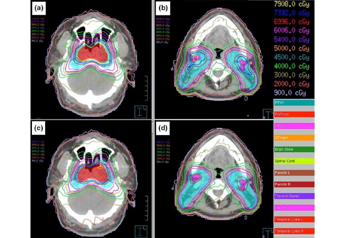
Treatment plan dose distributions for a locally advanced nasopharyngeal carcinoma case with three targets, using MLCs with maximum leaf speeds of 1.0 cm/s (a, b) and 3.5 cm/s (c, d). The 3.5 cm/s MLC achieved better plan quality. (Courtesy: CC BY 4.0/J. Appl. Clin. Med. Phys. 10.1002/acm2.13020)
Increasing the multileaf collimator (MLC) leaf speed may help improve the quality of volumetric modulated arc therapy (VMAT) plans, according to a study published in the Journal of Applied Clinical Medical Physics. Researchers in Beijing investigated seven MLCs with different maximum leaf speeds to investigate their influence on plan quality. They determined that VMAT plans for locally advanced nasopharyngeal carcinoma (NPC) and rectal cancer improved significantly when maximum MLC leaf speeds were increased from 1 to 3.5 cm/s.
VMAT delivers highly conformal radiation dose to a targeted tumour, sparing surrounding healthy tissue. MLCs, which consist of several sets of metallic leaves, are used to shape the radiation beam as it exits the linear accelerator (linac). In addition to improving the dose distribution, MLCs are highly efficient and reduce treatment time. However, dose leakage and transmission through MLC leaves can occur, negatively impacting treatment plan quality.
To achieve highly modulated dose distribution, MLCs need to move at a high speed when the gantry rotates. But what is the optimum speed required to achieve the highest quality VMAT plan for specific types of cancer?
To answer this question, the research team – from the National Cancer Center/National Clinical Research Center for Cancer/Cancer Hospital, Chinese Academy of Medical Sciences and Peking Union Medical College – investigated six NPC cases, representing complex clinical treatments, and nine rectal cancer cases, representing simple treatments. All patients had previously received VMAT at the National Cancer Center.
Principal investigator Jianrong Dai and first author Jiayun Chen configured seven treatment plans for each patient, representing seven linacs configured with maximum MLC leaf speeds (MMLS) of 1.0, 1.5, 2.25, 3.5, 5.0, 7.5 and 10.0 cm/s. Automated VMAT plans with identical initial optimization parameters were designed based on the centre’s clinical protocols, thereby eliminating interoperator variability. The VMAT plans were calculated using 6 MV photons, with a maximum variable dose rate of 600 MU/min. Gantry angle spacing, arc and collimator rotation direction and degrees, and maximum rotation time of each arc were identical for all plans.
The researchers evaluated the plans and scored them using the Plan Quality Algorithm (PQA) tool and the Plan Quality Metric (PQM) of the ESTRO QUASIMODO project. The PQA provided an objective method by which to quantify plan quality, while the PQM provided a fair comparison of plan results.
Plan scores for rectal cancer increased dramatically when the MMLS increased from 1 to 3.5 cm/s, but only grew slowly beyond that speed. The researchers believe that high leaf speeds helped smooth out variations in dose distributions, either through small fields or by the leaves blocking high-dose regions for organs-at-risk. Findings were similar, but not as dramatic, for NPC cases.
“Plan quality is greatly improved as MMLS increases from 1 to 3.5 cm/s; above that, the quality change is marginal,” write the authors. “It demonstrated that the MMLS influences plan quality regardless of the tumour size.”
It should be noted that actual MLC performance depends on a number of other considerations, such as leakage and reliability, for example, and further research is needed to evaluate how speed affects clinically deployed models.
The authors tell Physics World that the team has now studied the effect on treatment plan quality of two major MLC characteristics: MLC leaf speed, as examined in this study; and MLC transmission, for patients with advanced lung cancer, published in Medical Dosimetry. The team’s future research will incorporate the interactions of dose rate, gantry speed and MLC speed, to obtain a more comprehensive representation of their dosimetric effects.
The annual meeting of the European Society for Radiotherapy and Oncology (ESTRO), originally due to take place in April in Vienna, was one of the early casualties of the Covid-19 pandemic. Postponed once to August, the event organizers decided to further delay the meeting to the end of November in the hope of convening a reduced live congress along with enhanced online participation.
However, the resurgence of coronavirus in Europe has forced another rethink, and ESTRO 2020 will now be staged as a fully digital event. The main online congress will take place from 28 November to 1 December, with attendees able to access the scientific programme via a digital platform that includes Q&A and polling functionality to enable interaction with the speaker. Several pre-congress sessions are also available online, including three short courses on a dedicated education platform, while all registered participants will be able to view all sessions on demand after the congress has finished.
The theme for this year’s event is “Translating research and partnership into optimal health”. Meeting chair Umberto Ricardi from the University of Turin, Italy, points out that technologies and methodologies in radiation oncology are evolving rapidly, but that these advances will “only truly provide impact when the new findings can be translated into clinical applications that will result in optimal benefit for each individual patient in daily practice”.
ESTRO 2020 will also host Europe’s largest industrial exhibition in radiation oncology. This year a virtual exhibition will enable attendees to interact with industry leaders and to find more about the latest innovations in technology, techniques and oncology products – a few of which are highlighted below.
Innovating and evolving radiotherapy quality assurance
Quality assurance (QA) plays a fundamental role in any radiotherapy procedure. IBA Dosimetry is working to shape QA to advance patient safety in radiation therapy, proton therapy and medical imaging. The company predicts that its latest innovations will bring the accuracy and efficiency of QA to a new level. Future solutions, meanwhile, will significantly reduce QA times and further streamline the medical physics workload.
Independent QA is essential to ensure reliable, trustworthy and accurate QA, and has been assumed as a given in the radiotherapy community. But as radiotherapy systems increasingly offer built-in “self-check” QA, the need for independent QA becomes imperative. To raise awareness of this topic, IBA Dosimetry has teamed with radiotherapy QA equipment vendors worldwide to launch an “Independent Quality Assurance” initiative.
Convergence of machine QA and patient QA will unlock the potential of real risk-based radiotherapy QA. (Courtesy: IBA Dosimetry)
IBA Dosimetry also highlights the need for convergence of machine and patient QA. Today, QA applications for validating the treatment machine and those for verifying the patient-specific plan generally have little or no connectivity. Combining data from patient QA and machine QA will provide more precise outcomes and faster results.
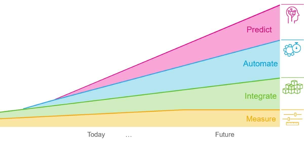
These QA innovations are based on four strategic pillars – implementation of measurements, integration, smart automation and prediction of QA results – that help save valuable time of the medical physicist and provide higher accuracy and increased confidence. IBA Dosimetry notes that while measurements will remain important for future QA, further integration, Monte Carlo-based predictive QA and automation will enable users to measure only where it really matters, leading to fewer measurements with better quality results.
IBA Dosimetry’s QA innovation strategy is based on four pillars: measure, integrate, automate and predict. (Courtesy: IBA Dosimetry)
Software platform standardizes treatment plan review
Peer review of treatment plans is a powerful strategy in radiation oncology clinics to ensure patient safety and plan quality, but time constraints in the daily routine can make it difficult to prepare and review patient cases in an optimal way. MIM Harmony is a dedicated software platform that provides rapid and reproducible treatment plan review, allowing medical teams to focus their efforts on a deeper, critical evaluation of patient cases and to improve the quality of their treatment plans.
MIM Harmony allows clinicians to prepare patient cases automatically, saving valuable time and ensuring that all the necessary information is available at the review meeting. Each review is guided by a disease site-specific worklist that can be fully customized to suit the needs of each clinical team, offering a flexible but standardized approach to patient care.
A dedicated platform from MIM Software is designed to improve both the efficiency and effectiveness of treatment plan reviews. (Courtesy: MIM Software)
MIM Harmony’s customizable worklist, along with flexible task automation, reporting tools, and advanced analytics, enhance multiple peer-review formats such chart rounds, contour rounds, and one-to-one consultations. Its robust data analytics platform moves beyond traditional peer review, compiling critical information after each meeting to help identify areas for improving quality or clinical practice.
Fast and filmless patient QA now extends to multitarget treatments
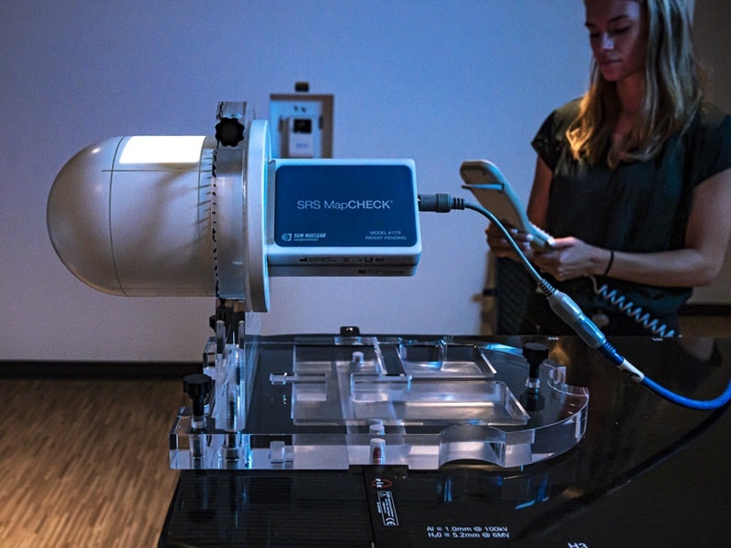
Quick and convenient patient-specific quality assurance (QA) is essential for delivering stereotactic radiotherapy safely and effectively. The SRS MapCHECK from Sun Nuclear is a patient QA solution that replaces conventional film with a high-density diode array, providing accurate dose measurements in minutes rather than hours. With nearly 400 devices in clinical use and a growing body of supporting literature, the SRS MapCHECK has been proven to efficiently detect the most common sources of errors in stereotactic treatments and has become the gold standard for time-sensitive patient QA.
The system comes with SNC Patient software, which corrects for angular dependence, field size and pulse rate to ensure accurate patient QA from any angle. A new update to the software now makes it possible to simplify QA workflows for single-isocentre multiple-target (SIMT) treatments, with a new QA Setup Tool providing guidance for the optimal set-up of SIMT plans and simplified shifts for larger field sizes.
The SRS MapCHECK from Sun Nuclear now provides enhanced support for single-isocentre multiple-target (SIMT) treatments. (Courtesy: Sun Nuclear)
This software update, described in more detail in this short video, complements Sun Nuclear’s MultiMet-WL Cube, designed for machine QA of SIMT treatments, and adds to a suite of StereoPHAN QA solutions for stereotactic radiotherapy. During ESTRO 2020, on 30 November at 13.00 CET, Sun Nuclear will host a satellite symposium entitled “Advancing QA: Latest Solutions from Sun Nuclear”. Presentations and demonstrations will showcase both SRS MapCHECK and the SunCHECK platform.
Tracking device promises more precision for prostate cancer treatment
New radiotherapy technologies such as image guidance, high-precision dose delivery and more accurate target definition are making it possible to administer higher radiation doses in fewer treatment sessions. For prostate cancer, an improved understanding of the biological mechanisms underlying the use of hypofractionation suggests that the delivery of fewer and larger fractions has the potential to improve the therapeutic ratio while also shortening the overall treatment time.
Hypofractionated radiotherapy delivered via stereotactic body radiation therapy (SBRT) offers a high dose per fraction within a small number of fractions, but the steep dose gradient demands a high level of reliability during the entire treatment delivery process. At even small distances from the target the radiation dose decreases rapidly, which means that prostate motion during treatment might result in spatial misses and the exposure of surrounding healthy tissues to radiation.
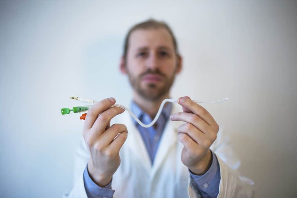
The RayPilot HypoCath system from Micropos Medical is a removable electromagnetic transponder that offers real-time tracking of the tumour throughout the treatment process. The electromagnetic tracking device is compatible with conventional linacs and requires no surgical intervention, with the transmitter integrated into a standard urinary catheter to enable localization of both the prostate and the urethra.
The RayPath HypoCath keep track of prostate motion during SBRT treatments. (Courtesy: Micropos Medical)
The system has already been trialled at the Ospedale San Gererdo in Monza, Italy, with the results reported in a recent white paper by Professor Stefano Arcangeli. Tests with four patients showed that the electromagnetic tracking device kept the average target motion within 2 mm throughout treatment delivery, with no impact on patient comfort. Arcangeli notes that further improvements in the accuracy could be achieved by fine tuning the workflow, which he believes would make SBRT well positioned to become the procedure of choice for patients with localized prostate cancer.
Upgraded software enhances daily testing regime
Quality assurance (QA) in radiation treatment planning is becoming ever more important as medical physicists seek to optimize image-guided radiotherapy (IGRT) protocols for the delivery of higher doses and emerging adaptive treatments. Imaging phantoms are essential to calibrate and commission IGRT systems on a daily basis, and the QUASAR Penta-Guide Phantom from Modus QA has become recognized globally as the preferred tool for efficient daily testing of key parameters such as system alignment and imaging performance.
Modus QA is now enhancing the capabilities of this phantom with the new Penta-Guide 2.0 software, a comprehensive daily QA solution that is free for all existing and new Penta-Guide users. Designed to improve the daily QA workflow, the software offers advanced features such as automated monitoring of system alignment, image quality analysis, and improved reporting and visualization tools.
Updated software for Modus QA’s Penta-Guide phantom is designed to improve the efficiency of daily testing protocols. (Courtesy: Modus QA)
An online presentation by product manager Rocco Flores will highlight the innovative features of the Penta-Guide phantom, review the daily QA user workflow, and explain the advanced utility of the Penta-Guide 2.0 software.
Researchers in the US and Israel have developed a way to make 3D superconducting nanostructures by combining DNA with niobium and silicon. This new technique might be used to make signal amplifiers that enhance the speed and accuracy of quantum computers as well as ultrasensitive magnetic field sensors for medical and geophysics applications.
While traditional nanofabrication techniques like electron-beam lithography can produce one-dimensional and two-dimensional superconducting nanostructures, their ability to produce three-dimensional structures is limited. For the past 15 years or so, researchers have instead turned to self-assembly techniques that use DNA to construct 3D nanoscale structures and integrate them with functional inorganic nano-components.
One such technique, known as “DNA origami”, uses the natural pairing of DNA’s four nucleotide bases – A, T, C and G – to produce a multitude of self-assembled engineered shapes. The process involves folding a long single strand of DNA with the help of shorter complementary strands at specific locations to make pre-defined nanoscale structures. These nanostructures can then be used as scaffolding for building 3D nanoscale architectures that can be “converted” into inorganic materials such as superconductors – as in this new work, which was led by Oleg Gang, a nanoengineer at Columbia University and the Brookhaven National Laboratory’s Center for Functional Nanomaterials.
DNA origami “frames”
In their experiments, Gang and colleagues at Columbia designed octahedral-shaped DNA origami “frames” using a computer software package called caDNAno. Each edge within the frames comprises a six-helix bundle of DNA that is 28.6 nm long and contains 84 base pairs. At each end of these bundles, the researchers added a single-stranded DNA chain that measured roughly 2 nm in length and was designed to complement the DNA chain of the opposing DNA origami.
To better visualize (and subsequently characterize) the structure, the researchers inserted 10-nm-diameter gold nanoparticles into each octahedral “cage”, where the particles were held in place by a structure made from DNA that is complementary to the inner strands of the frame. The researchers then assembled the cages into a simple cubic superlattice of octahedra made from two pairs of frames and designed to have specific DNA strands targeting four complementary counterparts in-plane and two counterparts out-of-plane. The resulting superlattice samples are flakes 5-10 microns long and 1-3 microns thick, and the researchers used small angle X-ray scattering at the Brookhaven National Synchrotron Light Source II to confirm their nanoscale structure.
Mechanically robust 3D architecture
The team then solidified their ensemble by using a wet chemistry technique to coat the DNA lattices with a layer of silicon dioxide. “In its original form, DNA is completely unusable for processing with conventional nanotechnology methods,” Gang explains. “But once we coat the DNA with silica, we have a mechanically robust 3D architecture that we can deposit inorganic materials on using these methods. This is analogous to traditional nanomanufacturing, in which valuable materials are deposited onto flat substrates, typically silicon, to add functionality.”
The next step was to use an evaporation technique to coat the silica-coated superlattices with a layer of niobium around 10 nm thick. This part of the work was done at the Institute of Superconductivity at Bar-Ilan University in Israel. There, researchers led by Yosi Yesurun carefully controlled both the temperature of the silicon substrate and the rate at which they deposited the niobium so that the niobium only coated the sample and did not penetrate all the way through it. They did this to prevent short circuiting between the electrodes used for later electronic transport measurements.
A periodic array of Josephson junctions
Once this step was complete, the researchers used scanning transmission microscopy with energy dispersive spectroscopy (STEM-EDS) to check the structure of their samples. This imaging technique revealed a porous superlattice structure made up of the silica-coated DNA and the gold nanoparticle “tracers”.
This structure forms a periodic array of Josephson junctions – thin non-superconducting barriers though which superconducting current tunnels – with the niobium in the superlattice being mainly confined to the top three pairs of octahedra layers (which have a total thickness of around 290 nm). As a final step, the researchers measured the current-voltage characteristics of their superlattices at temperatures between 1.9 and 3.7 K. The curves produced are typical of single Josephson junctions – that is, they show zero voltage for currents up to a certain temperature-dependent critical current (indicated by the appearance of resistance) and then a gradual voltage increase.
Gang and colleagues note that Josephson junctions are key components for leveraging quantum phenomena in practical technologies, with examples including the superconducting quantum interreference devices (SQUIDs) used to sense magnetic fields. The three-dimensional nature of the Josephson junctions created in this work means that more of them can be packed into the same small volume, which could increase the power of a device that uses them.
While DNA is not necessarily the most useful functional material for such work, the researchers say they have demonstrated that complex DNA organization can, in principle, be used to create highly nanostructured 3D superconducting materials. “This material conversion pathway gives us an ability to make a variety of systems with interesting properties – not only superconductivity but also other electronic, mechanical, optical, and catalytic properties,” they report. “We can envision it as a ‘molecular lithography’, where the power of DNA programmability is transferred to 3D inorganic nanofabrication.”
Spurred on by this possibility, the researchers, who report their work in Nature Communications, say they are now planning to apply the same strategy to create highly-structured 3D inorganic nanomaterials with a broad range of functions.
For nearly 50 years, researchers from around the world have converged in Boston for the Fall Meeting of the Materials Research Society. This year, however, as a result of the Covid-19 pandemic, the meeting will be convened as a fully digital event in combination with the Spring Meeting, which was due to take place in Pheonix, Arizona, back in April.
The joint 2020 MRS Spring/Fall Meeting & Exhibit, scheduled for 27 November ̶ 4 December, will include the scientific programmes from both meetings, with a combination of live and on-demand presentations. John Rogers of Northwestern University will give a plenary lecture on functional materials that enable small electronic devices to be integrated into the body, while a range of networking, Q&A sessions and professional development events are available to access before and after the meeting.
A virtual exhibit will enable attendees to interact directly with equipment vendors, with extended “booth hours” where delegates can chat with company representatives. It is also an ideal opportunity to up to date with the latest technology developments – a few of which are highlighted below.
Turnkey system delivers fast and precise Hall measurements
A fully integrated measurement platform from Lake Shore Cryotronics makes it quicker and more convenient to acquire high-precision Hall measurements. Ideal for anyone studying semiconductor materials, the MeasureReady FastHall Station incorporates Lake Shore’s M91 FastHall controller into a tabletop system that enables simplified Hall measurements and reduces the time needed for experimental setup.
Along with the M91 measurement controller, the all-in-one station includes a Windows-based computer, a 1 T permanent magnet, a high-precision sample holder, and all the necessary software and cabling to provide a comprehensive range of Hall measurements. The system can detect sample resistances up to 1 GΩ and mobilities as low as 0.01 cm2/V s –making it ideal for research into low-mobility materials.
The MeasureReady FastHall Station for Lake Shore Cryotronics provides an integrated solution for quick and easy Hall measurements. (Courtesy: Lake Shore Cryotronics)
Lake Shore’s patented FastHall method speeds up measurement times, while easy-to-use spring pin and solder sample holder cards accommodate up to 10 mm × 10 mm van der Pauw and Hall bar type samples. The station also features an electronically shielded, low-noise sample space with guarded contacts, making it quicker and easier to derive accurate measurements of the carrier type, carrier concentration, mobility, and Hall coefficient.
The system’s MeasureLINK-MCS software collects all the data, and provides standard sequences, charts, and test scripts that can be customized by the user. In addition to performing complete Hall analyses and outputting all measured and derived values, detailed reports can be generated that include all the supporting intermediate data, so a researcher can readily confirm the integrity of the results. Optional extras include a gate-bias instrument plus a liquid-nitrogen add-on to convert the standard room-temperature station to a cryogenically cooled sample space maintained at 77 K.
Large-sample AFM pinpoints polymer properties
Atomic force microscopy (AFM) has become a crucial tool in both research and industry for studying the properties and structure of many different materials and devices. In an online presentation that is now available to view on demand, Dr Vladimir Korolkov, a senior application scientist at AFM manufacturer Park Systems, highlights the use of AFMs to study the structure of polymer materials.
In the presentation, Korolkov explains how to exploit an AFM to resolve individual polymer chains in real space, which is particularly important because the properties of polymer materials are strongly influenced by the packing and conformation of individual macromolecules as well as their monomer composition. He also shows how an AFM can be used to acquire ultrahigh-resolution images of individual PTFE molecules on the semi-crystalline surface of a commercial Teflon tape.
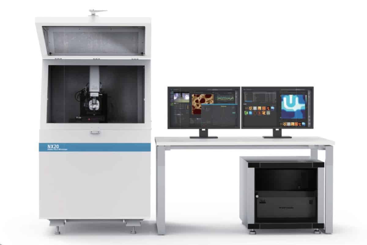
The NX20 AFM from Park Systems is designed for failure analysis and large sample research. (Courtesy: Park Systems)
Using these real-world polymer samples, Korolkov highlights the capabilities of Park Systems’ NX20 AFM for high-speed acquisition of high-resolution images. The NX20 is designed to study large samples and conduct multi-sample analyses, and comes with an automated interface that makes it easy for non-experts to acquire high-quality data. The instrument supports single-click imaging through the auto mode of Park’s SmartScan software, while automatic operation of the cantilever brings it safely into contact with the surface within just 10 seconds.
The NX20 also controls the scan speed based on the peaks and valleys of the sample surface, minimizing the scanning time while also acquiring a high-quality image. When moving to a neighbouring location or zooming into a target, the system automatically applies a new optimal condition.
Single instrument combines AFM with scanning microscopy
A unique atomic force microscope (AFM) from Quantum Design can easily be integrated into the high-vacuum environment of almost any scanning electron or scanning ion microscope (SEM/FIB). The AFSEM® Nano enables simultaneous operation of SEM, FIB, and AFM inside the vacuum chamber, allowing these complementary techniques to be combined without needing to transfer the sample or break the vacuum.
The AFSEM Nano directly correlates AFM functionality to SEM or FIB information at the same location on the sample. This correlated functionality allows users to, for example, measure the real 3D-topography of their sample with (sub)-nanometer resolution inside their SEM or FIB system, or easily identify the region of interest with the SEM and then use the AFM to measure the physical, electrical, or magnetic properties at the same location.
Best of both worlds: the AFSEM Nano from Quantum Design can be integrated into the vacuum chamber of almost any scanning microscope. (Courtesy: Quantum Design)
Combining the different techniques makes it easy to analyse samples with very different shapes or sizes. Users can also obtain in-situ correlation of chemical (EDX) and crystallographic (EBSD) information, and achieve 3D subtractive tomography by combining FIB slicing with the mapping of nanomechanical properties using the AFM.
The AFSEM Nano is a laser-free AFM that exploits a self-sensing cantilever technology to avoid the need for optical alignment inside the vacuum chamber. Its open design allows for easy integration with other optional add-ons, such as tensile stages, nano-indentors or nano-manipulators.
Materials matter for 2D layered perovskite research
2D layered perovskites have attracted huge research interest for their unique optoelectronic properties, helped enormously by the discovery that the most promising structures – usually hybrid organic–inorganic lead halides – can be prepared easily and cheaply using solution processing techniques. Photoluminescent 2D perovskites have a direct bandgap with a narrow emission peak that can be tuned by changing the layer thickness and the material composition, and have been widely studied for applications in light-emitting diodes, phototransistors, lasers, and solar cells.
MilliporeSigma supplies the precursor materials needed to create 2D layered perovskites with different compositions and properties. (Courtesy: MilliporeSigma)
Materials company MilliporeSigma offers a range of fully synthesized layered 2D perovskites, including some of the most popular organolead halides. Also available is a comprehensive portfolio of precursor materials, including lead halides, organohalides, and halide acids, as well as other key materials needed to fabricate 2D layered perovskites.


 Shawn Prince has an undergraduate degree in Business Administration from The Ohio State University and a Master’s of Business Administration degree from Arizona State University. Shawn is a Senior Director of Patient Access at Accuray. His previous professional experience includes reimbursement and sales for various pharmaceutical and biotechnology companies.
 Awais Mirza is the senior manager of patient access with Accuray. Within the patient access department, Awais works to secure appropriate coding, reimbursement coverage and payment from both Medicare and commercial insurers. He has an expansive background in clinical and administrative radiation oncology and has worked for a number of hospitals as a licensed radiation therapist and administrative manager/leader prior to joining Accuray.