Physicists in the US have reconstructed images of illuminated colourful objects that are hidden behind nearly opaque walls. Michael Gehm and colleagues at Duke University gathered the images by encoding the light that had passed through an unknown scattering object, and then separating it into different wavelengths. Their technique could be useful for capturing spectral information in astronomy, where speckle is often caused by light passing through the Earth’s turbulent atmosphere, and in healthcare.
When the light emanating from a colourful image passes through a translucent scattering object, the paths of its constituent photons change in complex, unpredictable ways. Once the light emerges, it is distorted into apparently random speckle patterns. Various techniques allow researchers to recover original images from these patterns, but many of these require access to the image or scattering object, or prior knowledge of it. Unfortunately, this renders them useless if either object or image can’t be accessed.
This problem can be overcome to an extent using so-called memory effect (ME) imaging, which can spot correlations in the speckle patterns originating from adjacent points on the image. Yet this technique has so far been restricted to narrow bandwidths. Since different wavelengths produce different speckle patterns, it is difficult to disentangle the patterns associated with different colours from a single speckle image.
In their study, Gehm’s team took ME imaging a step further by introducing an aperture that encodes the speckle signals by blocking out specific wavelengths. A prism then separates this encoded light into different spectral channels, teasing individual speckle patterns apart by shifting them by an amount proportional to their original wavelengths. Each pattern is then collected by a monochrome camera, where an image reconstruction algorithm uses the known positions of the blocked wavelengths produced by the coded aperture to reproduce the original colour image.
In contrast to previous ME imaging techniques, Gehm and colleagues only needed prior knowledge of the aperture and the prism, and not of the image or scattering object, to reconstruct their images. Their setup allowed them to reconstruct a letter ‘H’ containing complex patterns of yellows, pinks, and blues, by separating its original speckle image into five spectral channels. In simulations, they also reconstructed an image of a cotton plant cell, which spanned the entire visible spectrum and some of the infrared.
With further improvements, Gehm’s team believe their technique could allow researchers to collect accurate spectra in situations where either the object to be imaged, the scattering object, or both, cannot be accessed. This could allow astronomers to determine the chemical compositions of distant objects from speckle images, which are distorted after passing through Earth’s unpredictably turbulent atmosphere. In addition, biologists could more easily track biomolecules tagged with fluorescent markers. This would bring about important advances in healthcare, where non-invasive monitoring techniques can be critical.
With the start of the new academic year fast approaching at many universities around the world, now is the perfect time for final-year physics students to start thinking about what they’ll do after graduation. Many students will naturally have their eyes on a master’s or PhD, hoping to finally do some “proper” research after all those years studying textbooks. It’ll be your chance to perhaps be the next Curie, Hawking or Einstein.
But knowing what to do for at post-graduate level can be hard – there are almost too many options out there, from astronomy to Z-bosons. With that in mind, the bumper Physics World Careers 2019 guide is here to help, available free-to-read in both print and digital formats.
In the wake of the discovery of the Higgs boson at CERN’s Large Hadron Collider (LHC), particle physics is one of the most exciting areas of physics for many students. But the LHC won’t last forever and China is already planning its own ambitions accelerator – the Circular Electron Positron Collider. It’s being spearheaded by the Institute of High Energy Physics of Chinese Academy of Sciences, which is right now looking for scientists and engineers at all stages of the career.
If you’re thinking of a PhD in a more applied field, condensed-matter physics is where many physicists make their mark. A lot of exciting work is going on graphene – the 2D “wonder material” – and what better place to study it than the University of Manchester, where Andre Geim and Kostya Novoselov isolated the material in 2004, bagging a Nobel prize for their efforts in 2010. The university offers doctoral training in graphene, through its Graphene NOWNANO CDT.
Optics is another field where many PhD physicists find their fortunes. Physics World Careers 2019, for example, features ICFO – the Institute of Photonic Sciences in Barcelona, which has more than 400 researchers on both basic and applied research, using light to tackle challenges in health, energy, IT, security and industry.
Some physicists, of course, want to feel part of something big, so you might like to consider the Scottish University Physics Alliance. It brings together eight different physics schools – at Aberdeen, Dundee, Edinburgh, Glasgow, Heriot-Watt, St Andrews, Strathclyde and West of Scotland – to form a 1200-strong community of physicists, including 600 postgraduate students.
The School of Physics and Astronomy at Cardiff University offers graduate schemes and training. (Courtesy: Cardiff University)
Energy is another big sector where physicists end up and if you’re set on a career in the burgeoning nuclear industry, it helps to get formal training at master’s level before moving in to the field. A great option is the University of Birmingham’s School of Physics and Astronomy, which offers a fantastic MSc on the physics and technology of nuclear reactors. The course has an exceptional pedigree, having been running for more than 60 years since the very early day so of nuclear power.
A similar Master’s programme is available from the South West Nuclear Hub at the University of Bristol, giving students a grounding in the science and engineering of the global nuclear industry. Another option is the UK’s Nuclear Technology Education Consortium, which offers postraduate education in nuclear science and technology – both directly taught and via distance-learning formats.
We hope you find Physics World Careers 2019 useful. You can also sign up today for our brand new careers newsletter. Sent once every two months, the newsletter will bring together a variety of case studies, valuable careers advice, and information from leading employers you might be interested in working for. To sign up, simply sign into your Physics World account and tick the “Careers bimonthly” box.
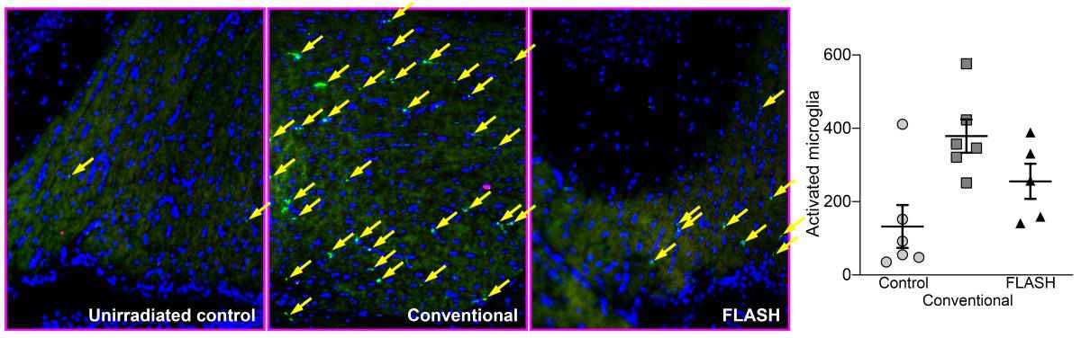
Representative sections of mouse hippocampus with fluorescent staining of activated microglia (bright green cells), which are mediators of neuroinflammation. FLASH induced less activation of microglia than conventional irradiation, leading to preserved neurocognitive function. (Courtesy: Billy W Loo Jr)
Delivering whole-brain radiotherapy in a single, ultrafast dose minimizes the cognitive impairment associated with damage to the hippocampus. This, at least, is the case for mice, whose performance in a series of object-recognition tests was measured after whole-brain irradiation administered in either one sub-second dose or extended over a conventional four-minute duration. The effect is due to the smaller inflammatory response provoked by more rapid treatments compared with conventional dose rates, and could one day lead to radiation therapies with reduced side effects.
It is easy to see the appeal of administering an entire course of radiotherapy in a single dose. Apart from being more convenient for the patient, ultra-rapid irradiation means that the effect of patient motion can be virtually disregarded. When dose delivery is near-instantaneous, even moving targets appear practically stationary. But even without these advantages, the technique – named “FLASH” in an in vitro study from 2014 – has intrinsic radiobiological benefits that are only just coming to prominence.
“Certain aspects of FLASH radiobiology have been discovered and rediscovered since the 1960s,” says Billy W Loo Jr of Stanford University School of Medicine, who conducted this latest research with a team from the departments of radiation oncology and neurology & neurological sciences. “The 2014 study was the first to gain general recognition, and even then it was slow, in part because it didn’t fit with accepted radiobiology paradigms, and also because people could not see a practical path to clinical implementation.”
That path has become clearer now that Loo and colleagues have started development of a linac that can deliver the high dose rates necessary to implement FLASH clinically. This has prompted more work on the radiobiological aspect of the technique, leading to the promising results reported by Loo and his team in this latest study (Radiother. Oncol. 10.1016/j.radonc.2019.06.006).
The researchers divided healthy mice into three groups. One group underwent a whole-brain electron radiotherapy procedure delivered at the standard dose rate; one group received the same overall dose but in a single fraction lasting less than a fifth of a second; a control group received no treatment at all.
Ten weeks after the treatments, the researchers observed the mice responding to unfamiliar objects placed within and moved around a testing environment. Mice in the control group and the FLASH group spent about the same time investigating these changes to their environment, whereas mice that had received the standard irradiation procedure appeared significantly less sensitive.
When the mice were sacrificed and their brains analysed, Loo and colleagues found two major differences linked to the groups’ varying cognitive performance. The first was an indication of inflammatory response, expressed by the presence of activated microglia and cytokines, especially in the conventional-dose-rate group. In the FLASH group, these indicators of brain damage appeared at a level intermediate between the control and the conventional-irradiation groups.
The second difference was seen in the density of dendritic spines in the mice’s hippocampi. These subcellular structures help pass signals between neurons, and play a role in memory formation. Spine density in the conventional-irradiation group was much lower than that seen in the control group but was largely preserved in the mice that underwent FLASH.
“There is evidence that the anatomic and functional changes are a result of the neuroinflammation,” says Loo. “So at one level, the mechanism of FLASH sparing of brain injury appears to be through reduced neuroinflammation compared to conventional-speed irradiation.”
Why the same radiation dose delivered at a much higher rate should have such an effect is not clear, however. “Ultimately, this is still a phenomenological observation. We need to do more work to understand specifically why FLASH triggers less neuroinflammation,” says Loo.
Whatever the mechanism, human patients will not be benefiting from whole-brain FLASH for some time. Until the technology behind the group’s dedicated FLASH platform matures, the technique will be limited in the near term to superficial tumours using electron beams, or very small embedded tumours using narrow, single-angle proton beams.
Fusion, with its promise of unlimited, safe, carbon-free energy, has for many years occupied a special place in the imagination of physicists. The process of combining small atoms into larger ones releases enormous amounts of energy (fusion fuel is more than a million times more energy dense than fossil fuels), and the goal of harnessing this energy to generate electricity has been pursued by scientists in laboratories around the world since the 1960s.
So far, a practical fusion power plant has proven elusive. This is perhaps not surprising, given the difficulty of replicating conditions similar to those in the centre of a star. Before fusion can take place, the electrical repulsion of the atomic nuclei that make up the fusion fuel must be overcome, so that atoms get physically close enough to fuse. To do this, the fusion fuel must be heated to temperatures of around 200 million K. At these temperatures, all matter exists as a plasma, in which electrons and nuclei have separated to form what can be thought of as an ionized gas.
The task of creating and containing these plasmas raised technical hurdles that were sometimes underestimated. For example, the role of turbulence in transporting heat out of plasma confinement systems was once poorly understood. Now, however, these hurdles are being overcome. Building on past progress, and enabled by a new generation of superconducting materials, fusion scientists and engineers are pushing the technology still further, with the goal of realizing a fusion power plant soon enough to make a difference on climate change.
Apart from this technological progress, one of the most exciting recent developments in the field has been the rapid growth of private investment in fusion. There are now more than a dozen companies around the world working toward the goal of fusion energy, each focusing on slightly different strategies to get there. The ability of these companies to attract investment is both an indication of the value placed on achieving fusion energy and a validation of the ongoing research in labs worldwide.
Commonwealth Fusion Systems (CFS) was founded in mid-2018 as a spin-off from the Massachusetts Institute of Technology’s Plasma Science and Fusion Center. Its scientists are now working closely with colleagues at MIT in order to design and construct SPARC, a new fusion device aiming to be the first to achieve net energy gain – that is, producing more energy than is required to heat the plasma and sustain the fusion reactions.
Smaller superconducting magnets
Multiple approaches to create and contain fusion plasmas have been pursued, but the most successful to date – and thus the one CFS has adopted – involves confining the hot fusion plasma in a device known as a tokamak. In this type of fusion device, strong magnetic fields produced by powerful electromagnets are used to contain the plasma in a toroidal (doughnut-shaped) vacuum environment, in order to insulate and isolate the plasma from ordinary materials. Radio frequency waves or one of a few other methods are then used to heat the plasma.
The highest-performing tokamak so far, in terms of the ratio of fusion power to the required heating power, is the Joint European Torus (JET) in the UK, which was constructed using copper magnets. A viable fusion power plant, however, will require superconducting magnets to ensure that the power expended in running the magnets is far less than the fusion power generated by the device itself.
In principle, magnets made from conventional low-temperature superconductors, such as Nb3Sn, can achieve this. However, these materials are severely limited by how much current can pass through them at high magnetic field, which in turn limits the strength of the magnetic field that they can produce. To overcome this and construct a tokamak that will produce a net energy gain, one must therefore make the machine very large, and thus very expensive. This limitation is what led to the design of the ITER tokamak, which is currently being constructed in France under the aegis of an international collaboration, to the tune of tens of billions of dollars. The enormous size of ITER and the complexity of organizing many international partners have put the project far behind schedule and over budget, with a current expected peak performance date somewhere around 2035.
What sets SPARC apart from ITER, JET, and other previous fusion tokamaks will be its use of a new type of high-temperature superconductor (HTS), yttrium barium copper oxide (YBCO). Current-carrying tapes made from YBCO remain superconducting at considerably higher magnetic fields than is possible with older superconductors. This is valuable because higher magnetic fields improve the thermal insulation of the plasma and thus allow for considerable improvement over the performance of previous tokamaks.
YBCO superconductors have existed for a number of years, but they have only recently become commercially available in the quantity and quality required for fusion devices. By using this new superconductor to develop high-field magnets – capable of producing fields of 12 T at the centre of the plasma, compared to 5 T in ITER – CFS and MIT hope to drastically accelerate the timetable to fusion energy and achieve net energy gain in a device that is roughly 2% the size of ITER. To make a comparison, we believe that YBCO superconductors will be an enabling technology for fusion in the same way that lightweight internal combustion engines were an enabling technology for powered flight.
The SPARC project’s first task, over the next few years, will be to build and test a full-scale prototype HTS fusion magnet
Material progress
Although high-temperature superconductors benefit the design of fusion power plants by reducing plant size, lowering capital costs, and allowing for higher operating temperatures, they also pose challenges and technical risks that must be overcome. These challenges include higher mechanical stresses in the magnets, larger heat loads from the fusion process, and the uncertainty of manufacturing and operating a first-of-its-kind HTS coil. To mitigate these technical risks and validate the magnet system design, the SPARC project’s first task, over the next few years, will be to build and test a full-scale prototype HTS fusion magnet.
The prototype HTS coil will demonstrate the operation of the magnet system and test the integrated cryogenic coolant system used to cool the superconductors to 20 K – the operating temperature required to generate the high magnetic fields in SPARC. Maintaining this temperature in SPARC will be challenging, since the compact nature of SPARC leads to higher power density and thus high heat fluxes generated by the fusion reactions in the core of the machine. Additionally, the properties of the mechanical structure around the conductor in the magnet depend strongly on the temperature of the materials, and therefore this structure must be kept cold in order to withstand the stresses generated in the magnet. The model HTS coil will therefore experimentally validate the heat removal capabilities in HTS magnet systems, and the lessons we learn from it will be critical in moving the SPARC project forward.
In addition to addressing structural and cooling challenges, one of the primary missions of the prototype HTS coil will be to investigate our ability to detect and mitigate a “quench” event, which is a sudden loss of superconductivity due to external events such as reaching critically high temperatures, current, or strain levels. A quench can lead to rapid, localized heating of the superconductor, potentially causing damage if the magnet is improperly designed or operated. Understanding how to detect and prevent quench events is of particular importance for the robustness and reliability of the magnet and fusion system.
Compared with older, low-temperature superconductors, HTS magnets are more stable in the face of local heating due to their higher operating temperature. For these same reasons, however, the quenched area of the magnet, which is no longer superconducting, can propagate more slowly in HTS magnets than in previous superconducting magnets and may be more likely to experience damage. Understanding how a quench event initiates and grows, and devising quench detection and mitigation techniques, is critical to the design of robust HTS magnets that are resilient to quench. Both of these systems will be proven on the prototype TF coil. After the successful completion of the prototype TF coil, MIT and CFS will go on to construct the SPARC device, and push toward practical fusion energy.
Considering the ambitions of the SPARC project, there is plenty of room for innovation and new ideas to address the various challenges in the development of fusion energy. CFS is looking for further improvement and innovation in the future with HTS joint technology, as well as with other technologies, such as power exhaust via advanced diverters and energy capture and conversion systems for next generation power plants like ARC. ARC plans to integrate the results of SPARC with these advanced technologies in order to be the first fusion power plant to demonstrate electricity generation. The passion for tackling technical challenges and developing innovative ideas to push these technological barriers is exactly what the fusion industry needs to reach our goal of limitless, clean energy.
A superconducting qubit can be used to reliably detect the presence of multiple phonons at the same time, US physicists have demonstrated. Patricio Arrangoiz-Arriola and colleagues at Stanford University built their “quantum microphone” using materials that minimized phonon losses, while narrowing the spectra of their qubit’s emissions to reduce uncertainties. The technology could allow for new capabilities in quantum computing, including modems that link together many quantum computers at different locations.
While the quantum properties of photons have been explored and exploited extensively, those of quantized mechanical vibrations, known as phonons, have remained much more difficult to study. Although phonons are important for explaining many properties in solid materials, the technologies required to measure and control them have faced significant challenges because – in contrast to photons – the quantized states of phonons do not have well-defined energies. Instead, they exist as collective excitations at equally spaced energies.
The most successful attempts to detect phonons so far have involved a technique named quantum acoustics, in which an artificial atom is coupled to a vibrating nanostructure. This atom can be in one of two quantum states, depending on whether or not it has absorbed a phonon. In their study, Arrangoiz-Arriola’s team devised a more sophisticated version of this setup – replacing the atom with a superconducting qubit to allow for stronger coupling with the nanostructure. Where the artificial atom would need to entirely absorb a phonon, this coupling allows the qubit to change states simply in the presence of one or more phonons.
To further improve their quantum microphone, Arrangoiz-Arriola and colleagues combined the qubit with a piezoelectric resonator, which produces a large voltage in response to mechanical deformation. This heightens the peaks of the energy spectra emitted by the qubit as it changed states. Shielding the hybrid qubit-resonator platform with a periodic crystal ensures that only the phonons produced by the nanostructure can interact with the qubit, while also minimizing losses of phonons to the surrounding environment.
The physicists then excited phonons through resonant vibrations of the nanostructures, and probed the peak positions of the qubit’s resulting transition spectra – which shifted to different degrees depending on the number of phonons present. They observed energy shifts around five times larger than the linewidths of each peak, revealing the presence of up to three phonons with a high degree of certainty.
In future studies, Arrangoiz-Arriola’s team hope to improve their setup to reveal phonon numbers without changing them, allowing for repeated measurements. Further developments could allow the quantum microphone to provide a basis for quantum modems, potentially creating networks of quantum computers in a variety of locations, and could also inform designs for novel architectures for quantum computers themselves.
If wasted food were a country, it would be the world’s third-largest emitter of greenhouse gases. A staggering one-third of all food produced goes to waste, accounting for an estimated 8% of global emissions – more than any country except China and the US. As well as an environmental disaster, food waste is also bad for the economy, costing close to a trillion dollars annually.
There are many strategies for reducing food waste. They include better storage and transportation in countries with less advanced infrastructure, and a range of measures – including more consistent labelling; less rigorous cosmetic standards for food among retailers; and education campaigns aimed at consumers – in more developed regions. But there are also big savings to be made by reducing waste higher up in the supply chain, before the food reaches either consumers or retailers. In 2018, the Boston Consulting Group reported that supply-chain improvements, together with increased efficiency, could reduce loss and waste by $270bn.
To realize those benefits, food producers urgently need accurate, non-invasive tools that enable them to assess the quality of food in real time, in a way that integrates seamlessly with existing production-line systems. This is where a technology known as hyperspectral imaging comes in. In effect, this type of imaging combines spectroscopy with computer vision, making it possible to measure both spatial and spectral characteristics reliably and simultaneously. Each pixel in a hyperspectral image contains information about how a corresponding spatial pixel absorbs or reflects more than 100 wavelengths of light. The higher information per pixel ratio allows a larger volume of data to be obtained than is possible with traditional RGB cameras, or even with multispectral imaging, which typically records light at fewer than 10 wavelengths.
Hyperspectral imaging was originally developed for remote sensing applications, but as the price and size of sensors has decreased, it is now being deployed in industries as diverse as criminal forensics, pharmaceuticals and manuscript analysis. Although the food industry has been relatively slow to digitize, the time is now ripe (forgive the pun) for hyperspectral imaging to be rolled out across food-supply chains, too.
Finding things that don’t belong
In a food production context, hyperspectral imaging can provide valuable insights about food quality. For example, it enables us to classify the freshness of fish into discrete categories, because the fish’s chemical composition determines how much light it reflects at different wavelengths. This type of imaging can also yield better estimates of a product’s shelf life or ripeness, and information about whether there are any contaminants present. Previously, such information could typically only be acquired via a range of destructive tests (such as sample-based testing for pH, a shear force test, the laboratory oven method, the refractometer and many others) and by time-consuming, costly and imperfect visual inspections. Hyperspectral imaging could therefore transform the way food supply chains operate by facilitating a shift towards 100% product coverage, resulting in less waste, higher yields and increased revenues.
One of the food industry’s biggest challenges – and a key focus for ImpactVision, the company I co-founded – is keeping non-food objects out of production lines. Foreign-matter contamination is the third leading cause of recalls in both the US and the UK, after biological contamination and mislabelling. Such recalls can be prohibitively expensive: a 2011 study from the Food Marketing Institute and Grocery Manufacturers Association estimated that a single foreign-matter-related food recall can cost a major producer up to $10m. The number of recalls is also on the rise. Between 2015 to 2016, recalls related to foreign bodies jumped 60 percent in the US, and recalls in the EU also rose. In 2018 alone, US food companies were subject to 30 high-profile recalls, amounting to hundreds of thousands of pounds of wasted food.
Technologies for detecting foreign matter do exist, but they serve specific purposes. X-ray machines can detect dense objects, but they miss lighter ones such as certain plastics, paper, cardboard and insects. Magnets and metal detectors will not pick up many of the most common foreign-object culprits – plastics, paper, stones, textiles – at all. (They also miss some more emotive contaminants, such as the live frog that one Californian woman decided to keep as a pet after she found it in a bag of salad.) Whenever such objects get into the production line, stoppages in the line, lost batches and missed deliveries are the inevitable result – not to mention lower production yields and eroded customer confidence and brand credibility.
ImpactVision’s system overcomes these limitations by using hyperspectral imaging and machine learning to detect, identify and eliminate foreign objects. By gathering information about the spectral and spatial characteristics of foreign objects, we can discern differences in their spectral signatures, which vary considerably based on chemical composition. Our hyperspectral imaging cameras cover wavelengths from visible to the near-infrared (400-1700 nanometres). This gives the system the information it needs to identify insects, wood, plastic, and organic matter – all of which of which can be similar in texture, colour and shape to the food products themselves, and thus very hard for human eyes to distinguish. The system also uses advanced machine learning techniques to help recognize contaminants. High-speed learning models can provide rapid answers in real time, making it possible to detect foreign objects within the production line. More advanced convolutional neural networks and deep learning techniques can provide more detailed analysis and qualitative assessments.
From lab to food processing plant
The challenge of foreign-object detection varies somewhat across different food industry sectors. For example, ImpactVision is working with the largest sugar processor in Mexico, Beta San Miguel, to introduce our hyperspectral imaging technology into their operations. One of the hurdles we had to overcome in that case involved mitigating the effects of sugar dust on our system. We are also developing applications for products such as leafy greens and potatoes, where producers have strict wash-down requirements for their machinery, and for spices, where dust is a key issue. Another big challenge will be to detect foreign objects hidden underneath opaque foods such as raisins and other dried fruits.
A watchful eye ImpactVision’s hyperspectral imaging cameras have been installed in the production line of a major sugar processor to monitor the line for contaminants. (Courtesy: ImpactVision)
Irrespective of the type of foodstuff involved, however, there are some common hurdles associated with taking a relatively new, lab-based technology and turning it into a commercially viable product that delivers benefits for the food industry. One hurdle is that hyperspectral images are enormous. Whereas a full-resolution colour image has three pieces of data per pixel, a hyperspectral image may have over 200 spectral values associated with each pixel, meaning that a single image file takes up 1 GB of storage space. This means that image processing must happen locally in order to deliver real-time information on production lines that move at speeds of 0.75 m/s and up.
Another hurdle was that certain elements on our equipment, such as camera casings and enclosures, needed to conform to strict US Food and Drug Administration (FDA) regulations before we could use them in a real food-production facility. We have done initial integration work with a major manufacturer of programmable logic controllers, Allen-Bradley PLC, to ensure that our systems are fully integrated with the industrial automation systems used to control equipment on food-processing lines.
Replacing visual inspection and destructive tests
Margins in the food industry are slim, and manufacturers are often slow to adopt new technologies. For companies like ours, it is therefore important to develop solutions to the most urgent, expensive problems first. In March 2019, we conducted market research with produce companies based in California and other US states. Along with foreign-object detection, all the companies we interviewed ranked quality improvements among their highest priorities. The products we are currently developing, therefore, include systems that can predict the shelf life of leafy green vegetables and analyse the ripeness of avocados without needing to remove them from the production line.
For produce companies, an extra day of shelf-life – combined with a greater ability to predict and communicate how long that shelf-life is – would be a unique selling point and a way to differentiate their brands. For supermarkets, understanding shelf-life more accurately would enable them to adapt their sell-by dates accordingly. At the moment, grocery retailers manage their inventory on a first-in, first-out basis. More accurate shelf-life readings could change this (especially if integrated with other automation technologies), promoting efficient distribution and reducing waste both in-store and at home. Meal kit delivery companies are another interesting and rapidly growing market for shelf-life prediction. For these firms, a key factor driving the need for more information is a desire to increase product quality, so that they can deliver consistent produce to consumers and reduce the number of complaints.
As for avocados, the US avocado industry alone is worth $1.26bn in retail sales annually, and the price of avocados has more than doubled in the last year – meaning that waste is even more costly. For avocado processors, distributors and exporters, uneven ripening (which affects the product’s consistency as well as its taste) is a major source of waste and cost. Hyperspectral imaging is an attractive solution because it enables distributors and ripening facilities to obtain accurate readings of “dry matter” content (the easiest of the reasonably accurate fruit maturity tests) non-invasively for every avocado on the line. This means they can sort and ripen the fruit more precisely. As one avocado distributor noted when we spoke, “Everyone in the industry is dreaming of ways to evaluate maturity non-invasively and predict fruit quality more accurately.”
Universal quality assurance
In many ways, detecting foreign objects and evaluating avocado ripeness represents the tip of the iceberg for hyperspectral imaging in the food industry. Knowing the quality of 100% of products early in the supply chain will also influence the way decisions are made about their ultimate use. Potato processors, for example, might use hyperspectral imaging data to sort potatoes into those best suited for potato mash and other value-added products, and those that should be sold whole. For shipping and logistics providers (such as Maersk, our partner and investor), objective pre-export data on food quality could feed into models of how much damage is likely to occur during shipment. This could, in turn, have a dramatic influence on the way goods are stored and transported, ensuring that they reach market in optimum condition with as little waste as possible.
At all stages along the supply chain, from farm to fork, non-invasive, continuous quality data will help businesses promote food security and efficiency, while ensuring that all stakeholders have an objective, time-stamped record of quality. Ultimately, we believe that technological innovations – alongside effective regulation, consumer awareness campaigns and greater standardization in how quality is defined – are all part of the solution to the trillion-dollar problem of food waste.
When Celeste Labedz – a geophysics PhD student from the California Institute of Technology – took to Twitter to upload a photo of herself on a field trip to Juneau Icefield in Alaska, she wasn’t quite expecting the level of attention it would garner. The photo shows Labedz standing on a glacier dressed as the fictional character Princess Elsa from Disney’s hit-film Frozen. To create “glaciologist Princess Elsa”, Labedz packed a blue cape in her backpack that was originally made for a Halloween costume. When the team were having a break from their work, she put it on and posed for the photo and then uploaded it to social media when she returned from the trip.
“I firmly believe that kids should NOT be taught that girly things & science-y things are mutually exclusive. Therefore, I packed a cape with my field work gear just to show what Glaciologist Princess Elsa would look like!” she wrote on Twitter. The Tweet soon racked up over 10,000 likes and over 2000 retweets and even prompted other scientists to showcase their pictures as well as parents to say how helpful the image was for children needing a role model. Let it go (viral)!
Physicists at Emory University in the US have studied the fluid mechanics of giant bubbles — those that have a volume of nearly 100 m3. The current record for a free-floating soap bubble is 96.27 m3, which gives a diameter of 5.7 m and a surface area of 101 m2 for a perfectly spherical bubble. But with a film thickness of just a few microns how are these giant bubbles created and why don’t they instantly pop?
The physicists have applied fluid mechanics to the problem, finding that the addition of long-chain polymer molecules such as those found in lubricants and guar gum – a thickening agent extracted from guar beans — can dramatically increase the lifetime of the film even under “high molecular weights”. Another technique – known as “pH buffering” – is to add baking power to the bubble solution, but the researchers say that this mostly extends the solution’s shelf life by stopping it from clumping.
Finally, Rhett Allain has looked at the physics of an “epic pitch” that was thrown by Oliver Drake of the Tampa Bay Rays in a match against the Chicago White Sox last month. Allain’s model shows the ball could have curved as much as 1 m horizontally, but looking at the video it seems like Drake manages even more than that.
New updates to the Very Large Telescope (VLT) in Chile have allowed a team of astronomers to detect elusive optical emissions in the remnants of three type Ia supernovae. The international team, led by Ivo Seitenzahl at the University of New South Wales in Canberra, used the improvements to observe Doppler shifts in the spectral lines emitted by highly ionized states of iron and sulphur in the gases.
Formed when white dwarf stars collapse in colossal thermonuclear explosions, type Ia supernovae are known to influence processes including star formation and galaxy evolution. Extensive optical surveys have provided astronomers with huge amounts of data about the events, allowing for reliable theoretical models to explain their formation and evolution.
Among the predictions of these models are that type Ia supernovae must be triggered by white dwarf stars above the Chandrasekhar mass limit, which can be reached by accreting material from companion stars. In addition, the models predict that different elements will be more abundant in different layers of the explosion due to the onion-like structure of the white dwarf’s progenitor star, in which heavier elements reside in layers closer to the core.
Yet despite their successes so far, these models remain plagued with uncertainties due to limitations in previous observations of the events. In the first year of a supernova, for example, its remnants are optically thick, which means that astronomers can only measure the composition of its outermost layers. While X-rays emitted by the superheated fronts of shockwaves in the remnants can reveal their composition to an extent, the limited spectral resolutions of current instruments makes them difficult to detect.
In their study, Seitenzahl’s team exploited a new spectrometer that has recently been added to the VLT to study the remnants of a supernova at visible rather than X-ray wavelengths. The MUSE spectrometer combines high spectral resolution with a wide field-of-view, which makes it possible to acquire spectra at thousands of positions at the same time.
The team used the spectrometer to search for visible wavelengths emitted by highly ionized states of iron and sulphur in the slocked, nonradiative remnants of three supernovae in the Large Magellanic Cloud. Though the light is extremely faint due to these transitions being optically forbidden, the VLT’s new setup had high enough spectral resolution to detect the characteristic transmission lines of several different ions.
Seitenzahl and colleagues then used a new technique, dubbed “supernova remnant tomography”, to relate the Doppler shifts of the spectral lines to the velocities of supernova remnants at different positions. This allowed them to test previous models of supernova explosions, and their subsequent evolution, more rigorously than ever before.
Their analysis revealed a clearly layered structure in one of the supernova remnants, with sulphur emission occurring in a region outside of one dominated by iron emissions. The team also found that one supernova appeared to have originated from a white dwarf with a lower mass than the Chandrasekhar limit. Though the dynamics of this event appeared consistent with current models, the observed spectral lines were less Doppler shifted than predicted.
Seitenzahl’s team believe that this new technique represents an important advance in supernova analysis. They now aim to use observed their observed shifts in spectral lines to update current models of supernova formation and evolution.
Amid the fanfare around the announced Mott insulating and superconducting states observed in magic-angle twisted bilayer graphene (TBG) in the spring of 2018, the real excitement was around the questions the results left unanswered – what were these states and what might they explain about other systems? By measuring the capacitance of magic-angle TBG Pablo Jarillo-Herrero (who led the research behind the observations announced in 2018) and Ray Ashoori, alongside colleagues at Massachusetts Institute of Technology (MIT) in the US and the National Institute of Materials Science (NIMS) in Japan, have now directly probed the density of states in these systems, as reported in Physical Review Letters. Fast on their heels three other collaborations reporting in three separate letters to Nature have studied these systems with scanning tunnelling microscopy and spectroscopy to provide the first microscopic images of what they look like.
Why worry about the density of states?
The energy states available to electrons dictate what momentum and wavelength and so on they can have, thereby determining the system’s electronic behaviour. Although not directly demonstrated in their experiments, when Jarillo-Herrero and colleagues reported Mott insulating and superconducting behaviour in magic-angle TBG a lot of people thought they were seeing signatures of “strongly correlated electron” behaviour, that is, interactions between electrons that are large compared to their kinetic energy. With its exceptional electron mobility, graphene might be the last place you would expect to find strongly correlated electrons. However, moiré potentials in TBG can alter the usual electron density of states.
Moiré patterns – a popular effect in fabrics – emerge wherever one mesh overlays another at a slight angle. Theory had suggested interesting effects from moiré bands at magic angles in TBG but it was not until Jarillo-Herrero’s group finessed the art of producing and studying these systems that the striking changes in behaviour with the magic angle of twist of 1.1° could be experimentally observed. By probing the density of states Jarillo-Herrero and Ashoori hope to understand the cause of these changes better.
Why capacitance measurements?
“Capacitance is a true thermodynamic measurement of the density of states,” Jarillo-Herrero told Physics World. “Earlier reports had measured some of the correlated gaps via transport techniques, but this is not a direct measurement.”
The capacitance experiments provide the first direct measurements of the density of states of magic-angle TBG, and give quantitative estimates of the gaps corresponding to two of the correlated insulator states, which are larger than transport measurements had suggested. In strongly correlated systems the energy of the electrons changes very little with momentum so that they appear to have a “flat dispersion band”. The capacitance measurements also indicate the width of this flat band and find it much higher albeit less flat than previously thought.
Producing devices sensitive enough for this kind of capacitance measurement is no mean feat but the researchers were able to draw on extensive expertise from Ashoori’s group. “When I was a postdoc at Bell Labs, I had the idea that we could use unpackaged commercial transistors to detect single electrons in a semiconductor,” Ashoori tells Physics World. “After testing out many transistors, I found the one with the least noise at low temperatures and in the frequency range where we make our measurements, and we still use that same kind of transistor. Other groups are now doing capacitance measurements with those same transistors.”
The expertise in Ashoori’s group means their future experiments could look at vertical structures where the charge tunnels into the 2D layer perpendicularly to the surface. “This has the advantage that we don’t need to have a path for charges to flow to any particular region of the graphene layer. Thus, in the vertical structures, we could detect charging even in the case where there is no lateral transport path to charge an area. These insulating regions may not have zero electronic density of states.”
Seeing is believing
Meanwhile several other groups have also been busy determining the true atomic and electronic structure of magic-angle TBG to help model and understand these systems better. In three simultaneously published reports, Jinhai Mao and Eva Andrei alongside co-workers at Rutgers University in the US, Ali Yazdani and colleagues at Princeton University in the US, and Angel Rubio and Abhay Narayan Pasupathy and researchers at Columbia University and the Flat Iron Institute in the US and Freie Universität Berlin and Max Planck Institute for the Structure and Dynamics of Matter in Germany – all in collaboration with Kenji Watanabe and Takashi Taniguchi at NIMS in Japan – have reported measurements of magic-angle TBG using scanning tunnelling microscopy (STM) and spectroscopy (STS).
The atomic structure of twisted bilayer graphene has a large impact on the electronic structure - Rubio and Pasupathy
These techniques hit the scene in the mid-1980s, providing the world with the first view of not just atomic structure but a glimpse of the electronic wave functions as well. They operate through measurements of the current that tunnels through the gap between a conducting tip and sample, and because this current is so sensitive to the topology and composition of the sample it provides exceptional spatial and energy resolution that subsequent developments of the technique have further enhanced.
“The ability to visualize electronic wavefunctions using STM/STS will be critical to understanding the properties of this [magic-angle TBG] system,” Yazdani told Physics World, although he concedes that only a few groups have the resources and expertise the experiments require. Rubio and Pasupathy also highlight the technical challenges STM measurements pose, such as the precision needed to control the angle between twisted layers and the electrostatic doping of the micrometre-sized samples on polymer substrates. “From a theoretical perspective, the atomic structure of twisted bilayer graphene is quite delicate and has a large impact on the electronic structure,” they add. “Secondly, including the effect of electronic interactions on the electronic structure is a difficult task.”
However in terms of successful STM/STS experiments on magic-angle TBG, much of the ground work had been done already. “Our foray into moiré physics began serendipitously in 2009 with our work on chemical vapour deposition (CVD) graphene suspended on a transmission electron microscope grid – it turns out that these CVD samples contained a wide range of TBG regions with many different twist angles that produced moiré patterns,” explains Andrei, whose group were the first to carry out spectroscopic studies to elucidate how the moiré patterns observed in graphite at the invention of the STM affected the electronic properties. The studies revealed peaks in the density of states, a flat band at the magic angle 1.1 ° where “electrons come to an almost dead stop” and a gap at partial filling of the band, which “provided a tantalizing clue to the emergence of a correlated-electron phase”.
Converging conclusions
The STM/STS reports give the first view of what these systems look like at the nanoscale. The team led by Rubio and Pasupathy state outright that their “experimental observations illustrate that MATBG [magic-angle TBG] is a highly interacting problem, the physics of which cannot be captured with weak-coupling theoretical approaches”. In addition, the teams led by Mao and Andrei and by Rubio and Pasupathy highlight a break in the rotational symmetry of the moiré superlattice and local density of states.
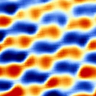
Scanning tunneling charge spectroscopy, a variant of STS, reveals a correlated electron state in “magic angle” twisted bilayer graphene characterized by alternating positive (blue) and negative (red) charge stripes. Credit: Yuhang Jiang
Andrei describes how her team’s moiré work to identify this symmetry breaking was spurred on by “the spectacular discovery in 2018 of superconductivity in TBLG and its uncanny resemblance to cuprate high-temperature superconductors”, as well as Jarillo-Herrero’s student Yuan Cao’s “invention of an ingenious “tear and stack” method” to fabricate twisted bilayer graphene samples with control. “This led us to the discovery of a correlated electron phase in which the charge carriers in the sample organize themselves into a formation with alternating stripes of positive and negative charge,” says Andrei. “This stripe phase, which appears to be a precursor of the superconducting phase, could provide an important clue to the mechanism driving the superconductivity in this system and possibly in the cuprate superconductors.” The bilayer graphene system, which comprises just carbon atoms, could potentially provide a simpler system for studies to get to the bottom of what causes high-temperature superconductivity in these cuprates.
All three papers also confirm the conclusions from the capacitance measurements that the bandwidth is larger than previously thought. “It was very nice to see different techniques converging on answers to all of this,” says Jarillo-Herrero. “These three papers are a fantastic addition to the set of experimental papers already published.”
Healthy competition
The reports come just weeks after observations of ferromagnetism in TBG, highlighting that experimenting with the twist in these systems remains a fertile breeding ground for discovering new physics. “It is a “control knob” which had no precedent in the history of materials science,” says Jarillo-Herrero. “In condensed matter systems, every time you “explore a region” which has not been explored before interesting surprises are bound to happen”.
The field has attracted the interest of a multitude of research groups, who have already reproduced and extended the initial results, which Jarillo-Herrero describes as “the best thing that can happen, as it shows that the effects are real, and that they represent the opening of a field.” This appreciation of the how the high level of competition and interest benefits the field is shared among the researchers. Yasdani adds that although competing now many of the STM groups have known and worked with each other for a long time, some having trained in his group.
The ability to perform a whole-brain MRI scan at the point-of-care for patients with mobility challenges could soon become a reality with a portable, lightweight device now under development by researchers at the Massachusetts Institute of Technology (MIT).
The low-field, helmet-like scanner could overcome large-scale financial investments and structural requirements of siting conventional, long-bore, 1.5- and 3-tesla MRI systems. What’s more, the developers believe the work-in-progress system will ultimately yield diagnostic-quality images and enable healthy outcomes for patients.
“This [device] is built on a large body of work, with innovation and a design that is focused on an appropriate size that fits around an adult’s head,” said co-developer Patrick McDaniel, a doctoral student in MIT’s electrical engineering program. “It is optimized for this application because it is a very large area of clinical MRI. That is where this particular work fits into this bigger picture of portable and lower-cost systems.”
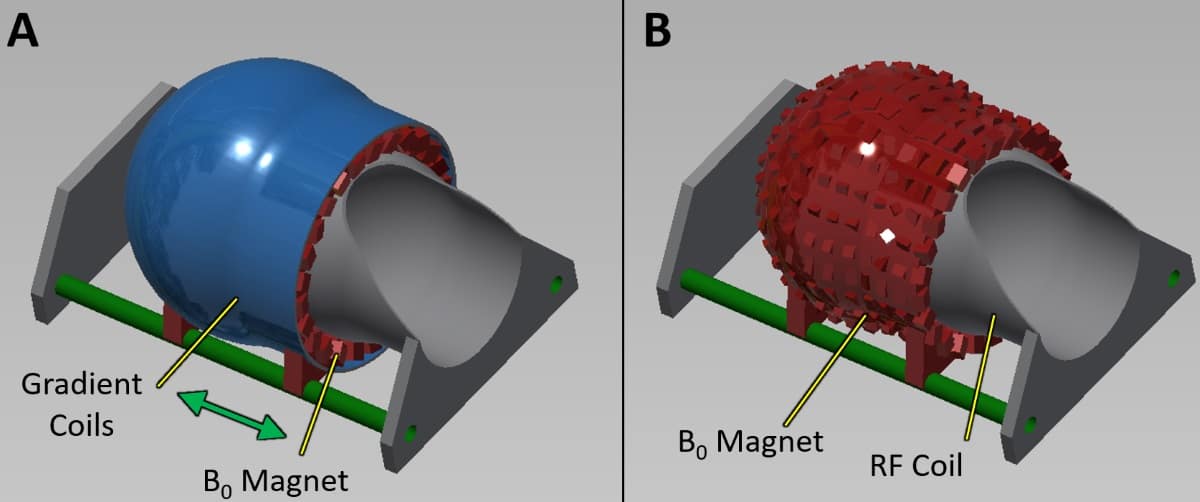
Design of a standalone, whole-brain MRI scanner and gradient assembly (A) being developed by researchers at MIT. A conceptual drawing (B) removes the gradient coils to show the head-only magnet and RF coil. (Courtesy: Patrick McDaniel and ISMRM)
MRI’s limitations
Stationary, conventional MRI scanners are not very accommodating for bedridden patients with mobility issues. To help alleviate this roadblock, researchers have long sought to develop smaller, portable devices to image extremities and other body regions. Redesigning components and reconfiguring hardware requirements can be a slow and arduous process, but the end result of a low-cost, portable system is worth the laborious effort.
MIT researchers began this project to scale down MRI technology back in 2010, led by principal investigator Larry Wald. Speaking with AuntMinnie.com at the 2019 International Society for Magnetic Resonance in Medicine (ISMRM) annual meeting in Montreal, Wald said he, McDaniel, and their MIT colleagues are attempting a “quantum leap to handheld devices” for MRI – similar to the evolution and redesign of other modalities, including ultrasound.
By 2017, the researchers developed a second iteration of their compact whole-brain magnet, complete with gradient and radiofrequency (RF) coils, as well as other components to perform scans on healthy human subjects.
The MR images produced by the second-generation device were encouraging. McDaniel compared the results to T2-weighted and turbo spin-echo images, adding that second-hand opinions from various radiologists declared the images were “pretty good.” He stopped short of making any claims regarding the diagnostic quality of the images, except to say they “internally were received very well.”
However, image artefacts did present in certain parts of the brain that the second-generation device could not rectify. The images were “squished together, stretched out or blurred,” McDaniel added. “So that was something we hope to be able to address and mitigate in this third iteration.”
Design modifications
A conventional, whole-body MRI scanner has a uniform magnetic field and gradient coils to handle MR image coding. Similarly, the current third iteration of this work-in-progress scanner is designed to have a more uniform magnetic field for image coding.
“The reason is the flexibility that we get when we do image coding with these gradient coils. It allows us to reconstruct and create better images,” McDaniel explained. “One issue that we ran into was imaging artefacts, sometimes due to the limitations of the second iteration. We hope that using this more flexible image coding approach will allow us to generate images with fewer artefacts.”
In addition, the technology’s third version is smaller and lighter than its preceding prototype and is designed to not sacrifice head imaging performance. The third-generation system is a standalone, helmet-shaped scanner that includes a magnet and coils just like the essential hardware on a conventional MRI scanner – only much smaller.
When completed, the device is expected to weigh approximately 75 pounds with an outer diameter of approximately 37 cm and length of 37 cm. The inner dimension is closer to 25 cm, which would accommodate a slightly above average-size human head. The device stays stationary, so a patient would slide into the helmet for scanning.
The magnet strength is 72 mT, which equals 0.072 T. That low-field magnet certainly is much less than current conventional MRI systems. So, what kind of images could clinicians reliably expect?
“Obviously, we would not get as strong an [MRI] signal at this field strength, and that limits the resolution we are able to image,” McDaniel said. “If we try to get 1 mm x 1 mm x 1 mm images, they would not look very good. If we can relax that resolution requirement, we can get MR images at a size of 2 mm x 2 mm x 5 mm as a reference resolution and working standard. That is what we have in mind, but there is a flexible trade-off between how noisy the images [are] and how high the resolution is.”
Currently, the researchers are still in the design and materials purchase phase of the project, but a working prototype could be available as soon as the end of this year. Assuming that target date is accomplished, McDaniel and colleagues hope to begin scanning phantoms or healthy subjects in early 2020.
How might this portable device help in the clinical setting? Based on the MR images acquired by the second-iteration magnet, the researchers could clearly see cerebrospinal fluid and ventricles in the brains of healthy subjects. That leads them to think the technology also could detect anomalies such as haematomas or hydrocephalus, a task that would not necessarily require high-resolution scans.
“We also see this [device] potentially used where it may be cost-prohibitive to install a full-size conventional MRI scanner,” McDaniel added, such as a “rural area where the economics do not support an expensive commercial system or a developing third-world setting.”