A couple of years ago, Margaret Harris – my predecessor as reviews editor at Physics World – and I promised one another that if we ever wrote a popular-physics book, we would not fall into the trap of starting the book with a chapter on Greek science before jumping to Kepler, Copernicus, Newton and finally fast-forwarding to Maxwell or Einstein. I was unsurprised to say the least when author John L Heilbron began his The History of Physics: a Very Short Introduction with a chapter titled “the Greek way”. Good then that I did read the book anyway, because I was pleasantly surprised.
The aforementioned “Greek way” states that physics in antiquity was much more philosophy and a liberal art than the rigorous experimental science we practise today. Physics was something between logic and ethics, rather than a hunt for first principles, and Heilbron refers to this as physica. “This short book describes some of the ways by which ancient physica became modern physics,” he writes in the introduction. “It does not ransack history to find items in ancient and medieval science that looks like physics, but sketches the place and the purpose of physica in the societies that supported it.”
Heilbron goes on from the Greek disciplines to the vast enterprise that was Islamic science. I particularly enjoyed reading about the House of Wisdom – a library-cum-academy set up in Baghdad in the early 9th century that quickly amassed an impressive collection of ancient manuscripts, but also sponsored scientific expeditions. The author then tackles the more well-known Western intellectuals such as Galileo, Copernicus, Kepler and Newton, before discussing the science done at the courts of the Renaissance and, finally, the scientific revolutions of the 18th century that eventually led to current-day physics.
Like most history and philosophy books, the language is formal and somewhat monotonous, and there are many names and dates to contend with, but as it is such a short book, I found it easy to get through. Pick up a copy to get to grips with the 2500-year history of physics.
The liver is one the most important organs for when it comes to assessing drug toxicity, but animal models are often very bad at predicting how humans will react to drugs or to toxic biothreat agents. Researchers at Harvard University recently succeeded in bioprinting a new liver-on-a-chip platform that offers a promising solution, as they reported recently in the journal Biofabrication. We spoke to Su Ryon Shin of Harvard Medical School about her group’s new bioreactor.
Could you tell us a little about your work and your new platform?
The liver plays a critical role in metabolizing drugs and detoxifying blood. Drug-induced hepatoxicity is one of the main reasons that drugs do not make it past clinical trials so researchers need to develop good in vitro models for evaluating hepatoxicity. Our perfusable bioreactor is an important step forward towards such a model and has proved to be excellent for evaluating drug toxicity. It can even be extended to make multiple organ-on-a-chip systems by connecting a second or even third bioreactor.
Bioprinting is an easy way to make precisely controlled 3D architectures that can be combined with miniaturized bioreactors to make next-generation organ-on-a-chip platforms. Such bioreactors contain tissue that has been engineered under continuous perfusion so that the cells being cultured receive a constant supply of nutrients and oxygen. It is easy to access tissue constructs within the structure and assess how they behave at different times during culture.
Among the various 3D cell culture models, multicellular spheroids, formed by cell aggregates, show promise. For one, the oxygen tension in the core of the spheroid is very different to that in the periphery, a situation that mimics the real-life in vivo oxygen gradient in hepatic lobules. Studying such cell spheroids instead of dispersed cells therefore allows us to better evaluate how cells respond to different drugs.
Another advantage of these spheroids is that we can encapsulate them with hydrogels that are compatible with rapid fabrication techniques, including bioprinting, which allows us to make complex architectures similar to those found in vivo. And this is exactly what we did in our platform for culturing 3D human HepG2/C3A spheroid cells.
How does your work fit in with your wider research programme?
My research team is actively involved in studying minimally invasive approaches to testing drug toxicity in general. These studies involve biomaterials research, tissue engineering, microfluidics, microfabrication, organs-on-chips, biosensors and bioprinting. Developing organs-on-chips is one of our central research areas for developing in vitro testing platforms.
Since the publication of our Biofabrication paper, my colleagues and I have also been working on developing a body-on-a-chip platform, which is a modular, scalable microfluidic system with multi-organ system integration (mimicking human organ function). We achieved this by creating resealable microfluidic devices capable of cell culture and analysis.
We have also recently developed multisensory-integrated organs-on-chips platforms for automated and continual in situ monitoring of organoid behaviour. This work was published in PNAS. Using the same technique, we are now developing bio-mimicked human organoids using patient-derived cells to further improve these systems.
How has the research community reacted to your work?
We are working with interdisciplinary scientists (clinicians and engineers, for example) from around the world and have received more than 12 National Institutes of Health (NIH) and Department of Defense (DOD) grants so far.
Especially relevant to the publication in Biofabrication is a NIH-U01 grant with Shiladity Sengupta, assistant professor at Harvard Medical School, who is developing novel therapeutic strategies for regenerative medicine.
We have also received an R01 grant with Jingyong Ye, associate professor at the University of Texas San Antonio to develop highly sensitive biosensors for real-time monitoring of cardiac organs-on-a-chip. Among our other projects is a collaboration with clinicians at the Mayo Clinic to build brain tumours-on-chips.
How is your research progressing, and what are your future plans?
Since our Biofabrication paper, we have been developing smart bioreactors in which we can control the amount of oxygen and the applied electrical signal. We have succeeded in building various types of organoids, including heart and liver, as well as breast cancer and brain tumour tissue using the bioprinting technique. We have also developed an immune-on-a-chip to evaluate nanoparticle toxicity.
Our future work will focus on developing efficient methods for accurately analysing both drug efficacy and toxicity in a personalized organ-on-a-chip system using patient-derived normal or cancer cells. This platform aims to reduce trial and error when treating real-world patients and will also improve clinical predictions of how they respond to different chemotherapeutics.
This article is one of a series of reports reviewing progress on high-impact research originally published in the IOP Publishing journal Biofabrication.
A round-up of the latest international patent applications in medical imaging.
Ultrasound offers three-in-one visualization Supersonic Imagine has devised an ultrasound technique that combines three types of images into a visualization image (WO/2018/046740). The method comprises emission and reception of sequences of temporally interleaved ultrasound waves, and a step during which the received sequences are processed to generate three images via three different process. The visualization image is then determined by combining the three images, for example by superimposing the second and third images over the first. In one aspect, the first process is B-mode ultrasound imaging, the second is elastography ultrasound imaging and the third is flow process imaging. This approach enables the user to identify the relationship between the three types of images, as well as reducing the examination time and improving diagnostic accuracy.
Radiomic features determine immunotherapy response
Researchers from Case Western Reserve University have developed apparatus that can predict a patient’s response to immunotherapy from CT images of a tissue region demonstrating non-small cell lung cancer (WO/2018/017355). In one example, the apparatus includes a series of circuits that: access the CT image of a tissue region demonstrating cancerous pathology; generate a tumoural surface boundary defining a tumour volume; generate a peri-tumoural region based on the surface boundary, and segment this region into annular bands; and extract a set of discriminative features from the tumour volume and at least one annular band. Finally, a circuit classifies the region-of-interest as a responder or a non-responder, based at least in part on the set of discriminative features.
D-Histo differentiates inflammation from solid tumours Washington University researchers have created D-Histo, a non-invasive diagnostic method for quantitatively distinguishing inflammation and injury from solid tumours (WO/2018/045066). One example utilizes diffusion-weighted MRI to provide an accurate diagnosis of prostate cancer, distinguishing it from prostatitis and enlarged prostate, which can be missed by current diagnostic methods. The D-Histo method can also provide metrics to reflect reversible versus irreversible damage in heart and central/peripheral nerves. For central and peripheral nerves, D-Histo also provides metrics to assess nerve functionality. The D-Histo biomarker(s) obtained using diffusion-weighted MRI have excellent test-retest stability, high sensitivity to disease progression and close correlation with current techniques.
Electrodes suit metal-sensitive neurological monitoring Rhythmlink has invented an electrode assembly for metal-sensitive neurological monitoring using methods such as CT and MRI (WO/2018/013751). Monitoring electrodes are employed to obtain signals from the brain. The assembly includes lead wires of at least 39-gauge copper electrical conductors, free of tin, in PVC insulation attached to electrodes. The electrodes, which may also be copper or made of metal-filled plastic, are attached to lead wires by either: crimping using copper or polymer crimps; shrink-wrap connections or ties; or radio frequency welding or heat staking. This electrode assembly reduces temperature increases during CT and MRI procedures, and minimizes the presence of artefacts in the resulting images.
Spectral CT fingerprints tissue types Philips has published details of a spectral CT fingerprinting technique (WO/2018/011321). The method includes generating spectral projection data of a subject, including at least one projection corresponding to a first energy range and one to a second energy range. The approach involves constructing an image from a combination of the spectral projection data, and constructing a set of basis images for the energy ranges. The filing also describes the construction of a multi-dimensional histogram from the set of basis images, which includes at least two axes, corresponding to each basis component. This multi-dimensional histogram includes a set of clusters, including one for each material or tissue type represented in the spectral projection data. The approach could enable identification of a number of tissues, including known normal and abnormal tissue of a particular type.
Transformable gamma camera maximizes efficiency
A transformable gamma camera is described by eV Products in patent application WO/2018/031584. The filing details how a large gamma camera can be subdivided into two or more smaller gamma cameras, each independently positioned for SPECT data acquisition. These transformable gamma cameras make more efficient use of the gamma photon detector area. Tiled arrays of semiconductor gamma detectors are especially suited for such transformation.
Researchers in Germany have developed a thermo-responsive hydrogel and loaded it with tendon stem/progenitor cells (TSPCs), developing a therapy for tendon injuries. This hydrogel allows TSPC survival, proliferation and metabolic activity, while being stable and biocompatible (Biomed. Mater.13 034107).
Tendon injury is common and can be debilitating, leading to deterioration of the patient’s life quality. Current treatments can provide some relief, but with a high recurrence of injury. Long-term solutions require the use of a tendon graft, usually taken from the patient.
Many strategies for tendon regeneration are under investigation and currently involve the use of cells and a scaffold. Two options in the choice of cells can be considered: the first is to implant tendon cells. To harvest those cells, a biopsy from a healthy part of the patient’s tendon is needed. The second option is to use stem cells, which will become tendon cells. Stem cells can be found in almost every tissue, including the tendon (TSPCs). Stem cells have the capacity to generate almost any type of cell, including cells of the bone, cartilage, tendon, brain, liver and more. Their capacity for self-renewal makes them of great interest in research.
In vivo, cells are supported physically, mechanically and biochemically by a scaffold known as the extracellular matrix (ECM). Scaffolds for therapeutic use are designed with dimensions that match the injury to repair. As a consequence, critical injuries would require big scaffolds and thus surgery for implantation. Invasive surgery can be avoided, however, by using an injectable scaffold that forms once it’s inside the body. Hydrogels are water swellable networks, which can be utilized for this purpose. Several approaches can be used for hydrogel gelation, including the use of a thermosensitive molecule; but this can be toxic for the cells, and thus for the patient.
Cell survival in hydrogel
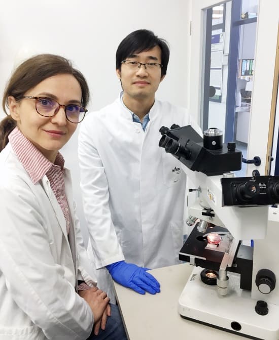
Senior author Denitsa Docheva and first author Heyong Yin.
This thermosensitive hydrogel hardens at 25°C and has shown low toxicity for TSPCs, with overall satisfying levels of TSPC survival, proliferation and metabolic activity. The hydrogel is made of butane diisocyanate (BDI) hydrogel and collagen type I. Collagen type I is the main component of the tendon ECM and is responsible for the biocompatibility of this hydrogel.
The hydrogel also demonstrated good stability. Indeed, the thermosensitive hydrogel didn’t change in shape and weight compared with a collagen type I hydrogel used as a control, making this new hydrogel more reliable.
Gene expression
The authors studied genes involved in the tendon ECM, and other tendon-related genes, as they are identifying features of the healing tendon. They observed that biomarkers for tendon were expressed. They also saw that some genes were down-regulated, but this seemed to be compensated by the up-regulation of genes with similar functions. One example is that a gene involved in crosslinking of collagen to form the hydrogel was down-regulated while an enzyme with this function was up-regulated. Understanding how and why, for similar genes, some are down-regulated and others are up-regulated is a highly interesting research subject.
The authors also observed that the thermosensitive hydrogel allowed blood vessel penetration (angiogenesis). This parameter is important as blood vessels supply nutrients, oxygen and immune cells (the immune system is also involved in the regeneration process), and also remove waste.
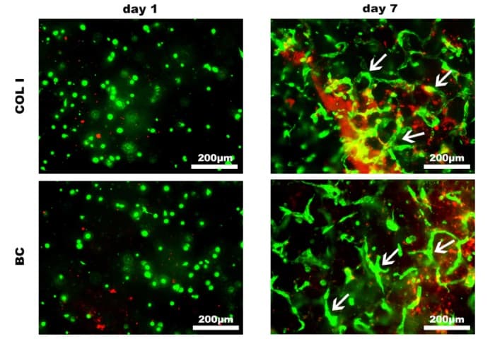
Vessel formation human umbilical vein endothelial cells.
These results are encouraging and the next steps aim to test this hydrogel scaffold in relevant in vivo models, with the aim of recognizing the therapeutic potential of this system in a clinical setting.
The US Department of Energy (DOE) has emerged as the biggest winner among agencies that fund science in the 2018 US budget. Despite not being finalized until almost six months into the financial year, which began on 1 October, the 2018 budget was passed by Congress late last week and signed by US president Donald Trump on 23 March.
In total, the US government will spend $176.8bn on R&D in the current financial year – an increase of 12.8% over last year’s figure. Funding for basic and applied research, meanwhile, will gain an extra 10.7% – its largest yearly increase since 2009.
[The budget increase] would go a long way to make sure our country remains a leader in scientific research and innovation
Illinois Democratic Representative Bill Foster
Within the DOE, the Advanced Research Projects Agency-Energy, which was created in 2009 to fund high-risk, high-reward research, will receive $353m – a 15% increase – rather than elimination as proposed in Trump’s budget request. The US contribution to the ITER fusion reactor that is currently being built in Cadarache, France, will almost double to $122m while every research programme in the department’s Office of Science will receive at least 10% more than last year’s budget. On top of that the Argonne National Laboratory’s Advanced Photon Source and Oak Ridge National Laboratory’s Spallation Neutron Source both receive sufficient funding to carry out long-planned upgrades.
‘Gratifying to see’
NASA fares equally well. Its $20.7bn budget represents $1.1bn more than in 2017. Trump’s 2018 budget request zeroed funding for the Wide-Field Infrared Survey Telescope (WFIRST) space mission, which is set to launch in the mid-2020s, as well as NASA’s Earth-science programme. In the 2018 budget, funding for WFIRST has been restored at $150m with NASA receiving $6.2bn for its science programme – a 7.9% increase. Funding for Earth science, meanwhile, remains unchanged at $1.9bn.
While the National Science Foundation only sees a 3.9% increase in its budget over 2017 it is a significant boost from the 11% decrease proposed by the Trump administration. And the Environmental Protection Agency, which was targeted by the administration for a reduction of 36% will receive $8.1bn – the same level as in 2017.
The budget has been met with relief from the US scientific community. “It was gratifying to see significant congressional support for funding that will enable greater scientific understanding, technological innovation, STEM education, US competitiveness and security, and ultimately, improvements in the human condition,” the American Physical Society commented in a statement. Illinois Democratic Representative Bill Foster, who has a PhD in physics, meanwhile, noted that the increase “would go a long way to make sure our country remains a leader in scientific research and innovation.”
Piezoresponse force microscopy (PFM) and related strain sensing atomic force microscope techniques are a good way to characterize materials at the nanoscale. However, they do suffer from unwanted artifact signals that make the cantilever vibrate more. To reduce these vibrations, researchers normally use very stiff cantilevers but this means that PFM cannot be used to probe fragile materials, such as biomaterials and ultrathin films. A new technique that makes use of higher-order contact resonance eigenmodes (or resonant vibrational modes) could allow for much softer cantilevers to now be employed.
In PFM and related techniques, an AC voltage is applied between an atomic force microscopy (AFM) probe, or cantilever, and the sample being studied. When the surface of the sample electro-mechanically deforms, this causes the AFM probe to vibrate and these vibrations can be detected and measured. Until now, measurements were made almost exclusively at or below the so-called first flexural contact resonance frequency.
Such techniques are ideal for studying piezoelectric materials, which convert electricity into mechanical force. These include ferroelectric materials, perovskite photovoltaic active layers and even some biological materials. However, these measurements are prone to a number of artifacts that can produce paradoxical results. These artifacts come from electrostatic forces between the tip and sample, and electrostatic forces between the cantilever body and sample.
Stiff cantilevers can reduce these artifacts because they vibrate less, but their downside is that they destroy fragile samples.
Out with excessively stiff cantilevers
A team led by Jason Killgore of the National Institute of Standards and Technology (NIST) is now saying that higher-order contact eigenmodes can be readily excited in PFM and that these can improve the technique’s overall sensitivity, reduce electrostatic artifacts and increase the proportion of total signal coming from wanted, as opposed to unwanted, strains. “This means that you no longer need to use excessively stiff cantilevers and their correspondingly high forces to achieve artifact-free measurements,” explains Killgore.
“AFM cantilevers can exhibit an infinite number of eigenmodes at increasingly higher frequencies,” he tells nanotechweb.org. “As we excite higher eigenmodes, we in fact increase the dynamic stiffness of the cantilever by producing a number of nodes, which act as virtual supports along the cantilever length. And by increasing the dynamic stiffness we can nearly eliminate the effect of electrostatic forces between the sample and cantilever body, which are major sources of error in today’s PFM measurements, as mentioned.”
The researchers say they are thus able to use cantilevers that are a thousand times softer than those typically employed, and with improved force control, which means a fragile sample can now be studied. “We also see enhanced sensitivity to small surface strains and can tune our measurements to preferentially sense in-plane versus out-of-plane ‘inverse’ piezoresponse with a single cantilever,” says Killgore.
More complex vibrational shapes are a problem
The NIST team tested out its technique on periodically poled lithium niobate as a test sample. This material was chosen since it is routinely used in PFM studies.
It is not all plain sailing though. The researchers say that higher eigenmodes show more complex vibrational shapes compared to sub-resonance vibrations. This makes it complicated to determine the relationship between AFM cantilever amplitude and true surface displacement. “We are working on resolving this problem,” says Killgore.
NASA has announced that it will delay the launch of the James Webb Space Telescope (JWST) to May 2020. The mission was expected to launch mid-2019 but continuing issues in testing and combining the observatory’s components have pushed it back a year.
The decision to delay the launch will likely mean that the project exceeds the $8bn budget mandated by Congress in 2011. NASA has said it will now establish an independent review board, chaired by NASA veteran Thomas Young, to investigate the impact, with the agency expected to send a revised budget to Congress in late June. “The primary issue will be getting congressional authorization or a funding measure passed in fiscal year 2019 to get relief for Webb,” says NASA spokesperson Bob Jacobs.
The [JWST] is a really complex machine and rigorous testing is required to have a high confidence of success
Thomas Zurbuchen, associate administrator for NASA’s science mission directorates
Developed by NASA along with the European and Canadian space agencies, the JWST will feature a mirror three times the diameter of the Hubble space telescope. Astronomers believe that the JWST will revolutionize their understanding of early star formation and exoplanets.
‘Failure is not an option’
The news of the delay comes after the US Government Accountability Office (GAO) released a report in late February stating that the JWST was unlikely to meet its 2019 launch date. One of the issues facing the JWST is the craft’s huge 21 x 14 m sunshield that will provide a cold and thermally stable environment for the mirror and science instruments. Deployment tests of the sunshield took much longer than anticipated and engineers discovered tears in the ultrathin fabric, which have now been fixed.
“Webb is the highest priority project for the agency’s science mission directorate, and the largest international space science project in US history,” says acting NASA Administrator Robert Lightfoot. “All the observatory’s flight hardware is now complete, however, the issues brought to light with the spacecraft element are prompting us to take the necessary steps to refocus our efforts on the completion of this ambitious and complex observatory.”
Indeed, it is crucial that there are no issues once JWST launches. Unlike Hubble, which is in orbit around the Earth, JWST will instead be placed out of reach for astronauts at Lagrange Point 2 – a gravitational balance point some 1.5 million kilometres beyond the Earth’s orbit around the Sun. “Webb is a really complex machine and rigorous testing is required to have a high confidence of success,” noted Thomas Zurbuchen, associate administrator for NASA’s science mission directorate. “We have one shot to get this into space. Failure is not an option.”
Targeted geoengineering to preserve continental ice sheets deserves serious research and investment, according to a comment in Nature.
“We understand the hesitancy to interfere with glaciers – as glaciologists, we know the pristine beauty of these places,” wrote John Moore of Beijing Normal University, China, and the University of Lapland, Finland, and colleagues. “But we have also stood on ice shelves that are now open ocean.”
The researchers propose stalling the fastest flows of ice into the oceans to buy a few centuries to deal with climate change and protect coasts. The ice sheets of Greenland and Antarctica will contribute more to sea-level rise this century than any other source, they say.
“There is going to be some sea-level rise in the 21st century, but most models say that the ice sheets won’t begin collapsing in earnest until the 22nd or 23rd centuries,” added team member Michael Wolovick of Princeton University, US, in a press release. “I believe that what happens in the 22nd or 23rd centuries matters. I want our species and our civilization to last as long as possible, and that means that we need to make plans for the long term.”
There are three potential ways to slow glaciers in these sheets: preventing warm ocean waters from reaching their bases; buttressing the ice shelves where they start to float with artificial islands in the sea; and drying glacier beds by draining or freezing the thin film of water they slide on.
“Many of the most important outlet glaciers in Greenland are about 5 km wide, and there are bridges that are longer than [that],” said Wolovick. “The important ice streams in Antarctica are wider, tens of kilometres up to 100 km, but their societal consequences are larger as well, because they could potentially trigger a runaway marine ice sheet collapse. The fast-flowing parts of the ice sheets – the outlet glaciers and ice streams – might be the highest-leverage points in the whole climate system.”
Engineers have already constructed artificial islands and drained water beneath a glacier in Norway to feed a hydropower plant, according to the press release. Raising a berm in front of the fastest-flowing glacier in Greenland – constructing an underwater wall 3 miles (4.8 km) long and 350 feet (107 m) high in Arctic waters – would be a comparable challenge, and could cost billions of dollars
Without coastal protection, the global cost of damages from sea-level rise could reach $50 trillion a year, the team wrote. And the sea walls and flood defences necessary to prevent those damages would cost tens of billions of dollars a year to build and maintain. Glacial geoengineering has potential risks, especially to local ecosystems, and the glaciers and their outflow channels need to be more precisely mapped and modelled, according to the team. What’s more, glacial geoengineering is not a substitute for climate mitigation through emissions reductions.
“Glacial geoengineering will not be able to save the ice sheets in the long run if the climate continues to warm,” Wolovick said. “In the long run, there are two possible routes that glacial geoengineering could take: on the one hand, it could be a stopgap solution meant to preserve the ice sheets until the climate cools enough that they are once again viable on their own; on the other hand, it could be a managed collapse meant to keep the rate of sea-level rise down while slowly letting the ice sheet waste away.”
At a symposium on nuclear medicine organized by the Association of Imaging Producers & Equipment Suppliers (AIPES), doctors, professors and other specialists argued passionately that the latest imaging techniques used to diagnose and treat cancers are already saving lives. The AIPES event was heavy on historic symbolism: it took place at the art nouveau Solvay Library, site of physics conferences for more than a century, attracting the likes of Albert Einstein, Marie Curie, Max Planck, Robert Oppenheimer and Ernest Rutherford.
For Johannes Czernin from the David Geffen School of Medicine at UCLA, the reluctance to embrace nuclear medicine was a moral failure. He lambasted medical authorities who drag their feet on PET imaging, calling it “a healthcare scandal” that it is so underused. “It is time to get angry – not using and not approving PET under many conditions should be considered malpractice,” he said.
He accused authorities of subscribing to “an evidence cult”, setting an impossible burden of proof for new imaging technologies. “But the evidence is absolutely overwhelming,” he said. “Let’s use common sense. It is not worthwhile policing everything, repeating everything country-by-country, and collecting evidence that takes years.”
Slow progress
Although some $1.6 billion has been devoted to the “war on cancer” since it was announced in 1971 by President Richard Nixon, it has not yet been won. “Not much has changed since then. Survival rates are not that different. We don’t detect it early enough,” Czernin said, as he called for complete deregulation of diagnostics. “It’s safe, you can inspect it at any time. If there is a problem, you just shut it down,” he said. “Radiation exposure is nothing – it is essentially homeopathic.”
Einat Even-Sapir Weizer
If the take-up is slow in Europe and the US, Einat Even-Sapir Weizer from the Tel Aviv Sourasky Medical Centre offered a model for rolling out new imaging techniques more quickly. She explained that Israel has led the adoption of novel PET imaging methodologies – such as Ga-PSMA for the early detection of prostate cancer – through a flexible and practical approvals mechanism.
Even-Sapir Weizer described Israel’s basket system for reimbursing the cost of drugs technologies and services, which is regulated by the country’s parliament. Every year the members of parliament decide which licences to give out, which requires them to examine the technology; assess the added value and the expected change in patient management; study the epidemiological data of the disease in Israel; and look at the budgetary impact. As a result, Israel has been quick to introduce technologies like DOTATATE PET-CT and Ga-PSMA, while fluoride is set to be added in the next basket as a tracer.
But even Israel faces barriers to nuclear medicine, Even-Sapir Weizer said. The country needs to build new cyclotrons, draft regulations on tracers, ensure that enough nuclear medicine physicians are trained in imagery. It will also be important to define precise clinical scenarios where the new technologies have a potential impact on patient management and outcome, and to predict the overall impact of any new technology on patients and the healthcare system.
Marcus Hacker, of the Medical University of Vienna, Austria – one of Europe’s leading nuclear cardiology specialists – predicted that the rise of nuclear medicine would not only provide safer and more efficient tumour treatments, but will also help doctors take tricky decisions on which patients to prioritize. Hacker believes that it would enable eventual treatment decisions to be based on more informed criteria.
“These technologies are available and increasingly cheap,” he said, pointing to the “complementary and incremental value” of combining molecular diagnostics and molecular imaging. There is also an evolution in strategies, as medicine enters a personalized approach, Hacker said, with treatments increasingly customized for individual patients. “We used to pour toxic drugs into the body to destroy any proliferating cells. Now we can be very targeted with our drugs,” he said. “Nuclear medicine has a high potential to link target identification with therapy and thus personalize treatment.”
Cost concerns
James Williams, CEO of Siemens Healthcare Molecular Imaging, argued that healthcare authorities often look at costs from a purely financial perspective. “Why do we beat ourselves up on costs? Accountants looking at a spreadsheet may view PET as the nail that sticks out – but that is the wrong way to look at it,” he said. “Ultimately, this is about giving people more time to live better. We can sometimes cure them.”
He said imaging would soon consign methods like deep biopsies to the history books, like “exploratory surgery” a generation ago. “With a combination of in vitro testing and in vivo imaging, will deep biopsy become as rare as exploratory surgery?” he asked. But Williams also warned of an impending resource threat as PET systems rely on “the rarest of the rare earth elements”, lutetium, which is almost entirely controlled by China.
Wim Oyen, professor of nuclear medicine at The Institute of Cancer Research (ICR) in London and the incoming president of the European Association of Nuclear Medicine (EANM), said the imaging community has a duty to reach out to stakeholders in the sector, including regulators and patient groups. “One of our main drivers is patient interest,” he said. “We should stop bickering with regulators, or else it will take way too long for our patients.” Oyen added out that, while nuclear medicine offered brilliant prospects, it still needed to cut its prices. “We need competitive pricing to get ahead. We cannot shoot ourselves in the feet by pricing too high,” he said.
The development of a total-body PET scanner could revolutionize PET imaging, both for clinical applications and medical research. The EXPLORER project, a multi-institutional consortium, has now reported the successful performance of the mini-EXPLORER, a total-body, long axial field-of-view (FOV) PET scanner for animal imaging (J. Nucl. Med. doi: 10.2967/jnumed.117.200519).
The EXPLORER project was officially launched in October 2015 with a five-year $15.5m Transformative Research Award from the US National Institutes of Health (NIH) to researchers at UC Davis Biomedical Engineering. The award is funding the development of the world’s first high-sensitivity total-body PET scanner that can image the entire human body simultaneously. This capability could increase image sensitivity by a factor of 40, or reduce radiation dose by a factor of 40, compared with current clinical PET scanners. It could also decrease whole-body scanning time from 10-20 minutes to 15-30 seconds. A 40-fold increase in dynamic range could extend the imaging of radiotracers by more than five half-lives.
The UC Davis research team, jointly led by principal investigators Simon Cherry and Ramsey Badawi, has been conducting research on the development of a total-body PET scanner since 2005. Initial work involved optimizing the system design with computer simulations and better understanding its expected performance.
System performance
The mini-EXPLORER, now in use at the California National Primate Research Center, is a precursor to and a small-scale version of the human EXPLORER. The scanner was built using detectors and electronics from a clinical system (a Biograph mCT). It has 192 detectors arranged into eight rings for a ring diameter of 43.5 cm and an axial length of 45.7 cm.
The researchers used NEMA NU-2 and NU-4 phantoms to measure sensitivity and count rate performance. They investigated the reconstructed spatial resolution by imaging a radially stepped point source and a Derenzo phantom. They compared the performance of maximum acceptance angles between 14° and 46° to determine the effect of the wide acceptance angle.
The authors reported that the NU-2 total sensitivity was 5.0% and the peak noise-equivalent count rate measured with the NU-4 monkey scatter phantoms was 1741 kcps with the maximum 46° acceptance angle. The NU-4 scatter fraction was 16.5%, and the reconstructed spatial resolution was about 3.0 mm at the centre of the FOV. Both the scatter fraction and the spatial resolution showed little dependence on the maximum acceptance angle used (from 14° to 46°).
In vivo study
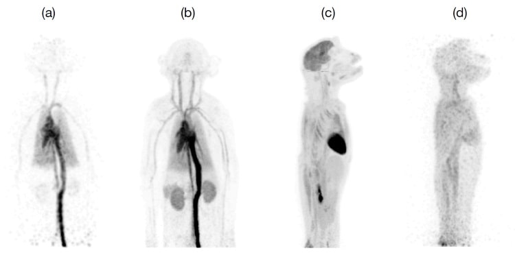
The team performed an 18F-FDG dynamic imaging study of a juvenile rhesus monkey with a single bed position. They verified that a wide acceptance angle can be used with a long axial FOV scanner to produce images with high sensitivity and overall excellent image quality. Early FDG distribution in major blood vessels and organs were visualized in one second frames shortly after injection. Reasonable image quality was obtained in a 40 minute scan at 18 hours post-injection, demonstrating the wide dynamic range of the mini-EXPLORER system and the ability to image very low activity concentrations.
Images from the FDG rhesus monkey study
Lead author Eric Berg, a postdoctoral researcher who received the College of Engineering’s award in 2017 for best doctoral dissertation on the development of the scanner, told medicalphysicsweb that the mini-EXPLORER was developed both to test some of the team’s hypotheses and to develop applications in non-human primates. He said that it is enabling the team to assess and study some of the effects of its much longer cylindrical geometry, to develop data correction and reconstruction algorithms, and to understand some of the features in the data expected to be encountered in the human EXPLORER scanner.
“The mini-EXPLORER is also allowing the team to develop applications in a relevant animal model that will ultimately be used in the human scanner,” Berg said. “These include studies of infection, inflammation and drug pharmacokinetics. We recently completed a study that demonstrated the distribution of 89Zr-labelled antibodies out to 30 days post-injection. We believe that this may be the longest time that a PET radiotracer has been successfully imaged after injection.”
Berg explained that the human EXPLORER scanner uses newer silicon photomultiplier-based detectors, as well as smaller scintillation crystals to achieve better spatial resolution. It will be much larger, with about 10 times more detectors and channels of electronics, with much greater, more sophisticated computing power and software for data acquisition and processing. The human scanner will also have an integrated CT for PET/CT studies.
The researchers are ahead of schedule to complete the human prototype scanner. “The underlying detector and electronics technology has been developed and tested, and it is operating at the required specifications,” said Cherry. “It has been incorporated in another small-scale prototype, the mini-EXPLORER II, which is being tested at the School of Veterinary Medicine. The first dog study was recently completed, and we have a good level of confidence in the scanner hardware. Remaining unknowns relate to whether the large volumes of data can be collected, sorted and reconstructed without problems. We also need to develop accurate data corrections for the many effects that can cause PET data to lose quantitative information if they are not properly handled.”