
One of the highlights in the Physics World calendar is the annual announcement of our Breakthrough of the Year. It’s always a tricky task to choose a winner, and it’s even harder this year as we have so many more articles to choose from. That’s because myself and two other new editors joined the Physics World team earlier this year, expanding the site’s coverage in three key research fields: medical physics and the biosciences; environment and energy; and materials science and technology.
To reflect this expanded scope in the 2018 award, each us will select our own top five shortlists from the research we have covered in 2018, based on three criteria:
- Significant advance in knowledge or understanding
- Importance of work for scientific progress and/or development of real-world applications
- Of general interest to Physics World readers
After all the shortlists have been published, we will get together to decide which of the breakthroughs will make it into the Top 10 – and which will be the overall winner. The final announcement of the Physics World 2018 Breakthrough of the Year will be made on Thursday 13 December.
In no particular order, here is my top five shortlist from the medical physics and biosciences sections:
EXPLORER PET/CT produces first total-body scans
The EXPLORER PET/CT scanner – the world’s first medical imaging system that can capture a 3D image of the entire human body simultaneously – has produced its first human images. Developed by UC Davis scientists and a multi-institutional consortium, EXPLORER can scan up to 40 times faster, or use up to 40 times less radiation dose, than current PET systems, making it possible to conduct repeated studies in an individual, or dramatically reduce dose in paediatric studies. The high-sensitivity scanner can also create movies that track radiolabelled drugs as they move around the body.
Artificial intelligence predicts cancer evolution
The ever-changing nature of tumours is a major challenge when treating cancer. If doctors could predict how a tumour will evolve, however, they could alter the treatment before the tumour has a chance to adapt and develop resistance. A team led by scientists at the Institute of Cancer Research and the University of Edinburgh has used artificial intelligence to predict how cancers will progress and evolve. They developed a technique called REVOLVER (repeated evolution of cancer), which picks out patterns in DNA mutation within cancers and uses this information to forecast future genetic changes.
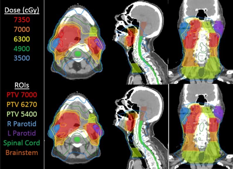
Compensator expands global access to advanced radiotherapy
Intensity-modulated radiotherapy (IMRT) is a precision treatment technique that uses complex multileaf collimators (MLCs) to shape the photon beam and spare more healthy tissue. But while IMRT is available in essentially all radiotherapy clinics in high-income countries, it is largely absent in vast regions of low- and middle-income countries. To address this shortfall, a team headed up at the University of Washington Medical Center developed a cost-effective alternative to the MLC, replacing it with a ring of compensators made from lightweight plastic moulds filled with attenuating beads such as tungsten beads. The proposed device can be retrofitted to existing linac and cobalt teletherapy units – allowing clinics to add IMRT without having to purchase a new treatment system.
Precise proton range detector gets ready for the clinic
The accuracy of proton therapy is limited by uncertainty in the beam range and, currently, a margin around the tumour is irradiated to ensure tumour coverage. To reduce the need for this margin, a team at MGH and Harvard Medical School is developing a proton range detector based on real-time detection of the prompt gamma rays produced when protons interact with tissue in the patient. Tests on phantoms showed the detector could predict each proton pencil-beam spot with a mean precision of 1.1 mm. The team is now working to integrate their detector into the clinical workflow, with a long-term goal of providing real-time feedback during proton therapy.
Activating retinal stem cells restores vision in mice
An international research collaboration has successfully restored vision in mice by activating retinal stem cells, a feat that has never been achieved before. The approach could one day transform treatment for patients with retinal degenerative diseases, which currently have no cure. To achieve this, the team performed a two-step gene transfer to reprogramme Müller glia cells in blind mice. Between four and six weeks after the reprogramming, the blind mice could sense light and regained their vision. The researchers note that further tests are needed to determine the degree of sight improvement.
- Take a look at the shortlists from Hamish Johnston and Anna Demming. And check back on Monday for Liz Kalaugher‘s top five breakthroughs in environment and energy.


